
Scoliosis: Physiotherapy Treatment
Table of Contents
Scoliosis (curvature of the spine)
Scoliosis is an abnormal curvature of the spine. In scoliosis, the spine curves to the side when viewed from the front, and each involved vertebra also twists on the next one in a corkscrew fashion. This twisting is known as rotoscoliosis. This may cause one shoulder to be higher than the other or one side of the ribcage or lower back to be more prominent (humpback).
The scoliosis spinal curve is usually “S”- or “C”-shaped over three dimensions.
If there is both a sidewise curvature and increase in the outward curvature of the upper back, this condition is called kyphoscoliosis. It is more common in children with neuromuscular diseases. A curve with a right-sided prominence is called dextroscoliosis, and a left-sided prominence is levoscoliosis. Generally, children with idiopathic (of unknown cause) scoliosis have two sidewise curves in opposite directions, but these may not be of the same size or severity.
ANATOMY OF SPINE:
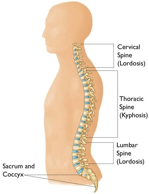
The normal anatomy of the spine is usually described by dividing up the spine into three major sections: the cervical, the thoracic, and the lumbar spine. (Below the lumbar spine is a bone called the sacrum, which is part of the pelvis). Each section is made up of individual bones, called vertebrae. There are 7 cervical vertebrae, 12 thoracic vertebrae, and 5 lumbar vertebrae.
An individual vertebra is made up of several parts. The body of the vertebra is the primary area of weight bearing and provides a resting place for the fibrous discs which separate each of the vertebrae. The lamina covers the spinal canal, which is the large hole in the center of the vertebra through which the spinal nerves pass. The spinous process is the bone you can feel when running your hands down your back.
The paired transverse processes are oriented 90 degrees to the spinous process and provide attachment for back muscles. There are four facet joints associated with each vertebra. A pair that face upward and another pair that face downward. These interlock with the adjacent vertebrae and provide stability to the spine. The vertebrae are separated by intervertebral discs, which act as cushions between the bones.
Each disc is made up of two parts. The hard, tough outer layer, called the annulus, surrounds a mushy, moist center, called the nucleus. When a disc herniates or ruptures, the soft nucleus spurts out through a tear in the annulus and can compress a nerve root.
The nucleus can squirt out on either side of the disc, or in some cases, both sides. The amount of pain associated with a disc rupture often depends upon the amount of nucleus that breaks through the annulus and whether it compresses a nerve. To help alleviate the pain, a laminotomy/microdiscectomy may be performed.
Type of scoliosis:
Idiopathic (cause unknown) scoliosis – this is the most common type of scoliosis, accounting for about eight cases out of every 10.
Infantile idiopathic scoliosis – usually develops before the age of three years. Boys are more commonly affected.
Juvenile idiopathic scoliosis – usually develops between three and 10 years of age. Girls are more commonly affected.
Adolescent idiopathic scoliosis – most cases of scoliosis tend to develop prior to adolescence and during the growth spurt. Girls are
more commonly affected.
Adult scoliosis – scoliosis is uncommon in adults. Adult scoliosis is sometimes caused by a degenerative joint problem. In most cases,
the scoliosis started in childhood but was not diagnosed until later in life.
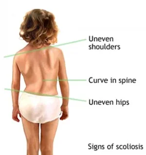
Signs and Symptoms:
Following signs and symptoms are seen in scoliosis back.
- Uneven shoulders
- One shoulder blade that appears more prominent than the other
- Uneven waist
- One hip higher than the other
- The head is not centered directly above the pelvis
- Changes in the look or texture of skin overlying the spine
- Leaning of the entire body to one side
- Rib prominence when bent over
- Back Pain
Causes of Scoliosis:
Nonstructural (functional): This type of scoliosis is a temporary condition when the spine is otherwise normal. The curvature occurs as the result of another problem (from one leg being shorter than another, muscle spasms due to a soft tissue injury, ruptured disc, or abdominal problems, such as appendicitis).
Structural: In this type of scoliosis, the spine is not normal. This may be due to abnormally shaped vertebrae or neuromuscular diseases. About 30% of children with idiopathic scoliosis have a family history of the condition, but the exact hereditary (genetic) association is not known at this time.
Pathology-related scoliosis can arise in people with a neuromuscular disease such as muscular dystrophy or in response to a severe injury to the spinal cord such as quadriplegia.
DIAGNOSIS:
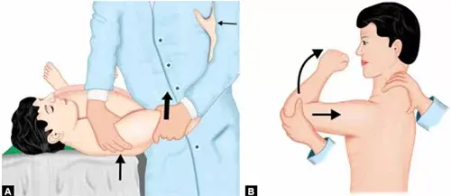
Forward Bend Test:
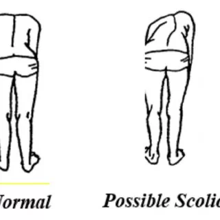
This test involves a healthcare professional observing the patient bending forward at the waist 90 degrees with arms stretched toward the floor and knees straight. From this position, most scoliosis signs that present as asymmetry are clearly visible in the spine and/or trunk of the body, such as :
- one shoulder or shoulder blade is higher than the other
- Rib cage appears higher on one side (also called a rib hump)
- One hip appears higher or more prominent than the other
- The waist appears uneven
- The body tilts to one side
- One leg may appear shorter than the other
Scoliometer to Measure Spine Rotation:
The scoliometer will be placed over the area of possible scoliosis to give the doctor a “reading”—a rough estimate of your spine’s curvature.
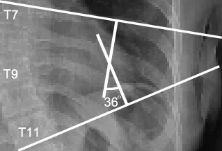
Cobb Angle Measurement:
The lateral curve of scoliosis is described by the Cobb angle. Using an X-ray of the full spine, the Cobb angle is found by drawing a perpendicular line from the spine’s most-tilted vertebra above the sideways curve’s apex and a second perpendicular line from the
most-tilted vertebra beneath the apex. The angle formed where those two lines meet is the Cobb angle.
A Cobb angle of at least 10 degrees is typically considered the borderline for a scoliosis diagnosis.
Scoliosis Treatment:
The doctor will recommend following up every 4 to 6 months to monitor the curve of the spine in the clinic and periodically with X-rays.
The following factors will be considered by the doctor when deciding on treatment options :
Sex: Females are more likely than males to have scoliosis that gradually gets worse.
The severity of the curve: The larger the curve, the greater the risk of it worsening over time. S-shaped curves, also called “double curves,” tend to worsen over time. C-shaped curves are less likely to worsen.
Curve position: A curve that is located in the center part of the spine is more likely to get worse compared with curves in the lower or upper section.
Bone maturity: The risk of worsening is lower if the person’s bones have stopped growing. Braces are more effective while bones are still growing.
Casting:
Casting instead of bracing is sometimes used for infantile scoliosis to help the infant’s spine to go back to its normal position as it grows. This can be done with a cast made of plaster of Paris.
The cast is attached to the outside of the patient’s body and will be worn at all times. Because the infant is growing rapidly, the cast is changed regularly.
Scoliosis Braces:
Thoracolumbosacral orthosis (TLSO) –
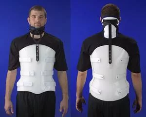
The TLSO is made of plastic and designed to fit neatly around the body’s curves. It is not usually visible under clothing.
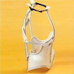
Milwaukee brace –
this is a full-torso brace and has a neck ring with rests for the chin and the back of the head. This type of brace is only used when the TLSO is not possible or not effective
Scoliosis surgery:
If Non-surgical treatment and Physiotherapy treatment are not beneficial than Surgical options are recommended.
Spinal fusion:
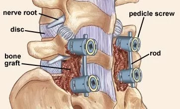
In a spinal fusion, the curved vertebrae are fused together so that they heal into a single, solid bone. This will stop growth completely in the abnormal segment of the spine and prevent the curve from getting worse.
All spinal fusions use some type of bone material, called a bone graft, to help promote the fusion. Generally, small pieces of bone are placed into the spaces between the vertebrae to be fused. The bone grows together — similar to when a broken bone heals.
Metal rods are typically used to hold the spine in place until fusion happens. The rods are attached to the spine by screws, hooks, and/or wires.
Exactly how much of the spine is fused depends upon your curve(s).
Bone graft surgery:
A bone graft is primarily used to stimulate bone healing. It increases bone production and helps the vertebrae heal together into a solid bone. In the past, a bone graft harvested from the patient’s hip was the only option for fusing the vertebrae. This type of graft is called an autograft. Harvesting a bone graft may require an additional incision during the operation. It increases the length of surgery and can cause increased pain after the operation because of an additional region of the pelvis being included in the procedure.
One alternative to harvesting a bone graft is an allograft, which is cadaver bone. An allograft is typically acquired through a bone bank.
Today, several artificial bone graft materials have also been developed.
PHYSIOTHERAPY TREATMENT:
Physiotherapy treatments are symptomatic pain reliever modalities like IFT (Interferential Therapy), TENS, and Hot Pack/Cold Pack are useful to relieve pain.
Stretching exercise of Tight muscle around Back muscles and Strengthening exercise of weak muscle as per assessment.
Exercise for scoliosis
Arm/leg raise exercise:
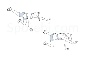
Get on your hands and knees. Keep your spine straight, with your hands directly below your shoulders and your knees aligned directly under your hips. Reach out with an arm and keep it straight and level. At the same time, extend the leg on your opposite side, keeping it straight and level. Hold for a few deep breaths, then gently lower your arm and leg to the starting position.
Repeat this exercise with your other arm/leg. Try for 10 to 15 repetitions on each side.
Pelvic tilt exercise:
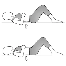
Lie on your back. Bend your knees so both feet are flat on the surface with toes pointed forward.
Pull your belly button in so your pelvis pushes toward the ceiling and your back flattens against the ground.
Hold this position for 20 seconds, and then relax. Try to do this exercise 10 times.
Cat/cow exercise:
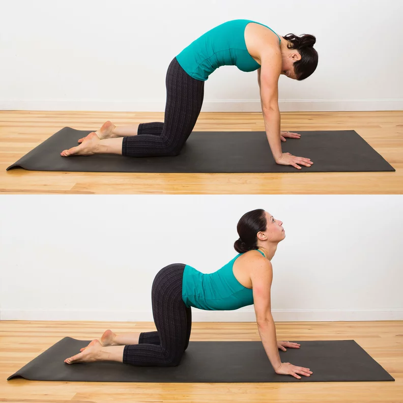
Start on your hands and knees. Align your arms straight under your shoulders and your knees under your hips. Look at the floor, keeping your head straight in line with your torso and spine. Round your back, lifting your spine toward the ceiling.
Your eyes should face your belly. Hold for a deep breath. Slowly lift your chest and tailbone toward the ceiling, letting your stomach sink toward the ground.
Your eyes will look up toward the ceiling. After another breath, gently round your back and lift your spine toward the ceiling again. Alternate between the poses.
Latissimus muscle stretching exercise:
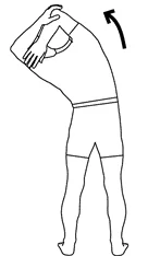
Stand with your feet shoulder-width apart, slightly bent at the knees. Reach overhead and grab your left wrist with your right hand.
Bend at your right side until you feel a stretch along your left trunk. Put most of your body weight on your right leg.
Hold for 5 to 10 seconds, then return to the starting position by pushing from your right foot.
Try this exercise on the opposite side.
Scoliosis Yoga – Tree Pose:
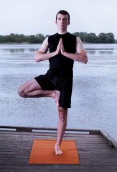
Stand straight with your feet shoulder-width apart.
Support your body on your right foot and flex your left knee. Grab your left knee with both your palms.
Now, put your left foot flat on your right inner thigh.
Join your palms together and put your hands up.
Hold this pose for 30 seconds to 1 minute.
Do this on the other side.
Repeat this 5 times.
Hip Bridging exercise:
lie supine on the floor with arms at the sides, knees bent, and feet flat on the ground.
Contract the abdominal and gluteal muscles and lift the pelvis so that the body maintains a straight line from the shoulders to the knees.
Hold this position for the recommended time.
Ball stretching exercise:
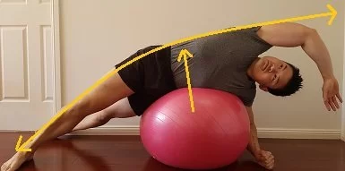
Situate yourself in a kneeling position on a workout mat. Rest a large exercise ball against your hip on the convex side of your curvature. Lean sideways over the ball until your side is resting against the ball between the hip and the bottom of the rib cage.
Balance yourself with both feet and your lower hand, while stretching your upper hand out to deepen the stretch.
Hold this position for 20-30 seconds, and do 2-3 reps. This exercise can be done daily.
Foam roller towel stretch:
Wrap a towel around a foam roller and lay it width-wise across the exercise mat. Lay across the foam roller so that it is perpendicular to your body. It should rest in the space between your hip and the bottom of your rib cage. Your top leg should be straight and your bottom leg bent at the knee behind you. Stretch your upper arm out until your hand touches the floor.
Try to hold the pose for 20-30 seconds, and do 2-3 reps. You can do this exercise daily.
Hamstring Stretching Exercise (Wall):
Start by lying on your back with your buttocks up against a wall or high-back chair. Place the foot against the wall or chair and make
the knee as straight as you can. As you progress, you’ll be able to get closer to your toes in the seated position or your knee
straighter while on the floor.
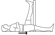
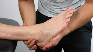
Hip Flexor Stretching Exercise :
kneeling on the floor. Holding onto a chair or other solid object, place one leg behind you and lean in slightly into the chair.
Then, lying on your back with knees bent and feet flat on the floor hip-width apart, flatten your back to the floor and exhale while raising the hips off the floor.
Tighten your glutes when you reach the top. Inhale and return to the starting position.
DO’S and DON’TS:
- Do Change Positions Frequently
- Do Use a Quality Mattress
- Don’t Carry Heavy Things
- Don’t Play Football
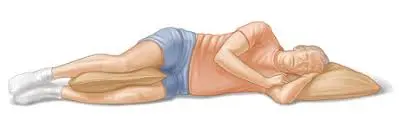
- Don’t Sleep on Your Stomach
- Don’t Run Long Distances on Hard Surfaces
FAQ
To fix scoliosis naturally:
Every morning, stretch. Daily stretching can help correct some of the imbalances caused by scoliosis and enhance spinal health.
Maintain Warm Joints.
Consume a diet low in inflammation.
Use vitamins as a supplement.
Use a Firm Mattress to Sleep.
Physical Therapy to Start Treating Scoliosis.
Scoliosis symptoms
Shoulder blades are not uniform.
There is a greater prominence or visibility of one shoulder blade than the other in the upper back.
One hip is higher than the other.
When the rib cage bends forward, one side is higher than the other.
The ribcage may press upon the heart and lungs in really severe cases of scoliosis, which can impair breathing and make it harder for the heart to circulate blood throughout the body. Additionally, this can raise the risk of lung infections like pneumonia and result in heart failure.
No, scoliosis cannot be completely cured, but treatment options such as bracing, physical therapy, and in some cases, surgery, can help manage and reduce the progression of the condition.


22 Comments