
Common Carotid Artery
The common carotid artery is a vital blood vessel in the human body, responsible for supplying oxygen-rich blood to the head, neck, and brain. It’s one of the major arteries of the neck, originating from the aortic arch in the chest.
Table of Contents
Introduction
The major blood supply to the head and neck region comes from the common carotid artery, a sizable elastic artery. Each side of the body has a single common carotid artery, and these arteries have different origins. The right common carotid artery emerges from the brachiocephalic trunk posterior to the right sternoclavicular joint, whereas the left common carotid artery originates from the aortic arch within the superior mediastinum.
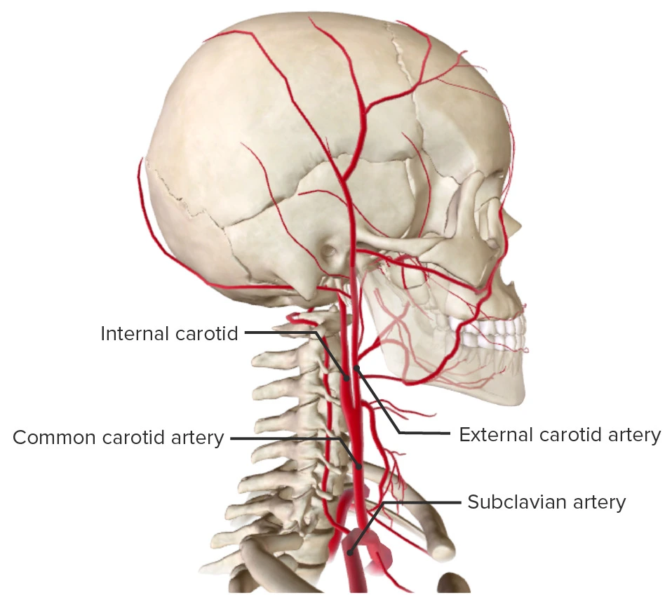
Blood is supplied to the head, face, and neck by the paired, branchless common carotid arteries (CCA). Internal and external carotid arteries split off from each common carotid.
The large, elastic common carotid artery supplies the majority of the blood supply to the head and neck. The carotid arteries are the principal blood vessels that supply blood to the face and brain.
From the brachiocephalic artery in the neck, the right common carotid artery (RCCA) emerges.
The aortic arch gives birth to the left common carotid artery (LCCA) in the thorax.
At the level of the carotid sinus, the right and left common carotid arteries split into the internal carotid artery (ICA), which nourishes the brain, and the external carotid artery (ECA), which supplies to the neck and face.
The carotid arteries are important to brain function because they facilitate blood flow to the brain and other parts of the head.
The arteries are essential for carrying blood enriched with oxygen from the heart to other regions of the body.
Anatomy of Common Carotid Artery
The body’s main artery, the aorta, from which the carotid artery extends, is responsible for carrying out blood from the heart.
Blood is transported to the brain via the carotid arteries, which go up the neck. The left and right carotid arteries are the two that exist.
Each of them divides into an external carotid artery and an internal carotid artery in the neck.
The area in the neck just under the jaw where a person feels their pulse is known as the branching carotid arteries.
The carotid arteries are divided into eight more important divisions. Blood is transported to various areas of the head and face using these divisions.
The common carotid arteries are located on the left and right sides of the body. These arteries have symmetrical paths but are descended from distinct arteries. The brachiocephalic trunk gives rise to the right common carotid and the aortic arch in the thorax to the left.
The brachiocephalic trunk, an upper branch of that artery that serves the right arm, head, and neck, is where the right originates. At the upper edge of the thyroid cartilage, roughly at the level of the fourth cervical vertebra, they are divided into the external and internal carotid arteries.
There are two components to the left common carotid artery: the cervical (in the neck) and thoracic (in the chest) segments.
The only artery with a thoracic section—which corresponds to the upper spine, below the neck—is the left carotid artery, which emerges straight from the aortic arch. This segment passes through the thoracic cavity’s superior mediastinum, or the area encircled by the ribs, to the sternoclavicular joint, which is the point at the top of the ribcage where the clavicle and sternum meet.
According to anatomy, the external and internal carotid arteries are formed by the division of the left and right common carotid arteries (carotids), which provide oxygenated blood to the head and neck.
The right common carotid arises in or close to the neck and contains only a small thoracic portion. Studies that have examined the geometric structure of the common carotid artery from both qualitative and quantitative (mathematical) perspectives may be found in the bioengineering literature.
Adult males and females have typical diameters of 6.5 mm and 6.1 mm for the common carotids, respectively.
Stroke can result from clots or narrowing of this blood vessel, a disease known as carotid artery stenosis. Moreover, a carotid aneurysm—a ballooning of a weak part of the vessel—can cause significant bleeding that could be fatal.
Course of Common Carotid Artery
The carotid arteries begin beyond the sternoclavicular joints and are enclosed in the carotid sheath in the neck, which is located behind the sternocleidomastoid muscle.
The common carotid arteries on the left and right have different origins, even though they travel the same path through the neck.
The origin of the CCA is located in the brachiocephalic trunk on the right, while it originates directly from the aortic arch on the left. The thoracic and cervical regions of the left CCA are two separate anatomical regions. Given that the proper CCA develops cranially, it only has a cervical portion.
The left CCA ascends through the superior mediastinum in the thoracic section to the level of the left sternoclavicular joint, which joins the cervical portion at that point.
Both CCAs’ cervical sections progress comparably. From behind the sternoclavicular joint, each vessel ascends obliquely to the level of the thyroid cartilage’s top border, or roughly at C4 level. The trachea divides the two CCAs in the lower neck from one another. The thyroid gland, the larynx, and the throat, however, cause the carotids to diverge as they travel up the neck.
The carotid sheath, which is made up of the deep cervical fascia’s three layers, houses the CCA. The internal jugular vein and vagus nerve are also located within the carotid sheath; the vein is situated lateral to the artery, and the nerve is situated between the two.
The common carotid arteries split into the ECA and ICA at the site of the upper border of the thyroid cartilage, which is usually at the level of the fourth or fifth cervical vertebra.
The “carotid body,” a chemoreceptor, and the “carotid sinus,” a baroreceptor, are located at this bifurcation point, which makes it clinically significant. The brain receives signals from the carotid body chemoreceptor, which alters the respiratory rate in response to changes in blood pH, PCO2, and PO2. Blood pressure is sensed and maintained by the carotid sinus baroreceptors, which react to variations in the blood vessel’s stretch.
Following division, the ICA stays in the carotid sheath to enter the carotid canal inside the temporal bone, while the ECA leaves the sheath to supply oxygenated blood to the face and neck.
Within the chest
The only artery that is significantly present in the thorax is the left common carotid. It starts at the aortic arch directly and ascends to the left sternoclavicular joint level through the superior mediastinum.
The left common carotid artery is connected to the following structures in the thoracic portion of its course: The sternohyoid and sternothyroid muscles, the front regions of the left pleura and lung, the left brachiocephalic vein, and the remnants of the thymus divide it from the sternum’s manubrium in front; behind, it rests on the trachea, oesophagus, left recurrent laryngeal nerve and thoracic duct.
Its left side contains the lung, left pleura, left vagus, and phrenic nerves; below it are the brachiocephalic trunk, and above are the trachea, inferior thyroid veins, and the remnants of the thymus. It is posterior to and slightly lateral to the left subclavian artery.
Around the neck
Arteries of the neck: The right internal carotid artery and right external carotid artery split off from the right common carotid artery. One description will cover both of the cervical parts of the common carotids because they are so similar to one another.
From behind the sternoclavicular joint to the level of the thyroid cartilage’s top border, each artery ascends obliquely before dividing.
The trachea lies in a tiny space that divides the two common carotid arteries at the lower neck; at the upper part of the neck, the thyroid gland, the larynx, and the pharynx separate the two arteries.
The internal jugular vein, vagus nerve, vein lateral to the artery, and nerve between the artery and vein on a plane posterior to both are all enclosed within the carotid sheath, which is derived from the deep cervical fascia and contains the common carotid artery. Each of these three structures has a distinct fibrous coating when the sheath is opened.
The common carotid artery divides into the internal carotid artery (ICA) and the external carotid artery (ECA) at the level of the fourth cervical vertebra (referred to as “bifurcates” in literature). The internal carotid takes a deeper, more interior route as it ascends, eventually entering the skull to supply the brain, while both branches continue upward. The external carotid artery supplies the face and neck through a number of branches that emerge closer to the surface.
The right side of the neck is superficially dissected to reveal the carotid and subclavian arteries.
The omohyoid, sternohyoid, deep cervical fascia, sternocleidomastoid muscle, integument, superficial fascia, platysma muscle, and sternohyoid all cover the deeply seated common carotid artery in the lower neck; in the upper part of its course, it is only covered by the integument, superficial fascia, platysma, deep cervical fascia, and medial margin of the sternocleidomastoid.
The carotid triangle, a triangular area, contains the artery when the sternocleidomastoid muscle is pulled backward. This area is bordered by the omohyoid’s superior belly below, the digastric muscle’s posterior belly above, and the sternocleidomastoid behind.
The sternocleidomastoid branch of the superior thyroid artery crosses this portion of the artery obliquely from its medial to its lateral side. The superior and middle thyroid veins, which terminate in the internal jugular vein, also cross this portion of the artery. The hypoglossal nerve’s descending branch descends in front of its sheath, and it is joined by one or two branches from the cervical nerves that also cross the vessel obliquely.
The hypoglossal nerve’s descending branch may occasionally be enclosed in the sheath.
The superior thyroid vein crosses the artery close to its end, and the middle thyroid vein is somewhat below the level of the cricoid cartilage, the anterior jugular vein crosses the artery just above the clavicle, but is kept apart from it by the sternohyoid and sternothyroid.
The sympathetic trunk is between the artery and the longus colli and longus capitis muscles, which divide it from the transverse processes of the cervical vertebrae behind. The bottom portion of the vessel is crossed by the inferior thyroid artery.
Higher up, it is related to the larynx and throat. Medially, it is related to the oesophagus, trachea, and thyroid gland (which overlaps it), with the inferior thyroid artery and recurrent laryngeal nerve acting as an intermediary. The internal jugular vein and vagus nerve are located lateral to the artery, inside the carotid sheath, along with the common carotid.
The right internal jugular vein splits from the artery at the lower neck region on the right side of the body, where the right recurrent laryngeal nerve crosses obliquely behind it. However, the lower portion of the artery is frequently approached and overlapped by the left internal jugular vein.
The oval, reddish-brown structure known as the carotid body is located behind the common carotid artery’s angle of bifurcation. The coccygeal body, which is located on the median sacral artery, and this structure are structurally identical.
Role of the artery in the circulatory system:
From that point on, the left and right carotid arteries’ paths—known as the cervical sections—are the same. Both sides proceed upward along a slanted path from the sternoclavicular joint to the upper border of the thyroid cartilage in the neck
The trachea, or windpipe, divides the two sides of the lower neck region. But as you progress upward, the throat’s structures—such as the larynx and pharynx—keep them more apart from one another.
The carotid sheath, which is composed of the three layers of deep cervical fascia—membranes that support and shield the neck’s deeper regions—is traversed by these arteries. The internal jugular vein, which is vital for returning blood from the head to the heart, and the vagus nerve, which is primarily responsible for transmitting brain signals that control breathing, heart rate, and digestion, are also located within this sheath.
The two carotid arteries terminating at the level of the fourth cervical vertebra are the sole significant branches of the carotid artery. These are known as the exterior and internal carotid arteries.
The internal carotid artery: The bigger of the two, this artery is mainly responsible for giving blood to the cerebral hemispheres and the hypothalamus, two forebrain regions.
The external carotid artery: The artery delivers blood to the teeth and gums, thyroid gland and other structures in the face and neck as it travels upward and to the back.
Running at the common carotid artery, taking her pulse
Branches of Common Carotid Artery
The carotid arteries split into two sections:
The brain receives blood supply via the internal carotid artery.
Blood for the face and neck is supplied via the external carotid artery.
NOTE: (The right common carotid artery arises cranially, therefore it only has a cervical portion; the left common carotid artery can be thought of as having two different parts: thoracic and cervical.)
Here are the descriptions of the two terminal branches that arise from the artery at the level of the superior border of the laryngeal thyroid cartilage.
External carotid artery
The external carotid artery ascends somewhat anteriorly before sloping posterolaterally, starting at the level of the intervertebral disc, between C3 and 4. It is anteromedial to the internal carotid artery in the carotid triangle. Eight major branches emerge from the external carotid, supplying the head and neck.
Internal carotid artery
The internal carotid artery splits into the anterior and middle cerebral arteries at the Circle of Willis after proceeding from its starting point at the carotid bifurcation to the anterior perforated material. It supplies the ipsilateral cerebral hemisphere, eyes, nose, and forehead.
Specialized structures:
The common carotid artery forms two specialized structures around its bifurcation; these are explained below.
Carotid sinus
The internal carotid artery’s base dilates to create the carotid sinus, which helps the hypothalamus receive information about arterial blood pressure. As a result, it is called a baroreceptor, and the glossopharyngeal nerve’s carotid branch innervates it.
Carotid body
The oval-shaped carotid body, which is situated behind the carotid bifurcation, is responsible for communicating with the brainstem’s respiratory centers based on the chemical makeup of the arteries. It is innervated by the glossopharyngeal nerve’s carotid branch, just as the carotid sinus. The carotid body is made up of many lobules divided by septa and is encased in a fibrous capsule.
Glomus (type I) and sustenacular (type II) cells are the two cell types found inside each lobule. Peptides like neurotensin and amines like dopamine, adrenaline, and noradrenaline are stored by the glomus cells. The glomus cells are isolated from a vast network of fenestrated sinusoids by the sustentacular cells. Chemoreceptors in the carotid body are activated by low pH, hypercapnia, and elevated hydrogen ion concentrations.
Through a reflex involving the respiratory centres in the brainstem, the carotid body adjusts the volume and rate of breathing in response to these changes.
Anatomical Variations
The origin of the left common carotid artery can differ, and it may develop alongside the brachiocephalic artery. The right common carotid artery arises above the sternoclavicular joint in 12% of cases.
There are several differences in this artery’s structure. One of the main arteries on either side of the neck, the vertebral artery, frequently originates as a branch of the common carotid artery rather than the central subclavian artery. This indicates that it does not emerge at the junction of the clavicle and upper spine, but rather further up in the neck.
Furthermore, several routes can be observed at its terminus, where it divides into the carotid and exterior branches.
The common carotid artery occasionally splits at a higher level close to the hyoid bone. Rarely, it bifurcates at the larynx’s level, which is lower than typical. The external and internal carotid arteries are absent when the common carotid artery fails to bifurcate.
This is an extremely rare occurrence. Nevertheless, arteries that emerge straight from the aorta may eventually take their place. The common carotid artery typically produces no more branches save these two terminal branches. But on rare occasions, it can result in ascending pharyngeal or superior laryngeal arteries, occipital, vertebral, inferior, and superior thyroid.
The common carotid artery is frequently the direct source of the superior thyroid artery, which nourishes the thyroid gland and certain neck muscles instead of the external carotid artery, as is customary. In other cases, the point at which it divides into the internal and external carotid branches is known as the bifurcation.
Anatomical Relations:
Within the lower neck
The linkages of the common carotid artery to other neck tissues are important in clinical practice. Within the lower neck, the two common carotid arteries are separated by the:
- Pharynx
- Trachea
- Larynx
- Thyroid gland
The common carotid artery ascends anteriorly to the vagus nerve and medially to the internal jugular vein within the carotid sheath.
The Omohyoid Muscle’s Level:
The muscles below the omohyoid that encircle the common carotid artery are as follows:
- skin
- platysma
- deep cervical fascia
- muscles of the sternohyoid, sternothyroid, and sternomastoid
- superficial fascia
At the level of the cricoid cartilage, the middle tendon of the omohyoid muscle crosses the artery anterolaterally. After that, the artery becomes more superficial, only being covered by the sternocleidomastoid, fascia, and platysma layers.
The middle tendon of the omohyoid muscle crosses the artery anterolaterally at the level of the cricoid cartilage. The artery thereafter becomes more superficial, covered only by the skin, layers of fascia, platysma, and sternocleidomastoid.
It is crossed above the omohyoid muscle by the superior thyroid artery’s sternocleidomastoid branch. The anca cervicalis, a loop of nerves that emerges from the cervical plexus, lies superficially to the common carotid artery within the carotid triangle.
Three veins pass superficially over the common carotid artery. The superior thyroid vein crosses the artery at the top of the thyroid cartilage, whereas the middle thyroid vein does so at the level of the cricoid cartilage. The anterior jugular vein, the last vein, crosses the artery above the level of the collarbone.
The inferior thyroid artery’s ascending cervical branch and sympathetic trunk are located behind the common carotid artery. The muscles known as the longus colli, longus capitis, and scalenus anterior are situated deep within these structures and are connected to the transverse processes of C4-6. The artery is situated behind C6, in front of the sympathetic trunk, vertebral vessels, inferior thyroid artery, subclavian artery, and between the scalenus anterior and longus colli muscles.
Neck vessels and nerves
Dissection of the carotid sheath in a cadaver: The left external carotid artery is always located medial to the internal jugular vein and before the vagus nerve.
Function:
The brain and head receive oxygen-rich blood from the heart via the carotid arteries. The following are some of the regions where carotid arteries assist in supplying blood:
- Brain
- Face
- Scalp
- Roof of the mouth
- Tongue
- Seventh cranial nerve
- Oral cavity
- Teeth
- Ears
- Supporting muscles in the face and neck
- Brain injury will arise from the death of brain cells in the absence of sufficient oxygen and blood flow.
External carotid branch: One of the main blood vessels supplying the head and neck with oxygen is the common carotid artery. It nourishes the face, head, tongue, upper and lower teeth, gums, sinus, external and middle ears, throat’s pharynx and larynx, and thyroid by its external carotid branch.
Internal carotid artery: Meanwhile, the forebrain, which contains the cerebral hemispheres (the location of language and cognition), the thalamus (important for sensory processing and sleep), and the hypothalamus (which controls hormones and metabolism), is supplied by the internal carotid artery.
Diagnoses:
Phase contrast magnetic resonance imaging (PC-MRI), CT angiography, or Doppler ultrasonography are typically used to assess the state and health of the common carotid arteries.
The common carotid artery’s blood flow velocities are commonly expressed as end-diastolic velocity (EDV) and peak systolic velocity (PSV).
The average PSV and EDV in a study of normal men, ages 20 to 29, were 115 and 32 cm/sec, respectively. PSV averaged 88 cm/sec and EDV averaged 17 cm/sec for men 80 years of age and older.
PSV and EDV measurements using Doppler ultrasonography reveal normal spectral flow in the right proximal common carotid artery.
Clinical Significance:
The pulse can be measured via the common carotid artery.
Carotid pulse: By palpating the common carotid artery in the groove between the trachea and the infrahyoid muscles on each side of the neck, one can detect the carotid pulse. This pulse is regularly monitored during cardiopulmonary resuscitation, and its absence may be a sign of cardiac arrest.
When a patient is in shock and their pulse cannot be felt in the body’s more peripheral arteries, the common carotid artery is frequently used to measure the pulse. By feeling the artery directly below the level of the superior border of the thyroid cartilage, deep at the anterior border of the sternocleidomastoid muscle, one can take the pulse.
The 50% percentile has been used to estimate that the presence of a carotid pulse indicates a systolic blood pressure of greater than 40 mmHg.
If only the carotid pulse is perceptible in the context of hypovolemic shock, this corresponds to a systolic blood pressure of 60 to 70 mmHg.
Since the brain receives its oxygenated blood from the carotid arteries, several problems, such as atherosclerosis leading to stenosis, carotid artery aneurysm, transient ischemic attack, and stroke, need to be monitored and treated, particularly if the patient is symptomatic.
The common carotid arteries play a vital function in supplying the head and neck, thus problems with them or damage to them can have a major clinical impact. The most notable ones are:
Cervical Artery Dysfunction (CAD): It is a condition that affects the cervical artery system as a whole, as well as a variety of diseases that affect it.
Carotid stenosis: Atherosclerosis, or plaque accumulation inside the arterial wall, is the cause of carotid stenosis, a constriction of the carotid arteries that lowers blood flow to the brain which may cause a stroke. Plaque is a viscous mass composed of calcium, fat, cholesterol, and other fibrous materials.
Pharmacological therapy, such as taking aspirin or warfarin, or surgical therapy are two possible treatment options. The most common surgical procedure, a carotid endarterectomy, involves removing material from the artery that has become atherosclerotic plaque. Usually, only patients with a stenosis greater than 50% should have this operation.
Carotid Sinus Hypersensitivity: An elevated response to carotid sinus stimulation is known as carotid sinus hypersensitivity, and it can be brought on by hypertension, coronary artery disease, or old age. Syncope or vertigo may result from bradycardia and hypotension brought on by external pressure on the carotid sinus. Therefore, it is not advised to palpate the carotid pulse in patients who have this condition.
Carotid artery aneurysm: A weak spot in the carotid artery causes a portion of the artery to balloon out with each heartbeat, which is known as a carotid artery aneurysm. Aneurysms run the danger of rupturing, which could cause serious bleeding, haemorrhage, or a stroke.
Carotid artery vasculitis: Vascular inflammation of the carotid arteries caused by an autoimmune disease or an infection. This may restrict healthy blood flow and result in a variety of symptoms, such as headaches and neck pain.
Summary:
The brain and other parts of the head receive blood supply from the carotid arteries.
When these blood vessels narrow over time, a person may get carotid artery disease.
A doctor should be consulted about therapies and lifestyle modifications if a person is worried about their risk of TIA or stroke.
FAQs
What is the origin of the carotid artery?
The carotid arteries begin beyond the sternoclavicular joints and are contained in the carotid sheath in the neck, which is located behind the sternocleidomastoid muscle.
Where is the carotid artery located?
There are two blood vessels called the carotid arteries. On either side of the neck, there is one. Blood is delivered to the brain and head by the carotid arteries. Plaques, or fatty deposits, clog the carotid arteries, which provide blood to the brain and head, causing the condition known as carotid artery disease.
What is the carotid artery’s purpose?
The carotid arteries’ job is to facilitate blood flow to the brain and other parts of the head, which makes them essential to brain function. When oxygen-rich blood is sent from the heart to various areas of the body, the arteries are essential.
What would happen if 100% of the carotid arteries are blocked?
A stroke may result from carotid artery constriction that is severe enough to obstruct blood flow. Additionally, if a fragment of plaque breaks off, blood flow to the brain may be impeded. Stroke can also result from this.
What degree of arterial blockage is typical?
Heart obstruction of less than 40% is usually classified as minor. Since these blockages do not restrict blood flow, it is highly improbable that they are the source of symptoms.
Reference:
- Barral, J. P. (2011, January 1). The common carotid artery. Elsevier eBooks. https://doi.org/10.1016/b978-0-7020-4351-2.00014-4
- Common carotid artery. (2023, October 10). Kenhub. https://www.kenhub.com/en/library/anatomy/common-carotid-artery
- Hacking, C., & Jones, J. (2008, October 17). Common carotid artery. Radiopaedia.org. https://doi.org/10.53347/rid-4795
- Common carotid artery. (2024, March 11). Wikipedia. https://en.wikipedia.org/wiki/Common_carotid_artery
- Common Carotid Artery. (n.d.). Physiopedia. https://www.physio-pedia.com/Common_Carotid_Artery#:~:text=The%20Common%20Carotid%20artery%20is,to%20the%20brain%20and%20face.
- Gurarie, M. (2023, July 13). The Anatomy of the Common Carotid Artery. Verywell Health. https://www.verywellhealth.com/common-carotid-artery-anatomy-4689581
- Common carotid artery | anatomy. (n.d.). Encyclopedia Britannica. https://www.britannica.com/science/common-carotid-artery
3 Comments