
Aorta
Table of Contents
Introduction:
The largest and most important artery in the human body is the aorta. It begins in the left ventricle of the heart, branches upwards right away, and moves down to the belly, where it divides at the aortic bifurcation into two smaller arteries (the common iliac arteries). All bodily parts receive oxygenated blood through the systemic circulation, which is facilitated by the aorta.
With an initial diameter of one inch, the aorta is the biggest artery in the human body. Through systemic circulation, it provides the body with oxygenated blood after receiving the cardiac output from the left ventricle.
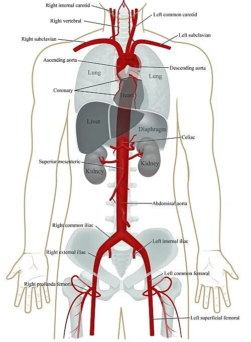
The four parts of the aorta are the abdominal aorta, thoracic (descending) aorta, ascending aorta, and aortic arch. It splits into the left and right common iliac arteries at the level of L4.
What is an aorta?
A cane-shaped artery makes up the aorta. Your body receives oxygen-rich blood from it. It begins in the ventricle, the lower-left chamber of your heart. It then extends a little way up toward your head before bending downward. The aorta terminates in your pelvis after passing through your chest and abdomen. Blood blood vessels leave the aorta along the route and travel to support tissue and organs.
Smaller blood arteries split off in pairs at different locations along the aorta. The aorta’s reach is expanded by these branches to include your body’s muscles, nerves, and organs.
The primary artery in the body, the aorta, serves the vital purpose of transferring oxygenated blood from the heart to the body’s other organs. It is significant to remember that the aorta supplies blood to every artery in the human body, with the exception of the pulmonary arteries, wherever they may be located. The several branches that the aorta produces throughout its path enable this.
The major artery known as the aorta is responsible for transporting blood rich in oxygen from the left ventricle of the heart to various regions of the body. The muscular pumping chamber of the heart, the left ventricle, is where the aorta starts. Through the aortic valve, the heart pumps blood from the left ventricle into the aorta. The aortic valve permits a one-way flow of blood by opening and closing its three leaflets with each heartbeat.
Anatomy:
This medical topic falls under the specialization of heart and vascular care. An aorta is the largest blood vessel in the human body. The blood that is rich in oxygen is transferred from your heart to the other parts of your body through this artery. The aorta begins in the left ventricle of the heart and ascends into the chest in the form of an arch. After that, it moves down into the abdomen, where it branches into the iliac arteries immediately above the pelvis.
It divides into the left and right common iliac arteries at the level of L4. The aorta is categorized as a big elastic artery
After that, it moves into the abdomen and divides into the iliac arteries a little above the pelvis.
Starting immediately from the left ventricle of the heart, the aorta is the initial section of the systemic arterial circulation. With three distinct segments, each with unique properties, particularly in terms of direction and orientation, it is the biggest artery in the body. The aorta starts off running uphill as the ascending aorta and soon after that it curves laterally to the left, forming the arch of the aorta. After that, the aorta descends as the descending aorta until its terminal branches split off.
What is the aorta’s size?
The biggest blood vessel in your body is the aorta. At its widest point, it measures an inch in diameter and more than one foot in length. The aorta’s diameter reduces to two centimeters as it approaches your pelvis.
What comprises the aorta?
Three tissue layers make up this intricate arrangement. Among them are:
- The inner layer, or tunica intima, is the tube that blood travels through. Endothelial cells, connective tissue, and smooth muscle tissue are all present. These unique cells allow blood to carry nutrients and oxygen to the appropriate location without being absorbed.
- The middle layer, also known as tunica medium, is composed of proteins called collagen, elastin, and smooth muscle tissue. Thanks to these compounds, the aorta can adapt to your body’s fluctuating demands for blood flow. When additional blood is needed, the aorta widens. It narrows if less blood is required.
- The outer layer, also known as tunica adventitia, serves as the aorta’s anchor. It is also connected to surrounding tissue and nerves.
Course:
Anterior view of the thorax showing the course of the aorta: anterior to the trachea, the oesophagus, and the right pulmonary arteries, posterior to the main pulmonary artery, and finally turning posterior to pass dorsally to these organs.
The aorta is typically sectioned into anatomical sources.
The thoracic aorta, or thoracic segment of the aorta, is divided into anatomical compartments where it travels from the heart to the diaphragm. From the diaphragm to the aortic bifurcation, the aorta descends further as the abdominal aorta or abdominal section of the aorta.
The aorta is divided by another system based on the direction and course of blood flow. The aorta in this system ascends initially, leaves the heart superiorly, and then turns into the aortic arch, which is a hairpin bend. The aorta then descends as the descending aorta after passing through the aortic arch. The aorta descends in two sections. The thoracic aorta is the name given to the aorta as it starts to descend in the thoracic chamber. The aorta is referred to as the abdominal aorta once it has passed through the diaphragm. The aorta splits at its terminus into the median sacral artery, a minor midline vessel, and the common iliac arteries, two main blood vessels.
The origin of the aorta, the first section of the systemic circulation, is the heart itself. It starts at the base of the left ventricle and at the aortic opening. The aortic valve, which is made up of the right, left, and posterior semilunar cusps, divides it from the ventricle. The aorta is nearly the whole length of the trunk and measures about 30 centimetres. This artery is the biggest in the body, having a maximum diameter of around 4 cm at the aortic root. Its diameter decreases with distance, reaching roughly 3.5 cm in the ascending aorta and 2.5 cm in the abdominal aorta. The aorta’s primary job is to carry oxygen-rich blood from the heart to the body’s other organs.
Aortic segmentation:
The aorta is a tube that is a little over an inch in diameter and about a foot long. Four sections comprise the aorta:
It is useful to divide the aorta into the following four portions because of the significant portion of the body it spans:
Ascending aorta:
The ascending aorta is two inches long and arises from the heart. The coronary arteries split off from the ascending aorta to feed the heart with blood.
When the aortic valve opens in the left ventricle of the heart, the ascending aorta starts. It passes through the pericardial sheath of the pulmonary trunk. The aorta begins posterior to the pulmonary trunk and ends by twisting to its right and anterior sides as a result of these two blood vessels twisting around one another. At the pericardial reflection on the aorta, the ascending aorta and aortic arch meet.
The aortic sinuses, also known as the sinuses of Valsalva, are tiny pockets in the lumen at the base of the ascending aorta that are located between the aortic valve cusps and the aortic wall. The left coronary artery originates in the left aortic sinus, and the right aortic sinus similarly produces the right coronary artery. These two arteries supply the heart together. A coronary artery does not originate in the posterior aortic sinus. Because of this, the terms “left-coronary,” “right-coronary,” and “non-coronary” are also used to refer to the left, right, and posterior aortic sinuses.
Starting from the sinotubular junction of the aortic root, the ascending aorta travels upward and outward from the heart until it meets the aortic arch.
- Branches:
Two arteries, however few, emerge from the ascending aorta and are important. These are the coronary arteries, the left and right of which supply the heart with blood. The origin point of the ascending aorta, also known as the aortic root, is where these arteries originate. Three aortic sinuses, or dilatations of the aorta wall located directly above the aortic valve cusps, are present in the aortic root. When viewed from above, the three distinct, spherical pockets that intersect in the middle of the aortic lumen are what these sinuses look like. The anterior, left posterior, and right posterior aortic sinuses are the names given to the aortic sinuses, sometimes referred to as Valsalva sinuses, based on their respective positions.
Anterior aortic sinus: The anterior aortic sinus is located between the right cusp of the aortic valve and the aortic wall. Because it gives rise to the right coronary artery, this sinus is also known as the right coronary sinus.
The left posterior aortic sinus is located between the left cusp of the aortic valve and the aortic wall. Because it gives rise to the left coronary artery, this sinus is also known as the left coronary sinus.
The region between the posterior cusp of the aortic valve and the aortic wall is known as the right posterior aortic sinus. Because it does not give rise to any vessels, this sinus is also known as the non-coronary sinus.
Pelvis
The cardiovascular system’s structure and operation (14 structures).
- Relations:
The ascending aorta and the heart are located in the inferior mediastinum. The pericardium and pulmonary trunk entirely encircle the ascending aorta. The pulmonary trunk’s infundibulum is located ahead of the ascending aorta when these two vessels are first running parallel to one another. Subsequently, the ascending aorta becomes anterior as the pulmonary trunk and ascending aorta wind around one another.
The right pulmonary artery and major bronchus are located on the right side of the ascending aorta, while the left atrium and pulmonary trunk are located on the left. The superior vena cava and the right atrium are located laterally to the right, while the left atrium and the pulmonary trunk are located laterally to the left. The sternum, the right pleura, the anterior margin of the right lung, the residual thymus gland tissue, and the ascending aorta are all connected superiorly.
The Aortic arch:
The curved section looks like a cane and gives the aorta its form. The descending and ascending aortas are connected by it. Blood is delivered to the head, neck, and arms by branches that emerge from the aortic arch, which curves around the heart
The section of the aorta that joins the descending and ascending aorta in the form of an arch is known as the aortic arch. The brachiocephalic artery, the left carotid artery, and the left subclavian artery are the three main arteries that emerge from the arch. Blood is supplied to the left side of the brain by the left carotid artery; the left subclavian artery supplies blood to the left arm; and the brain and right arm are supplied with blood via the brachiocephalic artery.
The ligamentum arteriosum, a piece of the fetal circulation that is destroyed a few days after birth, connects the aortic arch to the left pulmonary artery and the pulmonary trunk bifurcation, where it loops around them. The aortic arch also passes over the left major bronchus in addition to these blood veins. The cardiac, or aortic, plexus is a network of autonomic nerve fibers that runs between the aortic arch and the pulmonary trunk. The recurrent laryngeal nerve, a main branch of the left vagus nerve, originates anterior to the aortic arch and loops beneath the arch just lateral to the ligamentum arteriosum. After that, it returns to the neck.
The brachiocephalic trunk, left common carotid artery, and left subclavian artery are the three main branches of the aortic arch, arranged from proximal to distal. The right side of the head, neck, arm, and chest wall are supplied by the brachiocephalic trunk, whereas the left side of the same regions is supplied by the latter two together.
The level of the intervertebral disc between the fourth and fifth thoracic vertebrae is where the aortic arch finishes and the descending aorta begins.
The aortic arch ends about at the level of the sternal angle upon reaching the T4 vertebra, where it feeds off the left subclavian artery. The aortic arch develops the aortic isthmus, a tiny stricture, as it descends into the thoracic portion of the descending aorta. A dilatation follows this.
Branches:
The left common carotid artery, the left subclavian artery, and the brachiocephalic trunk emerge from the convexity of the aortic arch.
- Brachiocephalic trunk: The largest and first artery to emerge from the aortic arch is the brachiocephalic trunk, also known as the brachiocephalic artery. The right subclavian and right common carotid arteries are the two branches that emerge from this artery at the level of the right sternoclavicular joint, where they travel upward and slightly to the left. The artery that supplies blood to the right arm is the right subclavian, which runs laterally, above the clavicle, and then below it. The right side of the head, neck, and brain are supplied by the right common carotid artery, which runs upward.
- Left common carotid artery: The left common carotid artery originates straight from the aortic arch, in contrast to its right counterpart, which originates from the brachiocephalic trunk. About where the left sternoclavicular joint is, it splits off from the aortic arch. From there, it ascends laterally to the trachea and oesophagus inside the deep cervical fascia. Following the same path on opposite sides of the neck, the left and right common carotid arteries eventually split into the internal and external carotid arteries at the level of the laryngeal thyroid cartilage. The left side of the head and neck are subsequently supplied by the left common carotid artery.
- Left subclavian artery: Unlike the right subclavian artery, which emerges from the brachiocephalic trunk, the left subclavian artery is directly derived from the aortic arch. The left subclavian artery, which emerges at the level of the sternal angle and signifies the end of the aortic arch and the start of the descending aorta, is the final artery that the aortic arch releases.
Relations:
The aortic arch rests in the upper mediastinum over its whole length. There are numerous significant relationships between the aortic arch and the mediastinum’s neurovascular systems and organs.
It communicates with the left lung and pleura anteriorly and to the left. The aortic arch is crossed anteriorly by the left phrenic nerve, left vagus nerve trunk, left cardiac nerves (left inferior vagus nerve branch and superior cervical cardiac branch of the sympathetic trunk), and left superior intercostal vein. The recurrent laryngeal branch, which emerges on the other side of the aortic arch and wraps below it before climbing back towards the neck, is produced by the vagus nerve as it crosses it.
The aortic arch is connected to the trachea, esophagus, deep cardiac plexus, left recurrent laryngeal nerve, thoracic duct, and vertebral column from the posterior and right directions. The aortic arch is connected superiorly to the thymus, left common carotid artery, left subclavian artery, left brachiocephalic vein, and the branches of the brachiocephalic trunk. The bifurcation of the pulmonary trunk, left major bronchus, ligamentum arteriosum, superficial cardiac plexus, and left recurrent laryngeal nerve are all inferior.
Aortic valve
Three tissue flaps, or leaflets, snap open and closed to allow the heart to pump blood rich in oxygen out of the body.
Aortic root
The section of the aorta that is connected to the heart is called the aortic root. The aortic valve, which stops blood from flowing backward into the heart when closed and permits blood to flow from the heart to the rest of the body when it is open, is a significant component of the aortic root. The heart requires blood, much like the rest of the body, To supply the heart with the necessary blood, the aortic root branches into the left and right major coronary arteries.
Descending Aorta:
The descending aorta descends into the abdomen at the end of the aortic arch. The descending aorta is divided into two branches.
The diaphragm, which marks the boundary between the abdominal and chest cavities, is where the thoracic aorta ends at the aortic arch. It supplies blood to the spinal cord and chest wall muscles.
Thoracic Aorta:
Starting at the level of the T4 vertebra, the thoracic aorta descends via the posterior mediastinum. It is initially located left to the spinal column, but as it lowers, it slopes in the direction of the midline and ends up ahead of the bottom edge of the T12 vertebra’s body. At this point, it enters the abdominal aorta and travels through the diaphragm’s aortic aperture into the abdominal cavity.
Branches:
The thoracic aorta’s branches are classified as visceral branches, which are primarily meant for the mediastinum’s organs, and parietal branches, which are meant for the thoracic wall’s structures. The pericardial, bronchial, oesophageal, and mediastinal branches are the visceral branches, and the intercostal, subcostal, and superior phrenic arteries are the parietal branches.
Esophageal branches of the aorta
- Pericardial branches: A few arteries that supply the posterior part of the pericardium make up the pericardial branches.
- Bronchial artery: In most cases, there are two left and one right bronchial artery. The right bronchus, bronchopulmonary lymph nodes, pericardium, and oesophagus are all supplied by the right side artery. Blood is supplied to the parts of the bronchial tree below the level of the primary bronchi via the two bronchial arteries on the left side.
- Oesophageal branches: The four or five veins that supply the oesophagus with blood are called oesophageal branches.
- Mediastinal branches: A few branches that give blood to lymph nodes, nerves, arteries, and areolar tissue in the posterior mediastinum are known as mediastinal branches.
- Posterior intercostal arteries: The nine pairs of arteries that supply the intercostal gaps between the ribs are known as the posterior intercostal arteries.
- Subcostal arteries: The two arteries that go inferior to the twelfth ribs or inferior to the costal margin on either side make up the subcostal arteries, which supply blood flow to the subcostal space.
- Superior phrenic arteries: The musculophrenic and pericardiacophrenic arteries anastomose with the superior phrenic arteries, which supply the posterior section of the superior surface of the diaphragm.
Relations:
The posterior mediastinum is the only location for the thoracic aorta. It is connected to the left pulmonary hilum, the diaphragm, the lower portion of the oesophagus, and the pericardium that surrounds the left atrium from the anterior direction. The thoracic aorta is situated posteriorly, next to the hemiazygos and accessory hemiazygos veins, as well as the spinal column. The upper portion of the oesophagus, the thoracic duct, the azygos vein, the right lung, and the pleura are located laterally to the right. The left lung and pleura are located laterally to the left.
To avoid confusion, the oesophagus passes through the mediastinum, which explains why the thoracic aorta has many relationships with the oesophagus. As the oesophagus descends, it crosses the thoracic aorta anteriorly after initially lying laterally to the right of it.
Abdominal Aorta:
The diaphragm is the starting point of the abdominal aorta, which finishes just above the pelvis where it splits into the iliac arteries. The renal arteries, iliac arteries, inferior mesenteric artery, superior mesenteric artery, and celiac artery are the five arteries that emerge from the abdominal aorta.
Blood flows to the stomach, liver, and pancreas through the celiac artery; the small and large intestines receive blood from the superior and inferior mesenteric arteries; the kidneys receive blood from the renal arteries; and the muscles of the abdominal wall and lower spinal cord receive blood from the renal arteries. The iliac arteries, which transport blood to the legs and other organs in the pelvis, are the branches of the abdominal aorta.
At the level of the twelfth thoracic vertebra, the diaphragm’s aortic hiatus is where the abdominal aorta starts. It produces the renal and middle suprarenal arteries, the lumbar and musculophrenic arteries, and the visceral arteries (the inferior mesenteric artery, the superior mesenteric artery, and the celiac trunk). It splits into the left and right common iliac arteries at its terminus. The median sacral artery, a lesser branch, also emerges at the bifurcation point.
Branches:
The abdominal aorta’s branches can be categorized as visceral and parietal, just like the thoracic aorta’s branches. On the other hand, a more widely used classification divides the branches into anterior, lateral, and dorsal groups according to where they emerge from the aorta.
The anterior set of branches includes the superior and inferior mesenteric arteries, as well as the celiac trunk;
- Celiac trunk: A Celiac trunk is a single, short vessel that emerges at the level of the lower border of the T12 vertebra on the anterior part of the aorta, directly below the diaphragm’s aortic opening. The left gastric artery, common hepatic artery, and splenic artery are the three branches that emerge from the stem shortly after it originates. These arteries supply the foregut, or the portion of the digestive tract below the diaphragm and up to the proximal duodenum. The stomach, proximal duodenum, liver, gallbladder, pancreas, spleen, and abdominal portion of the oesophagus are among the foregut organs supplied by the celiac trunk.
- Superior mesenteric artery: The superior mesenteric artery is an unpaired artery that emerges at the level of the L1 vertebra, approximately 1 centimetre behind the celiac trunk. It provides blood to the midgut, the portion of the digestive tract located between the distal third of the large intestine’s transverse colon and the proximal segment of the duodenum. As a result, it supplies most of the small intestine, appendix, cecum, ascending colon, and first two-thirds of the transverse colon.
- Inferior mesenteric artery: The inferior mesenteric artery is an unpaired artery that emerges at the level of the L3 vertebra, approximately 4 cm above the abdominal aorta’s bifurcation. Blood is supplied to the hindgut, which is the term used to describe the remaining sections of the digestive tract, which comprise the rectum, the sigmoid colon, the distal third of the transverse colon, the descending colon, and the superior part of the anal canal.
The lateral group of branches of the abdominal aorta includes the gonadal, renal, and suprarenal arteries:
- Middle suprarenal artery: The middle suprarenal artery is a pair of arteries that emerges close to the superior mesenteric artery’s origin on either side of the abdominal aorta. These arteries provide blood to the suprarenal glands, along with the superior and inferior suprarenal arteries.
- Renal artery: The renal artery is a sizable pair of arteries that arises at a right angle from the lateral margins of the aorta, directly beneath the superior mesenteric artery. In addition to supplying the inferior suprarenal artery, which serves the suprarenal glands, it also supplies blood to both the left and right kidneys.
- Gonadal artery: The gonadal artery is a pair of arteries that emerge from the aorta,’s anterior aspect, immediately below the renal arteries. This artery, which nourishes the testes in men, is known as the testicular artery; its female counterpart is called the ovarian artery, which supplies the ovary.
The lumbar, median sacral, and inferior phrenic arteries are part of the dorsal group of branches.
- Inferior phrenic artery: The inferior phrenic artery is a pair of arteries that emerge from the aorta’s posterolateral aspect just below the diaphragm’s aortic hiatus. In addition to supplying the superior suprarenal artery, which supplies the suprarenal glands, this artery also nourishes the inferior surface of the diaphragm.
- Lumbar arteries: The lumbar arteries are a group of four pairs of arteries that emerge from the aorta’s posterior surface. They supply the spinal cord and the posterior wall of the abdomen.
- Median sacral artery: The posterior part of the abdominal aorta, immediately above the bifurcation, gives birth to the medial sacral artery. The sacrum and lower lumbar vertebrae are supplied with blood via this artery.
- The origin of the left and right common iliac arteries, which are the terminal branches of the abdominal aorta, is the bifurcation that occurs in front of the L4 vertebra’s body, around 1.25 cm to the left of the median plane. The pelvic viscera, the gluteal region, and the lower leg are supplied by the common iliac arteries.
Relations:
Within the retroperitoneal area of the abdominal cavity, the abdominal aorta runs. It is connected to the anterior longitudinal ligament and the vertebral bodies of the top four lumbar vertebrae posteriorly.
The abdominal aorta is connected to the abdominal cavity’s organs anteriorly. It is related to the pancreas, the small intestine, the mesentery root, and the descending portion of the duodenum from above to below. The abdominal aorta is also associated with other neurovascular structures: the renal and splenic veins are related to it anteriorly; the inferior vena cava is related to it laterally and to the right; and the sympathetic trunk is related to it laterally
Function:
- The aorta plays a vital role in providing the body with the oxygen-rich blood necessary for survival.
- The arteries that emerge from the ascending aorta supply oxygen to the heart.
- Arteries from the aortic arch supply oxygen to the head (including the brain), neck, and arms.
- Arteries that emerge from the abdominal aorta supply oxygen to the stomach, intestines, kidneys, and other essential organs.
Clinical Conditions:
- Aortic atherosclerosis: A build-up of plaque that hardens and obstructs blood flow through the aorta and weakens the walls of the aorta. Aortic aneurysms, arterial thrombosis, strokes, and angina are possible outcomes.
- Aortic stenosis: The aortic valve is connected to the interior of the ascending aortic wall, while not being a component of the ascending aorta. When the aortic valve, which joins the left ventricle and aortic root, becomes excessively narrow, aortic stenosis develops. To pump blood into your aorta, your heart must work harder. The cardiac muscle may enlarge as a result. This disease requires the heart to pump harder in order to push blood through the aortic valve and into the aorta because it prevents the valve from opening fully as it should. Diastolic heart failure, diastolic dysfunction, and left ventricular hypertrophy (LVH) are among the consequences that may result from it. Aortic stenosis in picture
- Aortic regurgitation: reverse blood flow through the aortic valve is caused by insufficient valve closure during diastole. Aortic dissection in the ascending segment and infectious endocarditis are the causes of its acute form. Aortic valve deterioration, thoracic aortic aneurysm, rheumatic fever, infective endocarditis, and trauma are the causes of the chronic form, which usually has no symptoms for a long period of time. Arrhythmias, cardiac failure, pulmonary edema, and left ventricular hypertrophy (LVH) can result from it. Another name for it is aortic insufficiency.
An abnormal growth or bulging of the aortic wall is known as an aortic aneurysm. Anywhere in the vascular tree can experience an aneurysm—for instance, the abdominal aorta. Depending on the extent of the aneurysm, therapy may be required after it is diagnosed. To stop the bleeding from ruptured aneurysms, immediate surgery is necessary.
- Aortic dissection: Three layers make up the aorta. A tear that appears in the aorta’s inner layer and allows blood to pass between the layers is known as an aortic dissection. After that, the layers split apart, stopping the blood supply and perhaps rupturing the artery wall. Aortic dissection can be a potentially fatal emergency in certain cases, necessitating immediate surgery to replace or repair the injured part of the aorta.
- Aortic aneurysm:
A localized swelling of the aorta wall is known as an aortic aneurysm. An arterial aneurysm is a bulge that develops from a weak point in the wall of the vessel. The ascending aorta is the site of more than 50% of all thoracic aortic aneurysms (TAAs). Both the tubular ascending aorta and the aortic root may experience them. Aortic aneurysms are potentially fatal disorders. Aortic dissections, which are rips in the blood vessel walls between its layers, may result from them. Alternatively, they may burst entirely, causing significant internal bleeding.
The ascending aorta, aortic arch, and abdominal aorta are the next most typical locations for its development. When the aorta wall diameter increases by 1.5 times its normal size, an aortic aneurysm is diagnosed. The diagnostic criteria for an aneurysm include a diameter of >5 cm for the ascending aorta and >4 cm for the abdominal aorta, while the normal diameters of the ascending and abdominal aorta are, respectively, approximately 3.5 cm and 2.5 cm.
When an aortic aneurysm affects surrounding structures, it can occasionally cause signs and symptoms, but these are rare. An aortic aneurysm is often asymptomatic. Aortic arch or ascending aortic aneurysms can compress the lungs, resulting in coughing and dyspnea, or strain the recurrent laryngeal nerve, leading to hoarseness in speech. Similarly, numbness in the lower limbs may result from compression of the spinal nerve roots caused by an abdominal aortic aneurysm.
- Bicuspid aortic valve (BAV): In certain individuals, the aortic valve has two flaps at birth rather than three. There may be a connection between these valves and ascending aortic aneurysms. Aortic regurgitation, aortic stenosis, or both are frequently caused by BAV.
- Connective tissue diseases: Conditions like Ehlers-Danos syndrome, Loeys-Dietz syndrome, and Marfan syndrome are examples of connective tissue illnesses.
- Inflammatory disorders: These encompass ailments such as penetrating atherosclerotic ulcers and giant cell arteritis.
FAQs
What is the aorta’s purpose?
The primary blood conduit that carries oxygen-rich blood from the heart to the rest of the body is the aorta. Hormones and minerals are also delivered via it. The branches of the aorta guarantee that these materials get to the surrounding supporting tissue and internal organs.
What happens if the aorta is damaged?
A rip (dissection) in the wall of the aorta, the body’s principal artery, is known as an aortic dissection. The body receives blood from the heart through the aorta. Blood can enter between the three layers of the aorta due to a tear. As a result, your body’s organs can receive less oxygen and nutrients.
What is an aorta weakness?
The main artery in the body is called the aorta. A weak spot in the upper section of the aorta, the main blood conduit that supplies the body with blood, is known as a thoracic aortic aneurysm. The aorta can develop aneurysms anywhere. A weak spot in the body’s major artery in the chest is called a thoracic aortic aneurysm.
Is aortic disease curable?
Procedures and Treatments for the Aortic Valve. It is possible to replace or fix aortic valves. Replacement is typically the sole course of action for people with aortic valve stenosis. The valve can be replaced or fixed if it is leaking.
Which organs are supplied by the aorta?
Numerous branches of the aorta emerge into the abdominal cavity, forming a vast network that supplies blood to various organs such as the kidneys, reproductive glands, small and large intestines, liver, pancreas, and stomach.
Reference:
- Professional, C. C. M. (n.d.). Aorta. Cleveland Clinic. https://my.clevelandclinic.org/health/body/17058-aorta-anatomy
- Aorta. (2024, April 24). Wikipedia. https://en.wikipedia.org/wiki/Aorta
- The Aorta – Branches – Aortic Arch – TeachMeAnatomy. (2024, April 21). TeachMeAnatomy. https://teachmeanatomy.info/abdomen/vasculature/arteries/aorta/
- Aorta Anatomy – UF Health. (n.d.). https://ufhealth.org/conditions-and-treatments/aorta-anatomy
- Aorta. (n.d.). Physiopedia. https://www.physio-pedia.com/Aorta
- Aorta. (2023, October 30). Kenhub. https://www.kenhub.com/en/library/anatomy/aorta
3 Comments