Common Iliac Artery
The common iliac artery is a vital component of the vascular system, playing a crucial role in supplying blood to the lower regions of the body. It originates from the bifurcation of the abdominal aorta at the level of the fourth lumbar vertebra. The common iliac artery extends bilaterally, with each artery dividing into the internal and external iliac arteries. These branches further distribute blood to the pelvic organs, gluteal region, and lower limbs.
The blood supply to the pelvis and buttocks is provided by the internal iliac arteries, whereas the external iliac arteries supply the blood to the legs.
Traumas or disorders affecting the common iliac arteries may result in life-threatening medical conditions. An illustration would be a common iliac artery aneurysm, which causes the artery to enlarge and may eventually burst.
Because the common iliac artery is a paired structure, the body has one on the left and one on the right.
Table of Contents
What is a common iliac artery?
Blood channels called the iliac arteries supply blood to the pelvis, legs, reproductive organs, and other organs located in the pelvic region. Located somewhat above the point where your legs join at the hips, the pelvis is the bottom region of your body. The iliac arteries split off from the main artery that emerges from the apex of the heart, the aorta, at its base.
Peripheral arteries include the iliac arteries. Peripheral refers to the fact that they supply blood to bodily areas that are further from the heart.
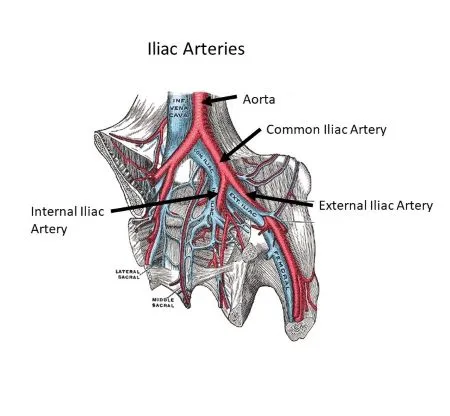
Large abdominal arteries paired on either side are called the common iliac arteries. It starts at the level of the fourth lumbar vertebra, where the aortic bifurcation occurs. It finishes in front of the sacroiliac joint, where it splits into the internal and external iliac arteries on either side.
Anatomy:
The aorta and the arteries of the pelvis and lower limbs are connected via the common iliac arteries.
The fourth lumbar spine vertebra is where the aorta ends. It splits into the left and right common iliac arteries at that point. These two arteries go for approximately five centimetres on either side of the body, down to the edges of the pelvis. It starts as an aortic branch. This is situated at the fourth lumbar vertebral level. It follows the medial edge of the psoas muscles inferolaterally. At the pelvic inlet, which is the point where the pelvis and abdomen meet, they are each divided once again into internal and external iliac arteries.
Blood is supplied to the pelvic organs by the internal iliac artery, which includes the woman’s uterus and vagina, the man’s prostate gland, and the bladder. Blood supply to the leg is mostly supplied via the external iliac artery. It develops into the femoral artery and splits into the anterior and posterior tibial arteries as well as the popliteal artery. Blood is supplied to the thigh via the femoral artery, the knee by the popliteal artery, and the area below the knee, which includes the toes and foot, by the anterior and posterior tibial arteries.
All of the branches that make up the common iliac artery are paired structures; that is, there is a left and a right branch.
The pelvis and lower limb (as the femoral artery) on the appropriate side are essentially the distribution of the common iliac artery.
Course:
The abdominal aorta, which transports blood from the heart, divides into two common iliac arteries. One turns left, while the other turns right.
At the L4 level, the aortic bifurcation gives rise to the common iliac artery. On the medial part of the psoas major muscle, the left and right common iliac arteries split as they descend and enter the pelvis.
Every common iliac artery and vein are parallel to one another (common iliac veins).
In front of the sacroiliac joint, the point where the sacrum and ilium bones of the pelvis meet, the common iliac artery divides into the external and internal iliac arteries, which are the two main ending branches.
Because the aortic bifurcation is located somewhat to the left of the midline, the right common iliac artery is about 1 cm longer than the left. It extends anteriorly to the distal portion of the right common iliac vein and the right sympathetic trunk. It is crossed by the ureter when it splits into the internal and external iliac arteries.
The left sympathetic trunk is located anteriorly and the left common iliac artery is situated lateral to the left common iliac vein. Along with the ureter, it is crossed distally by the superior rectal artery.
Compared to the right, the left CIA is shorter. The right common iliac vein (CIA) travels anteriorly to the left common iliac vein before continuing anteriorly and parallelly to the right CIA. Running laterally and parallel to the left common iliac vein, the left CIA path is less complicated.
At the level of the pelvic brim, anterior to the sacroiliac joint, the CIA bifurcates into the internal and external iliac arteries, which are its terminal branches where the ureter crosses it anteriorly.
Branches:
Before dividing, each common iliac artery descends approximately 1 inch (3 centimeters) from the aorta. There, it divides into the exterior and internal iliac arteries. To cover a larger area of your lower body, the iliac arteries’ smaller channels split into even smaller arteries.
External Iliac Arteries:
The external iliac artery, which originates at the sacroiliac joint as well, descends the pelvis to the groin (inguinal) ligament and splits into two branches. A primary source of blood supply to the lower limbs, the external iliac artery is termed the femoral artery after the separation.
The external iliac artery has two branches, which are the following:
- Deep Circumflex Arteries: The oblique and transverse abdominus muscles (core muscles) of the stomach receive blood supply from the deep circumflex arteries.
- Inferior Epigastric Arteries: The rectus abdominus muscle, which makes up the six-pack muscles that run vertically on either side of the stomach, receives blood supply from the inferior epigastric arteries.
Internal Iliac Artery:
This artery travels down the body with its matching vein in front of it, running behind the duct that permits urine to flow from the kidneys to the bladder (ureter) in its upper part. The artery delivers blood to several muscle groups, bones, nerves, and organs in and around the pelvis. It branches in the front (anterior) and back (posterior) of the body.
Nine anterior (front) and three posterior (rear) iliac arteries split from the internal iliac arteries. Only in the umbilical cord of pregnant women are two of the anterior iliac arteries present.
These arteries send blood to the various organs:
- Back muscles.
- Pelvic floor and organs.
- Rectum.
- Reproductive organs.
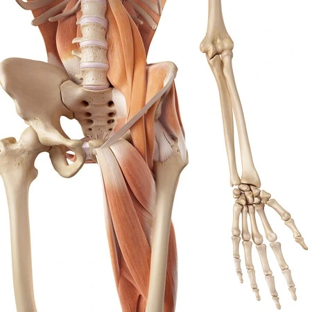
- Thighs, hips and rear.
Frequently, the left common iliac artery is somewhat shorter than the right. The former follows the left common iliac vein in a parallel fashion. The latter then travels parallel to the right of the right common iliac vein after passing in front of the vein.
Anatomical Relations:
The two common iliac veins, which are located posteriorly and to the right, follow the common iliac arteries as they travel along. The ureters cross their terminal bifurcation anteriorly. The bifurcation of the common iliac artery is the second point of restriction in the ureters, hence this is important.
Anteriorly (at its bifurcation): ureters
Posteriorly: common iliac veins, psoas major muscle
Anatomical Differences:
The internal iliac branch has the most frequent changes in the common iliac arteries.
The artery usually starts at the base of the spine, at the level of the lumbosacral joint, but occasionally it emerges at a higher point, at the fifth lumbar vertebra. In others, it happens at the tailbone’s tip (also known as the sacrum or S1).
The obturator artery, the first main branch of the internal iliac artery, has been noted by medical professionals to have multiple origins. It may also originate from the vesical or inferior epigastric inferior arteries farther down the artery.
The iliolumbar artery, which delivers blood to the abdomen, may also emerge sooner than usual at the internal iliac artery trunk.
Apart from the internal and external iliac arteries, the common iliac artery branches out to the peritoneum, the psoas major muscle, the ureter, and surrounding tissues and lymph nodes. The lower limbs receive blood supply through the external iliac artery, whereas the pelvis and its viscera are supplied by the internal iliac artery.
Function:
In the body’s circulatory system, the iliac arteries transport blood to the following areas:
- Muscles in the stomach and back.
- Lower extremities, including the thighs, feet, hips, and buttocks.
- Either the female or male reproductive systems.
- Other pelvic organs, such as the pancreas, liver, intestines, and bladder.
The common iliac artery’s main function is to supply oxygen-rich blood to the lower limbs and pelvis. Via its branches, the internal iliac artery delivers blood to the pelvic region, groin, and surrounding muscles and bones.
The internal branch ensures that there is a steady flow of blood to the genitalia, uterus, vagina, and prostate, as well as the buttocks muscles (gluteus maximus and minimus).
Blood is supplied to the legs’ muscles, nerves, and bones by the external iliac artery. The femoral artery supplies blood to the femur, tibia, and other lower limb bones. It is the external iliac artery that emerges from the pelvis.
The external iliac artery branches into the anterior and posterior tibial arteries, which deliver blood to the knee region, lower leg, feet, and toes.
Significance for clinical practice:
Constrictions:
It’s possible for the common iliac artery to narrow. Most frequently, this occurs at the aortic bifurcation.
Dilatation
The following classifications apply to dilatation of the common iliac artery:
- Normal : Diameter ≤ 12 mm
- Ectasia: Diameter 12 to 18 mm
- Aneurysm: Diameter ≥ 18 mm
Clinical Condition:
A common iliac artery injury or medical condition might have major repercussions.
An illustration of this is a common iliac artery aneurysm, which happens when a portion of the artery swells or “balloons” because its walls are weak. Between 10 and 20 percent of abdominal aneurysms are of this type.
Moreover, the illness may result in compression of the sciatic nerve, which travels from the base of the spine through the pelvis to the lower limbs.
Ruptures of a common iliac artery aneurysm can cause shock and severe abdominal discomfort, though they are usually asymptomatic.
A specialized, minimally invasive surgical technique called endovascular aneurysm repair (EVAR) is used to fix damaged or enlarging arterial segments. An implanted device known as an endovascular stent, or “endograft,” is designed to enlarge and seal an arterial leak or rupture.
Little incisions are made during the surgery, and fluoroscopy (an x-ray tool) is used to guide the incisions. Generally speaking, the recovery time is shorter than it would be for open surgery.
Surgeons must use extreme caution while doing any operation close to an artery, even if it is minimally invasive, as doing so may result in serious consequences.
When undergoing abdominal or pelvic surgery, these arteries are particularly susceptible to damage (such as a hysterectomy to remove the uterus). Because the common iliac artery is essential for supplying blood to the lower limbs, injury to the iliac arteries, if left untreated by a vascular surgeon, might result in bleeding, death, or limb amputation.
The following ailments can have an impact on the iliac arteries:
Atherosclerosis: Atherosclerosis is the constriction and slowing of arteries due to the accumulation of fat and cholesterol deposits (plaque) inside the arterial walls.
PAD: Depending on the degree of the iliac artery atherosclerosis, walking may become uncomfortable due to a reduction in blood supply to the feet and legs. Heart attack, stroke, and amputation risk are all increased by PAD. Of the nearly 10 million Americans affected by PAD, the majority are over 65.
Iliac artery stenosis: The condition known as iliac artery stenosis is caused by fibromuscular dysplasia (FMD), a rare blood vessel disease that affects a small number of people. Slim and rigid, the iliac arteries develop. Aneurysms, or weak, protruding areas in the arterial wall, and tears (dissections) are common in them.
May-Thurner syndrome: Also known as iliac vein compression syndrome, this condition is brought on by the left common iliac artery pressing against the spine by the pressure of the right common iliac artery. The illness increases your risk of developing a blood clot in your leg, known as deep vein thrombosis (DVT).
FAQs
Which arterial branches comprise the iliac common branch?
Divisions. The common iliac arteries supply branches to the peritoneum, psoas muscle, ureter, and nerves in addition to their terminal branches. The iliolumbar and auxiliary renal arteries are sporadic branches.
What is the iliac artery’s primary purpose?
The legs and feet receive blood flow via the external iliac arteries. The groin, hips, buttocks, rectum, bladder, and reproductive organs are among the tissues and organs in and surrounding the pelvis that receive blood supply from the internal iliac arteries.
What is the iliac artery’s normal diameter?
The average diameter of the common iliac artery is about 1 cm. When it comes to the threshold for AAA, the traditional threshold for surgical intervention has been around 3 cm.
What happens in the case of an iliac artery injury?
These iliac artery injuries might show up as fistulas, aneurysms, pseudoaneurysms, ischemic limbs, or ruptures that result in the production of hematomas. For such injuries, endovascular or surgical exploration with bypass treatments are accepted therapy modalities.
What is the iliac artery bifurcation?
The abdominal aorta splits into the left and right common iliac arteries at the aortic bifurcation. Typically, the aortic bifurcation is visible at L4, directly above the point where the left and right common iliac veins converge.
Reference:
- Baba, Y., & Bashir, O. (2012, January 7). Common iliac artery. Radiopaedia.org. https://doi.org/10.53347/rid-16293
- Professional, C. C. M. (n.d.). Iliac Artery. Cleveland Clinic. https://my.clevelandclinic.org/health/body/21681-iliac-artery
- Common iliac artery. (2024, May 6). Wikipedia. https://en.wikipedia.org/wiki/Common_iliac_artery
- Common iliac artery. (2023, October 30). Kenhub. https://www.kenhub.com/en/library/anatomy/common-iliac-artery
- Gurarie, M. (2022, October 20). The Anatomy of the Common Iliac Artery. Verywell Health. https://www.verywellhealth.com/common-iliac-artery-anatomy-4690926
- Common Iliac Artery | Complete Anatomy. (n.d.). www.elsevier.com. https://www.elsevier.com/resources/anatomy/cardiovascular-system/arteries/common-iliac-artery/18830