
Nervous System
Table of Contents
What is a Nervous System?
The human nervous system guides almost everything you do, think, say, or feel. It controls complicated activities like body movement, thought & memory. It also plays an essential role in the things the body does without thinking, such as breathing, blushing, blinking & digestion.
The nervous system affects every aspect of health including:
- Thoughts, memory, learning, & feelings.
- Movements, such as balance & coordination.
- Senses, including how the brain interprets what you see, hear, taste, touch & feel.
- Sleep, healing & aging.
- Heartbeat & breathing patterns.
- Reactions to stressful situations.
- Digestion, as well as how hungry & thirsty you feel.
- Processes of the body, such as puberty.
This nervous system is the command center of the body. It regulates the body’s systems & allows you to experience the environment. A vast network of nerves sends electrical signals to & from other cells, glands, & muscles all over the body. These nerves get information from the world around you. Then the nerves interpret the information & control the response. It’s almost like an enormous information highway running throughout the whole body.
Function of the Nervous System
The nervous system uses specialized cells known as neurons to send signals, or messages, all over the body. These electrical stimulus travel between the brain, skin, organs, glands & muscles. The messages help to move the limbs & feel sensations, such as pain. The eyes, ears, tongue, nose & nerves all over the body take in information about the environment. Then nerves carry that data to & from the brain.
Different kinds of neuron cells send different signals. Motor neurons tell the muscles to move. Sensory neurons take information from the senses & then send alerts to the brain. Other types of neurons control the things the body does automatically, like breathing, shivering, having a regular heartbeat & digesting food.
The main parts of the human nervous system include:
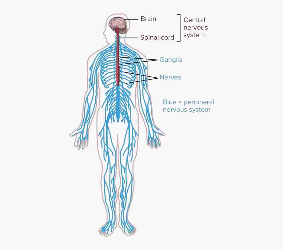
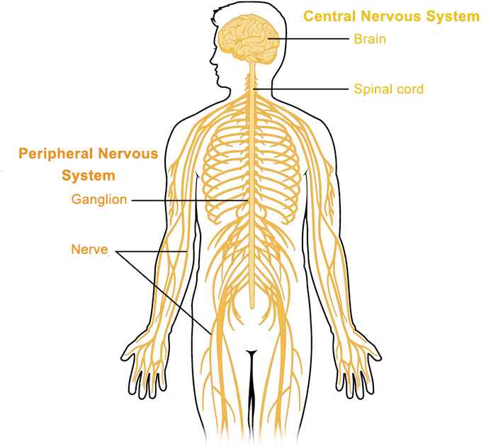
1. Central nervous system (CNS): The brain & spinal cord make up the CNS. The brain uses the nerves to send messages to the rest of the body. Each nerve has a protective outer layer called the myelin sheath. The myelin sheath insulates the nerve & helps the messages get through.
2. Peripheral nervous system(PNS): The peripheral nervous system consists of many nerves that branch out from the CNS all over the body. This system relays information from the brain & spinal cord to the organs, arms, legs, fingers & toes. The peripheral nervous system contains:
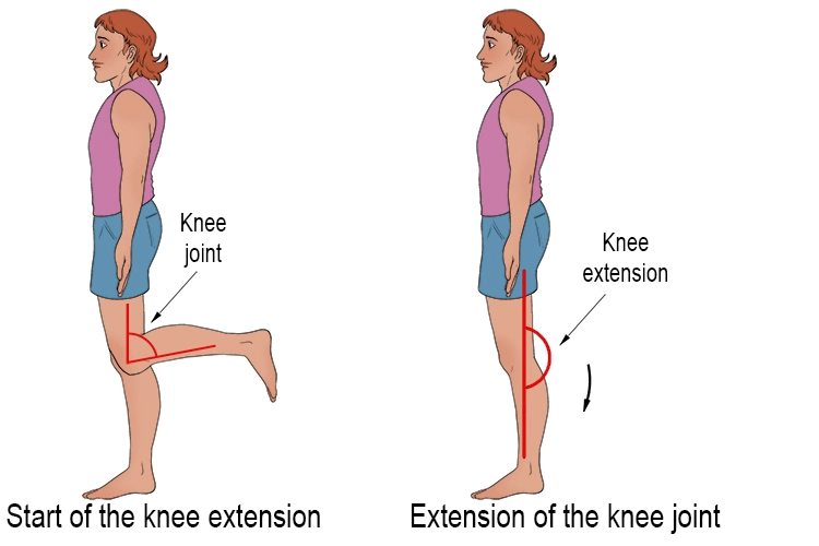
The somatic nervous system controls voluntary movements.
The autonomic nervous system controls all involuntary activities of the body.
Structure of Nervous System
Neurons
Neurons, or nerve cells, are the main structural & functional units of the human nervous system. Every neuron consists of a body (soma) & a number of processes (neurites). The neuron body contains the cellular organelles where action potentials are generated. The processes stem from the body, they connect neurons with each other & with other body cells, enabling the flow of action potential. There are two types of neural processes that vary in structure & function;
Axons are long & pass impulses away from the neuronal body.
Dendrites are short & act to receive impulses from other neurons, conducting the electrical signal toward the neuronal body.
Every neuron contains a single axon, while the number of dendrites varies. Based on that number, there are four structural types of neuron cells;
- multipolar,
- bipolar,
- pseudounipolar
- unipolar.
The morphology of neuron cells makes them highly specialized to work with neural impulses; they generate, receive & transfer these impulses onto other neurons & non-neural tissues. There are divided into two types, named according to whether they transfer an electrical signal towards or away from the central nervous system;
Efferent neurons (motor or descending) transfer neural impulses from the CNS to the peripheral tissues, instructing them on how to function.
Afferent neurons (sensory or ascending) receive impulses from the peripheral tissues to the CNS.
The site where an axon connects to another neuron cell to conduct the neural impulse is known as a synapse. The synapse doesn’t connect to the next neuron cell directly. Instead, the stimulation triggers the release of chemicals known as neurotransmitters from the end of an axon. These neurotransmitters bind to the effector cell’s membrane, causing biochemical events within that cell according to the orders transferred by the central nervous system.
Glial cells
Glial cells are called neuroglia or simply glia. glial cells are smaller non-excitatory cells that act to support neuron cells. They don’t propagate action potentials. Instead, they myelinate neurons, maintain homeostatic balance & provide structural support, protection & nutrition for neurons throughout the nervous system.
This set of functions is provided for by four different types of neuroglia;
Myelinating glia produces the axon-insulating myelin sheath. These are known as oligodendrocytes in the CNS & Schwann cells in the peripheral nervous system.
Astrocytes (CNS) & satellite glial cells (PNS) both share the function of supporting & protecting neurons.
The other two glial cell types are found in the central nervous system exclusively; microglia are the phagocytes of the CNS & ependymal cells which line the ventricular system of the central nervous system. The peripheral nervous system doesn’t have a glial equivalent to microglia as the phagocytic role is performed by macrophages.
Most axons are wrapped by a white insulating substance known as the myelin sheath, produced by oligodendrocytes & Schwann cells. Myelin encloses an axon segmentally, leaving unmyelinated gaps between the segments known as the nodes of Ranvier. The neural impulses propagate through the nodes of Ranvier only, skipping the myelin sheath. This significantly increases the speed of neural impulse transfer.
White & gray matter
The white color of myelinated axons of neurons is distinguished from the gray-colored neuronal bodies & dendrites. Based on this, nervous tissue is classified into white matter & gray matter, both of which have a specific distribution;
White matter: white matter comprises the outermost layer of the spinal cord & the inner part of the brain.
Gray matter: grey matter lies in the central part of the spinal cord, the outermost layer of the brain (cerebral cortex) & in several subcortical nuclei of the brain deep into the cerebral cortex.
Nervous system divisions:
The nervous system is structurally divided into two divisions;
Central nervous system (CNS) – this system consists of the brain & spinal cord.
Peripheral nervous system (PNS) – gathers all neural tissue outside the central nervous system.
Functionally, the peripheral nervous system is further subdivided into two divisions;
Somatic nervous system (SNS) – this system is described as the voluntary system
Autonomic nervous system (ANS) – this system is described as the involuntary system.
Although classified structurally into central & peripheral parts but interconnected with each other. Axon bundles transfer impulses between the brain & the spinal cord. These bundles within the CNS are known as afferent & efferent neural pathways or tracts. Axons that extend from the CNS to join with peripheral tissues belong to the PNS. Axon bundles within the PNS are called afferent & efferent peripheral nerves.
Central nervous system
CNS consists of the brain & spinal cord. These are found within the skull & vertebral column respectively.
The brain consists of four parts; cerebrum, diencephalon, brainstem, & cerebellum. Together these parts process the incoming information from peripheral tissues & generate commands; telling the tissues how to respond & function. These commands tackle the most complex voluntary & involuntary body functions, from breathing to thinking.
The spinal cord continues from the brainstem of the brain. its major function is to pass information between the CNS & periphery.
Cerebral Cortex
The cerebrum is the largest part of the brain, & it handles a wide range of responsibilities. Situated at the front & top of the skull.
It is instrumental in everything you do in day-to-day life, ranging from thoughts to actions. In essence, it’s responsible for the brain functions that allow us to interact with our environment & make us who we are.
Function
The cerebrum handles much of the brain’s “conscious” actions.
There are five senses: The cerebrum manages & processes everything senses take in. That includes vision, hearing, smell, taste & touch.
Language: Various parts of the cerebrum control the ability to read, write & speak.
Working memory: it is a type of short-term memory. An example of this memory is when you remind yourself to pick up something from the grocery store.
Behavior & personality: Part of the cerebrum is the frontal lobe, which manages personality & behavior. It’s part of the brain that acts as a filter to stop you from doing or saying things you might later regret.
Movement: Certain areas of the cerebrum transfer signals that tell the targeted muscles what to do when you need to use them.
Learning, logic & reasoning: Different areas of the cerebrum work together when you need to learn a new skill, make a plan of action or puzzle out a problem.
Anatomy
Where is the cerebrum situated?
The cerebrum is inside of the skull, at the top and front of the head & makes up the largest part of the brain. The outer surface of the cerebrum, the cerebral cortex, is mostly smooth but has many wrinkles, making it look something like a walnut without its shell. It’s broken down lengthwise into two halves, the left & right hemispheres, by a deep groove. The two hemispheres connect using a structure known as the corpus callosum. a collection of nerve tissue that transfers signals from one side of the brain to the other.
The two hemispheres of the brain also have five main lobes each:
Frontal (at the front of the head): The frontal lobe handles things like attention, behavior control ( the sense of what’s appropriate & what’s not), the ability to speak & certain types of muscle movements.
Parietal (at the top of the head): partial lobe handles touch, temperature & pain signals. It also helps in judging the distance from & the size of objects. It also plays a role in processing sound, languages you speak, the ability to use numbers & count, and how the organize information & make decisions.
Temporal (at the side of the head): temporal lobes help in understanding the language when other people are speaking. It also helps in recognizing people & objects. This part also helps in connecting emotions with memories.
Occipital (at the back of the head): This area manages much of the eyes’ sensory input, including the ability to see movement & colors.
A few structures that are part of the cerebrum stand out because they have very specific purposes. These are:
- Cerebral cortex: cerebral cortex is a thin layer of brain tissue on the surface of the cerebrum.
- Thalamus: This part of the brain acts like a relay station, sorting input from the senses & sending it to various parts of the cerebrum.
- Hypothalamus: This part of the brain manages functions in the nervous & endocrine systems, both of which help with controlling other systems & processes throughout the body. An example of this is how the hypothalamus helps manage the body’s temperature, heart rate & blood pressure.
- Hippocampus: This structure with the temporal lobe helps manage & store memories in areas of the cerebrum and fetches them when you need them.
Condition & Disorder
Which are the common conditions and disorders that affect the cerebrum?
Any condition affecting the brain can affect the cerebrum, including mental health conditions. Some main examples include:
- Alzheimer’s disease
- Anxiety disorders.
- Attention-deficit hyperactivity disorder (ADHD).
- Stroke
- Concussion & traumatic brain injuries.
- Congenital disorders
- Dementia
- Depression.
- Dizziness.
- Epilepsy
- Immune & inflammatory conditions
- Genetic disorders
- Infections
- Parkinson’s disease.
- Post-traumatic stress disorder (PTSD).
- Schizophrenia
- Vitamin deficiencies & nutrition problems
Which are common signs or symptoms of body organ conditions?
Many symptoms are possible when you have a condition that affects the cerebrum. Some of the main common symptoms include:
- Aphasia: Problems with the speech centers in the cerebrum can affect the ability to speak or understand others who are speaking.
- Ataxia: Loss of coordination. It can make you clumsy, causing balance problems or trouble in using your hands for common tasks.
- Behavior changes & confusion.
- Dizziness.
- Headaches & migraines.
- Memory problems.
- Paralysis. This can affect various parts of the body.
- Shaking or tremors.
- Trouble concentrating or thinking.
- Vision problems. The cerebrum plays a role in controlling the eyes and how the brain processes what you see.
Cerebellum
The cerebellum, which stands for ‘little brain’, is a hindbrain structure that controls balance, coordination, movement, & motor skills of the body.
Anatomy
It lies at the back of the brain. It is also classified into two hemispheres, like the cerebral cortex. Each hemisphere is associated with each side of the body within the cerebellum, there are thought to be three anatomical lobes which are classified by two fissures – the primary fissure & the posterolateral fissure:
The anterior lobe
The posterior lobe
The flocculonodular lobe – This part of the cerebellum is responsible for balance & spatial attention, also receiving visual input.
The cerebellum can also be classified into three functional areas:
Cerebrocerebellum – this is the largest area of the cerebellum, responsible for planning movements, motor learning, regulate the coordination of muscle activation & eye movements.
Spinocerebellum – this area is responsible for regulating body movements by allowing for error corrections.
Vestibulocerebellum – this area is involved in controlling the balance of the body& flexes of the eyes.
Functions:
- Coordination of voluntary movement.
- Balance.
- Posture.
- Motor-learning.
- Sequence learning.
- Reflex memory.
- Mental function.
- Emotional processing.
Brainstem
It is the stalklike part of the brain that connects the brain to the spinal cord It lies toward the bottom of the brain. It regulates some body functions such as breathing & heart rate. the brainstem also controls balance, coordination & reflexes of the body.
Function
The brainstem helps coordinate the messages that regulate:
- Balance.
- Blood pressure.
- Breathing.
- Facial sensations.
- Hearing.
- Heart rhythms.
- Swallowing.
- The brainstem also contains 10 of the 12 cranial nerves that start in the brain.
Anatomy
The brainstem consists of three parts:
Midbrain: it is the upper part of the brainstem regulating eye movements.
Pons: the middle portion of the brainstem helps coordinate facial movements, hearing & balance.
Medulla oblongata: it is the lower part of the brainstem that regulates breathing, heart rhythms, blood pressure & swallowing.
Diencephalon
The diencephalon is the caudal part of the forebrain (prosencephalon) that occupies the central region of the human brain. The diencephalon consists of the:
- Epithalamus
- Thalamus
- Subthalamus
- Metathalamus
- Hypothalamus
Function
Each of the four structures situated within the diencephalon plays a major role in normal brain functioning.
Thalamus: Thalamus also receives sensory & motor signals from the body & sends this information to the cerebral cortex of the brain. the thalamus is responsible for regulating the consciousness & alertness of the body.
Epithalamus: It contains the pineal gland which secretes melatonin hormone. Melatonin hormone helps in the sleep-wake cycle in the human body.
Subthalamus: It is home to many groups of nerves that connect different parts of the brain. It is a control center for the peripheral nervous system & connects the endocrine system to the nervous & limbic systems. The subthalamus has different parts including:
- The zona incerta: it helps in stimulates the thalamus.
- The reticular nucleus: this is regulating the thalamocortical pathway & Consciousness
- The peri geniculate nucleus: it plays an important role in vision
- The subthalamic nucleus: it is responsible for somatic motor function
Hypothalamus: It is known for maintaining homeostasis. homeostasis can be described as internal stability. It regulates homeostasis by regulating parts of the autonomic & somatic nervous systems. Hypothalamus regulates oxytocin & ADH hormones.
some of the physiological functions directly related to this part of the brain include;
- Body temperature
- Appetite
- Metabolism
- Emotions, behavior, memory
- Circadian rhythms
- Growth
- Sex drive
Peripheral nervous system
The peripheral nervous system consists of 12 pairs of cranial nerves, 31 pairs of spinal nerves & a number of small neuronal clusters throughout the body known as ganglia. Peripheral nerves can be sensory, motor, or mixed nerves. Depending on which structures they innervate, peripheral nerves can have the following modalities;
Special – innervating special senses & is found only in afferent fibers
General – this supplies everything except special senses
Somatic – this supplies the skin & skeletal muscles (e.g. biceps brachii)
Visceral – this supplies internal organs.
Cranial nerves
These nerves are peripheral nerves that emerge from the cranial nerve nuclei of the brainstem & spinal cord. They innervate the head & neck. Cranial nerves are numbered 1 to 12 according to their order of exit through the skull fissures. Namely, they are:
- olfactory nerve (CN I),
- optic nerve (CN II), oculomotor nerve
- (CN III), trochlear nerve (CN IV),
- trigeminal nerve (CN V),
- abducens nerve (VI),
- the facial nerve (VII),
- vestibulocochlear nerve (VIII),
- the glossopharyngeal nerve (IX),
- the vagus nerve (X),
- the accessory nerve (XI),
- the hypoglossal nerve (XII).
Spinal nerves
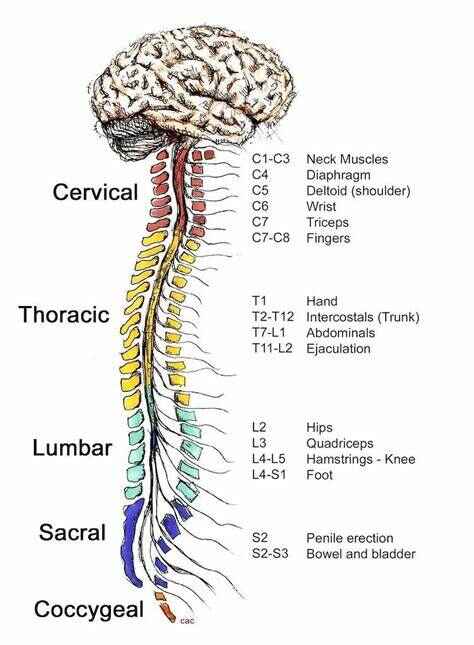
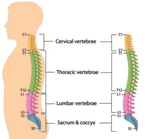
These nerves emerge from the segments of the spinal cord. They are numbered according to their segment of origin. Hence, the 31 pairs of spinal nerves are classified into 8 cervical pairs, 12 thoracic pairs, 5 lumbar pairs, 5 sacral pairs, & 1 coccygeal spinal nerve. All spinal nerves are mixed, made up of both sensory & motor fibers. Spinal nerves supply the entire body, except the head. They do so by either directly synapsing with their specific organs or by interlacing with each other & forming plexuses. There are four major plexuses that innervate the body regions;
Cervical plexus (C1-C4) – supplies the neck.
Brachial plexus (C5-T1) -supplies the upper limb.
Lumbar plexus (L1-L4) – supplies the lower abdominal wall, anterior hip & thigh.
Sacral plexus (L4-S4) -supplies the pelvis & the lower limb.
Ganglia
Ganglions are clusters of neuronal cell bodies outside of the central nervous system, meaning that they are the peripheral system equivalents to subcortical nuclei of the central nervous system. Ganglia can be sensory or motor (autonomic) & their distribution in the body is clearly defined. Dorsal root ganglia are clusters of sensory neuron cell bodies located adjacent to the spinal cord, They are a component of the posterior root of a spinal nerve.
Autonomic ganglia: these are either sympathetic or parasympathetic.
Sympathetic ganglia are found in the thorax & abdomen, grouped into paravertebral & prevertebral ganglia. Paravertebral ganglia locate at either side of the vertebral comprising two ganglionic chains that extend from the base of the skull to the coccyx, known as sympathetic trunks. Prevertebral ganglia lie anterior to the vertebral column closer to their target organ. They are further grouped according to which branch of abdominal aorta they surround the celiac, aortic renal, superior & inferior mesenteric ganglia.
Parasympathetic ganglia are found in the head & pelvic region. Ganglia in the head are associated with relevant cranial nerves & are the ciliary, pterygopalatine, otic & submandibular ganglia. Pelvic ganglia locate close to the reproductive organs comprising autonomic plexuses for innervation of pelvic viscera
Somatic nervous system
This system is the voluntary component of the PNS. It consists of all the fibers within cranial & spinal nerves that enable us to perform voluntary movements of the body (efferent nerves) & feel sensation from the skin, muscles, and joints (afferent nerves). Somatic sensation relates to touch, pressure, vibration, pain, temperature, stretch, and position awareness from these three types of structures. Sensation from the glands, smooth muscles & cardiac muscles is transferred by the autonomic nerves.
Parts of the Somatic Nervous System:
This system transfers information back & forth between the CNS & throughout the body.
this system contains two main types of nerve cells:
Sensory neurons: it is also known as afferent neurons sensory neurons are responsible for carrying information from the body to the central nervous system.
Motor neurons: it is also known as efferent neurons. motor neurons are responsible for receiving information from the brain& spinal cord to muscle fibers throughout the body.
Diseases of the Somatic Nervous System:
Somatic nervous system diseases are those that damage the peripheral nerves fiber outside of the brain & spinal cord. Diseases that impact the peripheral nerve fibers of the somatic nervous system can cause peripheral neuropathy. This nerve damage can be caused by any physical injury or trauma, diabetes, blood or vein issues, or autoimmune diseases.
Some somatic nervous system diseases are:
Brachial plexus neuropathy: it leads to damage to nerves in the upper shoulder region that results in pain in the shoulders or arms.
Guillain-Barre syndrome: in this disease, the immune system attacks the nerve fibers.
Myasthenia gravis: it is an autoimmune condition that results in weakness of the muscles & fatigue.
Nerve compression syndromes: it results in pain, weakness of the muscle, numbness, or tingling due to a pinched nerve.
Trigeminal neuralgia: it is a neuropathic condition causing shock-like pain or burning sensation in the face.
Autonomic nervous system
The ANS is the involuntary part of the PNS. Further classified into the sympathetic (SANS) & parasympathetic (PANS) systems, It consists exclusively of visceral motor fibers. Nerves from both these divisions supply all involuntary structures of the body.
- Cardiac muscle
- Glandular cells
- Smooth muscles
Sympathetic nervous system
SANS adjust our bodies for situations of raised physical activity. Its actions are mainly described as the “fight-or-flight” response as it stimulates responses such as faster breathing, increased heart rate, increased blood pressure, dilated pupils & redirection of blood flow from the skin, kidneys, stomach & intestines to the heart & muscles. Sympathetic nerve fibers have a thoracolumbar origin which stems from the T1-L2/L3 spinal cord segments. They synapse with the prevertebral & paravertebral ganglia, from which the postsynaptic fibers travel to innervate the target viscera.
Parasympathetic nervous system:
The PSNS adjusts our bodies for energy conservation, activating “rest & digest” or “feed & breed” activities. The nerves of the PSNS slow down the actions of CVS, divert blood away from muscles & increase peristalsis & gland secretion. Parasympathetic fibers have craniosacral outflow which originates from the brainstem & S2-S4 spinal cord segments. These fibers travel to thoracic & abdominal organs, where they synapse in ganglia situated close to or within the target organ.
Enteric nervous system:
The enteric nervous system consists of the SANS & PANS fibers that regulate the activity of the GI tract. This system is made of parasympathetic fibers of the vagus nerve (CN X) & sympathetic fibers of the thoracic splanchnic nerves. These fibers form 2 plexuses within the wall of the intestinal tube and are responsible for modulating intestinal peristalsis, i.e. propagation of consumed food from the esophagus to the rectum.
The submucosal plexus is found in the submucosa of the intestines & contains only parasympathetic fibers. Myenteric plexus situated in the muscularis externa of the intestines, containing both sympathetic & parasympathetic nerve fibers.
FAQ
Why is the brain important?
The brain controls & coordinates actions and reactions, allows us to think & feel, and allows us to have memories & feelings—all the things that make us human. While the brain only weighs about 3 pounds, the brain is a highly complex organ made up of many parts.
What is the function of the midbrain?
Its functions are the movement of the body & head. It is a channel for the spinal cord to transfer sensory information from the head & body directly to the brain.
What do you know about the nervous system?
The human nervous system comprises the brain, spinal cord, sensory organs & all of the nerves that connect all organs of the body with the rest of the body
What unit makes up the nervous system?
The unit which makes up the nervous system are known as neurons. D. The brain & the spinal cord along with nerves constitute the human nervous system.
What is the difference between CNS & PNS?
The CNS is made up of the brain & spinal cord & the PNS consists of nerves. We can say that the brain is the boss of the human body; the brain controls everything that you do.
24 Comments