
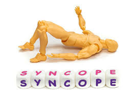
Syncope
Table of Contents
What is a Syncope?
Syncope is a medical term for fainting where sudden onset, brief loss of consciousness with loss of postural tone followed by spontaneous revival. The person is motionless and limp and generally has cool extremities, a weak pulse, and shallow breathing. Sometimes brief involuntary muscle jerks happen, resembling a seizure.
Near-syncope is slight-headedness and a sense of an impending faint without any kind of loss of consciousness. It is generally classified and discussed with syncope because the causes are the same.
- Seizures may cause onset loss of consciousness but are not considered syncope.
- However, seizures must be considered in person presenting for apparent syncope because history can be unclear or not available, and some seizures do not cause tonic-clonic convulsions.
- Furthermore, a brief (< 5 seconds) seizure sometimes happens with true syncope.
- Diagnosis is based on a careful history, eyewitness accounts, or fortuitous examination at the time of the event.
Pathophysiology of Syncope
Most syncope outcomes are from insufficient cerebral blood flow. Some cases include adequate flow but with an insufficient cerebral substrate (oxygen, glucose, or both).
- Insufficient cerebral blood flow
- Most deficiencies in cerebral blood flow outcome from the reduced cardiac output (CO).
Decreased CO may be caused by:
- Cardiac disorders that obstruct outflow
- Cardiac disorders of systolic dysfunction
- Cardiac disorders of diastolic dysfunction
- Arrhythmias (too quick or too slow)
- Conditions that reduce venous return
- Outflow obstruction may be aggravated by exercise, vasodilation, and hypovolemia (particularly in aortic stenosis and hypertrophic cardiomyopathy), which can precipitate syncope.
- Arrhythmias cause syncope when the heart rate is too quick to permit adequate ventricular filling (eg > 150 to 180 beats/minute) or too slow to give adequate output (eg, < 30 to 35 beats/minute).
- A venous return may be reduced by hemorrhage, improved intrathoracic pressure, improved vagal tone (which may also reduce heart rate), and loss of sympathetic tone (for example, from drugs, carotid sinus pressure, autonomic dysfunction).
- Syncope including these mechanisms (except for hemorrhage) is frequently termed vasovagal or neurocardiogenic and is usual and benign.
- Orthostatic hypotension, a usually benign cause of syncope, outcome of the failure of normal mechanisms (for example, sinus tachycardia, vasoconstriction, or both) to compensate for the temporary reduction in venous return that happens with standing.
- Cerebrovascular disorders (for example, strokes, and transient ischemic attacks) infrequently cause syncope because most of them do not involve the centrencephalic structures that must be affected to cause loss of consciousness.
- However, basilar artery ischemia, because of transient ischemic attack, stroke, or migraine, can cause syncope.
- Infrequently, patients with severe cervical arthritis or spondylosis generate vertebrobasilar insufficiency with syncope when the head is moved in certain positions.
Insufficient cerebral substrate
- The central nervous system (CNS) needs oxygen and glucose to function.
- Even with usual cerebral blood flow, a significant deficit of either will cause loss of consciousness.
- In practice, hypoglycemia is the first cause due to hypoxia infrequently developing in a manner causing abrupt loss of consciousness (other than in flying or diving incidents).
- Loss of consciousness because of hypoglycemia is seldom as abrupt as in syncope or seizures because warning symptoms happen (except in patients taking beta-blockers); however, the onset can be unclear to the physician unless the event was witnessed.
Symptoms of syncope

A person can experience several symptoms shortly before a person faints. Common symptoms may involve:
- Feeling dizzy or lightheaded
- Cold or clammy skin
- Feelings of weakness or unsteadiness
- Headache
- Nausea
- Changes in vision, like blurry vision, tunnel vision, or seeing spots
- Ringing in the ears
- Yawning or feeling tired
- Blacking out
Causes of Syncope
Causes are generally classified by the mechanism. The most usual causes are:
- Vasovagal (neurocardiogenic)
- Idiopathic
- Many cases of syncope never have a firm diagnosis but lead to no apparent damage.
- A little number of cases have a serious cause, generally cardiac.
Different types of syncope
There are many types of syncope, each with a different cause. Sometimes, though, the cause of fainting may not be identified. It is approximate that 10 to 40 percent of fainting cases have an unknown cause. Let’s take a nearby look at some of the most usual types of syncope or fainting episodes.
Reflex syncope
Reflex syncope, also identify as neurally mediated syncope is the most usual type of fainting. It occurs when certain reflexes are not perfectly regulated. This may cause the heart to decrease and a drop in blood pressure. In turn, this may reduce the flow of blood to the brain.
There are 3 types of reflex syncope:
Vasovagal:
- This occurs when the body overreacts to a trigger.
- There are many types of triggers, which may involve things such as intense pain, distress, or standing too long.
- Vasovagal syncope accounts for fifty percent of all cases of fainting.
Situational:
- This type of fainting occurs when a person performs certain actions, like laughing, coughing, or swallowing.
Carotid sinus:
- This kind of fainting occurs when pressure is placed on the carotid artery, located in the neck.
- Fainting may happen because of particular neck motions, wearing shirts with a tight collar, or shaving.
- In individuals with reflex syncope, fainting is frequently preceded by symptoms like:
- Lightheadedness
- Nausea
- Feelings of warmth
- Tunnel vision
- Visual blackout or “gray out”
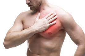
Cardiac syncope
Cardiac syncope is fainting caused by a problem with the heart. When the heart is not working quite as it should, the brain receives less blood. It is approximated that cardiac syncope causes about fifteen percent of fainting episodes. Several factors may cause cardiac syncope, involving:
- Structural problems with the heart, like ischemic cardiomyopathy, heart valve disorders, and dilated cardiomyopathy
- Electrical problems with the heart, like arrhythmias, and conditions such as Brugada syndrome
- Other conditions, like a pulmonary embolism or aortic dissection
Usual characteristics of cardiac syncope involve:
- Experiencing chest pain or heart palpitations prior to fainting
- Having fainting symptoms at the time of exercising or exerting yourself
- Fainting while people are lying down
Risk factors for cardiac syncope involve:
- Being older than 60
- Being male
- Having heart disease
- Having a hereditary history of heart conditions or fainting
Orthostatic syncope
Orthostatic syncope occurs because of a drop in blood pressure when a person stands up. The drop in blood pressure happens because of the effects of gravity. usually, the brain works to stabilize this. But in orthostatic syncope, this does not take place. As a result, it may lead to fainting. There are too many possible causes for this kind of fainting. They may involve :
- Dehydration, because of not drinking sufficient fluids, or from conditions such as vomiting or diarrhea
- Blood loss
- Medications, like some blood pressure medications, antidepressants, and diabetes drugs
- Alcohol use
- Underlying health conditions, like diabetes, Parkinson’s disease, or multiple sclerosis
- Symptoms are generally consistent with the warning signs that are usually experienced prior to a fainting episode.
- However, orthostatic syncope can also occur suddenly, without warning.
Cerebrovascular syncope
This kind of syncope occurs because of a problem with the blood vessels in and around the brain that may prevent the brain from getting sufficient blood. There are too many different types that may cause this kind of fainting, but they are not the usual causes of syncope. They may involve:
- Injury from the cerebrovascular disease may involve things such as stroke, carotid stenosis, and aneurysms.
- Basilar artery disease is a condition that may decrease the blood flow through the basilar arteries in the brain.
- Steal syndrome, is a reversal of blood flow in the subclavian arteries that supply blood to the arms.
Some symptoms that can happen with cerebrovascular causes of fainting involve:
- Feeling dizzy or lightheaded
- Headache
- Uncoordinated movements
- Trouble hearing
- Confusion
Risk factors for this type of fainting can involve:
- Older age
- Cardiovascular diseases, like atherosclerosis, high blood pressure, or high cholesterol
- Cerebrovascular disease
Diagnosis of Syncope
Evaluation should be done as early as possible after the event. The more faraway the syncopal event, the more difficult the diagnosis. Information from witnesses is quite helpful and best obtained as early as possible.
History
- History of current illness should ascertain events leading up to the syncope, involving the patient’s activity (for example, exercising, arguing, in a potentially emotional situation), position (such as, lying or standing), and, if standing, for how long.
- Important connected symptoms immediately prior to or after the event involve whether there was a sense of impending loss of consciousness, nausea, sweating, blurred or tunnel vision, tingling of lips or fingertips, chest pain, or palpitations.
- The length of time to recover should also be increased. Witnesses, if any, should be sought and asked to describe events, specifically the presence and at the time of any seizure activity.
- Review of systems should ask about any part of pain or injury, episodes of dizziness or near-syncope upon arising, and episodes of palpitations or chest pain with exertion.
- Patients should be asked about symptoms suggesting possible causes, involving bloody or tarry stools, heavy menses (anemia); vomiting, diarrhea, or excess urination (dehydration or electrolyte abnormalities); and risk factors for pulmonary embolism (current surgery or immobilization, known cancer, previous clots or hypercoagulable state).
- Past medical history should ask about past syncopal events, known cardiovascular disease, and known seizure disorders.
- Drugs utilize should be detected (particularly antihypertensives, diuretics, vasodilators, and antiarrhythmics).
- Hereditary history should note the presence at a young age of heart disease or sudden death in any family member.
Physical examination
- Vital signs are important. Heart rate and blood pressure are measured with the patient supine and after three minutes of standing.
- The pulse is palpated for irregularity.
- The general examination notes the patient’s mental status, involving any confusion or hesitancy suggesting a postictal state and any signs of injury (for example, bruising, swelling, tenderness, tongue bite).
- The heart is checked for murmurs; if present, any change in the murmur with a Valsalva maneuver, standing, or squatting is noted.
- Particular evaluation of the jugular venous waves at the time of palpating the carotid or auscultating the heart can allow diagnosis of arrhythmia if an ECG is not available.
- For example, cannon “a” waves happen when the atria contraction takes place against a closed tricuspid valve and indicate atrial-ventricular dissociation.
- The abdomen is palpated for tenderness, and a rectal examination is performed to check for gross or occult blood.
- A neurologic examination is performed to identify any focal abnormalities, which recommends a central nervous system cause (for example, seizure disorder).
Red flags
Few findings recommend a more serious cause:
- Syncope at the time of exertion
- Multiple recurrences within a short time
- Heart murmur or other findings suggesting structural heart disease (For example, chest pain)
- Older age
- Significant injury at the time of syncope
- The familiar history of sudden unexpected death, exertional syncope, or unexplained recurrent syncope or seizures
- Interpretation of findings
- Although the cause is frequently benign, it is important to identify the occasional life-threatening cause (for example, tachyarrhythmia, or heart block) because sudden death is a risk.
- Clinical findings help suggest a cause in 40 to 50% of cases. A few generalizations are useful.
Benign causes frequently lead to syncope.
- Syncope is precipitated by unpleasant physical or emotional stimuli (For example, pain, fright), generally happening in the upright position and frequently preceded by vagally mediated warning symptoms (For example, nausea, weakness, yawning, apprehension, blurred vision, diaphoresis), suggests vasovagal syncope.
- Syncope that happens most frequently when assuming an upright position (particularly in elderly patients after prolonged bed rest or in patients taking drugs in certain classes) recommends orthostatic syncope.
- Syncope that happens after standing for long periods without moving is generally because of venous pooling.
- Dangerous causes are recommended by red flag findings.
- Syncope with exertion recommends cardiac outflow obstruction or exercise-incorporate arrhythmia.
- A few patients sometimes also have chest pain, palpitations, or both.
- Cardiac findings can help identify a cause.
- A harsh, late-peaking, basal murmur radiating to the carotid arteries recommends aortic stenosis; a systolic murmur that improves with the Valsalva maneuver and disappears with squatting recommends hypertrophic cardiomyopathy.
- Syncope that starts and ends suddenly and spontaneously is typical of cardiac causes, most usually an arrhythmia.
- Syncope at the time of sleeping down also evaluates an arrhythmia because vasovagal and orthostatic mechanisms do not cause syncope in the recumbent position.
- Syncope accompanied by injury at the time of the episode improves the likelihood of a cardiac cause or seizure, and therefore the event is of greater concern.
- The warning signs and normal loss of consciousness that accompany benign vasovagal syncope somewhat decrease the likelihood of injury.
- Loss of consciousness at the time of a seizure or postictal confusion may sometimes be confused with syncope, but muscular jerking or convulsions that last more than a few seconds, incontinence, drooling, or tongue biting, if present, generally point to a seizure.
Testing
Testing typically is done.
- ECG
- Pulse oximetry
- Sometimes echocardiography
- Sometimes tilt table testing
- Blood tests only if clinically indicated
- Central nervous system imaging infrequently indicated
- In general, if syncope outcomes in an injury or is recurrent (particularly within a brief period), a more intensive diagnosis is warranted. Cardiac and brain imaging is not done unless suggest by clinical findings (suspected cardiac etiology or neurologic deficits).
Patients with suspected arrhythmia, myocarditis, or ischemia could be diagnosed as inpatients. Others can be diagnosed as outpatients.
ECG
- It is done for all patients. The ECG may recognize arrhythmia, a conduction abnormality, ventricular hypertrophy, pre-excitation, QT prolongation, pacemaker malfunction, myocardial ischemia, or myocardial infarction.
- If the evaluation is questionable after this basic evaluation, measuring cardiac markers and obtaining serial ECGs to find out MI in older patients plus ECG monitoring for at least twenty-four hours is prudent.
- Any detected arrhythmia must be connected with altered consciousness in order to be implicated as the cause, but most patients do not feel syncope at the time of monitoring.
- Furthermore, the presence of symptoms in the absence of rhythm disturbance helps rule out a cardiac cause.
- An event recorder (which can record cardiac rhythm for longer periods) can be useful if warning symptoms precede syncope.
- A signal-averaged ECG can identify a predisposition to ventricular arrhythmias in patients with ischemic heart disease or in post-myocardial infarction patients.
- If syncopal episodes are rare (eg, < 1/month), an implantable loop recorder may be used for longer-term recording.
- This device continuously records the rhythm and may be interrogated by an external machine that permits the cardiac rhythm to be printed.
Pulse oximetry
- It should be performed at the time of or immediately after an episode to identify hypoxemia (which can suggest pulmonary embolism).
- If hypoxemia is present, CT angiography is indicated to find out pulmonary embolism.
Laboratory tests
- These tests are done depending on clinical suspicion; reflexively obtained laboratory panels are of little use.
- However, all females of childbearing time should have a pregnancy test.
- Hematocrit is done if anemia is suspected.
- Electrolytes are measured only if an abnormality is clinically recognized (For example, by symptoms or drug use). Serum troponin is done if acute myocardial infarction is suspected.
Echocardiography
- It is suggested for patients with clinically unexplained syncope, exercise-induced syncope, cardiac murmurs, or suspected intracardiac tumors (For example, those with positional syncope).
- Tilt table testing can be performed if history and physical examination suggest vasodepressor or another reflex-induced syncope.
- It is also used to diagnose exercise-induced syncope if echocardiography or exercise stress testing is negative.
Stress testing
- It is (exercise or pharmacologic) perform when intermittent myocardial ischemia is suspected.
- It is frequently done for patients with exercise-induced symptoms.
- Exercise testing is not that much valuable unless physical activity precipitated syncope.
Invasive electrophysiologic testing is considered if noninvasive testing does not recognize arrhythmia in patients with any of the following:
- Unexplained recurrent syncope
- Unexplained red flag findings
- Ischemic cardiomyopathy, non-ischemic cardiomyopathy, adult congenital disease, or unexplained syncope that does not otherwise consider criteria for an ICD used for primary prevention
- A negative response defines a lower-risk subgroup with a greater rate of remission of syncope.
- The use of electrophysiologic testing is controversial in other patients.
EEG
- It is warranted if a seizure disorder is suspected.
CT and MRI
- These two of the head and brain are suggested only if signs and symptoms suggest a focal CNS disorder.
Differential Diagnosis
The underlying causes of syncope may be classified into 4 categories:
Reflex-mediated:
- It has 3 different variations which are vasovagal, carotid sinus, and situational.
- This kind of syncope has no improved risk of morbidity and mortality. Vasovagal syncope happens because of an inciting event or stimulus like stress, pain, and prolonged standing.
- The stimulus is frequently identifiable and lying down or removing the stimulus generally aborts the syncopal episode.
- Carotid sinus syncope is connected with syncope after head-turning, shaving, and wearing a tight collar and may be relieved with a carotid sinus massage.
- Situational syncope is connected with micturition, defecation, coughing, and gastrointestinal stimulation.
Cardiac-induced:
- Coronary artery disease, congestive heart failure, and myocarditis all are precursors of syncope.
- A person with cardiac disease is at greater risk for developing recurrent syncopal episodes than another person with syncope.
- Patients with cardiac-induced syncope have a greater risk of all-cause mortality and an improved risk of fatal and nonfatal cardiovascular events.
Orthostatic causes:
- Orthostatic hypotension is the declination in blood pressure within three minutes of standing which leads to tachycardia and blood volume depletion and thus syncope.
- It is usually found in elderlies and exacerbated with dehydration and a few medications such as anti-hypertensives, anti-depressants, and diuretic agents.
Cerebrovascular causes:
- Although syncope from cerebrovascular disease is very infrequent, transient ischemia can outcome from vertebrobasilar insufficiency and can cause syncope.
- Headaches, vertigo, dysarthria, and diplopia are recognizable signs of syncope from neurologic causes.
Treatment of Syncope
- In witnessed syncope, pulses are checked straight away.
- If the patient is pulseless, CPR is started.
- If pulses are available, severe bradycardia is treated with atropine or external transthoracic pacing.
- Isoproterenol may be utilized to maintain an adequate heart rate while a temporary pacemaker is placed.
- Tachyarrhythmias are treated; a direct-current synchronized shock is faster and safer than drugs for an unstable person.
- Insufficient venous return is treated by keeping up the patient supine, raising the legs, and giving IV normal salines.
- Tamponade is decreased by pericardiocentesis.
- Tension pneumothorax needs the insertion of a pleural cannula and drainage.
- Anaphylaxis is treated with parenteral epinephrine.
- Putting the person in a horizontal position with legs elevated typically ends the syncopal episode if life-threatening disorders find out.
- If the person sits upright too quickly, syncope can recur; propping the person upright or transporting the patient in an upright position can longtime cerebral hypoperfusion and prevent recovery.
- particular treatment based on the cause and its pathophysiology.
- Driving and use of machinery should be restricted until the cause is determined and treated.
Geriatrics Essentials: Syncope
- The most usual cause of syncope in elder adults is postural hypotension because of a combination of factors.
- Factors involve rigid, noncompliant arteries, decreased skeletal muscle pumping of venous return because of physical inactivity, and degeneration of the sinoatrial node and conduction system because of progressive structural heart disease.
- In older adults, syncope frequently has more than one cause.
- For example, the combination of taking several heart and blood pressure drugs and standing in a hot church at the time of a long or emotional service can lead to syncope even though no single factor can cause syncope.
Vasovagal Syncope:
- In avoiding situations that trigger a syncopal attack, tilt training which includes exercise sessions with prolonged upright posture and the use of salt and fluid would help prevent the decline in blood pressure which in return permits bringing enough oxygen to the brain tissue and thus prevent reoccurrence of syncope and in case conservative measures fail, drug therapy using beta blockers and other medications may be used.
- Pacemaker implantation is an invasive, permanent procedure, it should be considered only in person with the greatest benefit-to-risk ratio especially patients older than forty years of age with recurrent syncope causing injury and reduced quality of life.
Orthostatic Hypotension:
- Avoid rising rapidly from a supine or sitting position, avoid medications that trigger orthostatic hypotension like diuretics and vasodilators, and use compression stockings in order to improve venous return.
Cardiac-induced Syncope:
- The treatment is based on the underlying cardiac condition.
Prevention of fainting
There are many steps a person can take that may help prevent fainting:
- Do not skip meals. A person may want to eat smaller, more frequent meals throughout the day.
- Drink plenty of fluids. This can help prevent fainting because of dehydration.
- Understand if there are outside factors or triggers that may cause a person to faint.
- This could involve the sight of blood, getting an injection, or intense pain.
- If possible, try to avoid situations that can trigger a fainting episode.
- Take your time when standing up.
- Standing up too fast may cause a drop in blood pressure and prevent sufficient blood from flowing to the brain.
- Avoid shirts with tight collars. This may help prevent carotid sinus syncope.
FAQ
In most cases, syncope is not a sign of a life-threatening problem, although some individuals with syncope do have a serious underlying medical condition. In young individuals and children, most cases of syncope are not connected with an underlying medical problem.
Syncope is caused by a provisional global failure of cerebral perfusion. When cardiac, the brain is not perfused due to the heart is failing to create sufficient cardiac output to send its freshly oxygenated blood to the brain.
Syncope is the medical term for fainting or passing out. It is caused by a provisional drop in the amount of blood that flows to the brain. This leads to loss of consciousness and muscle control. An individual then falls down or over, which permits blood flow to return to the brain.
Syncope may also be a symptom of heart disease or other heart problems. It also can show a greater risk for some neurological conditions such as neuropathy. The symptoms of syncope that generally occur prior to someone losing consciousness involve: Feeling dizzy.
Syncope must be differentiated from other states of altered consciousness, like cardiac arrest, coma, seizures, vertigo, dizziness, lightheadedness, and presyncope. Cardiac arrest is an onset loss of consciousness without spontaneous recovery and needs electrical or pharmacologic cardioversion.

One Comment