
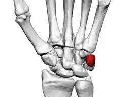
Pisiform Bone
Table of Contents
Introduction
The pisiform bone, also known as the pisiform or os pisiforme, is a small, pea-shaped bone located in the human hand. It is one of the eight carpal bones that make up the wrist joint, specifically positioned on the ulnar (pinky finger) side of the hand. The word “pisiform” is derived from the Latin term “pisum,” meaning “pea,” which aptly describes its size and shape.
Its main function is to provide support and stability to the wrist, as well as aid in the movement of the hand and fingers. Despite its small size, the pisiform bone plays an important role in the overall functioning of the wrist and hand.
Anatomy of Pisiform Bone
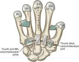
The pisiform bone is a small, pea-shaped bone located in the proximal row of the carpal bones in the wrist. It is situated on the ulnar side of the wrist, just below the triquetrum bone. The pisiform bone is named after its resemblance to pea (Pisum in Latin).
Anatomy:
Shape and Size: The pisiform bone is typically described as a rounded, oval-shaped bone. It is the smallest bone in the carpal region, measuring about 1 cm in length and width.
Articulations: The pisiform bone articulates with several neighboring bones:
- Triquetrum: The pisiform bone articulates with the triquetrum bone through a synovial joint, forming a pivot-like connection.
- Hamate: The pisiform bone also forms a fibrocartilaginous joint with the hamate bone, providing additional support and stability to the carpal region.
Surfaces: The pisiform bone has two main surfaces:
- Palmar Surface: The anterior surface of the pisiform bone faces the palm of the hand. It is smooth and slightly concave, allowing for the attachment of ligaments and tendons.
- Dorsal Surface: The posterior surface of the pisiform bone faces the back of the hand. It is rougher and provides attachment sites for muscles and ligaments.
Ligamentous Attachments: The pisiform bone serves as an attachment point for several important ligaments and tendons:
- Flexor Retinaculum: The pisiform bone is connected to the flexor retinaculum, a strong fibrous band that forms the roof of the carpal tunnel.
- Pisohamate Ligament: A fibrous band extends from the pisiform bone to the hamate bone, forming the pisohamate ligament.
- Abductor Digiti Minimi: The abductor digiti minimi muscle attaches to the pisiform bone, aiding in the abduction and flexion of the little finger.
Articulation of Pisiform Bone
The pisiform bone articulates with several neighboring bones and structures in the wrist and hand. Let’s delve into each articulation in detail:
- Triquetrum Bone: The pisiform bone forms a synovial joint with the triquetrum bone, which is located on the ulnar side of the wrist. This joint is known as the pisiform-triquetral joint or the pisiform-triquetrum articulation. It is a gliding joint, allowing limited movement and providing stability to the wrist. This articulation is supported by a joint capsule, ligaments, and surrounding soft tissues.
- Flexor Retinaculum: The pisiform bone is positioned on the palmar side of the wrist, contributing to the formation of the carpal tunnel. A small opening called the carpal tunnel is found on the front of the wrist. The flexor retinaculum, a band of tough connective tissue, extends across the wrist, forming the roof of the carpal tunnel. The pisiform bone acts as an attachment point and helps to anchor the flexor retinaculum.
- Muscles and Ligaments: The pisiform bone serves as an attachment site for several muscles and ligaments, which play important roles in wrist and hand movements. The flexor carpi ulnaris muscle, one of the forearm muscles, arises from the medial epicondyle of the humerus and attaches to the pisiform bone. This muscle aids in the flexion of the wrist and adduction of the hand.
Additionally, the ulnar collateral ligament of the wrist, also known as the radial collateral ligament, attaches to the pisiform bone. This ligament provides stability and prevents excessive sideways movement of the wrist joint.
Ligament attachment of the Pisiform Bone
The pisiform bone has several ligamentous attachments that contribute to the stability and function of the wrist joint. These ligaments include the pisohamate ligament, the ulnar collateral ligament, and the transverse carpal ligament.
- Pisohamate Ligament: The pisohamate ligament connects the pisiform bone to the hamate bone, which is located next to it in the wrist. This ligament helps stabilize the pisiform bone and prevents excessive movement or displacement. It also aids in transmitting forces from the hand to the forearm, contributing to grip strength and control.
- Ulnar Collateral Ligament: The ulnar collateral ligament attaches to the pisiform bone on one end and to the triquetrum bone on the other end. This ligament provides stability to the ulnar side of the wrist joint, preventing excessive deviation or movement of the hand towards the pinky finger side. It also assists in maintaining proper alignment of the carpal bones and supports the overall stability of the wrist.
- Transverse Carpal Ligament: The transverse carpal ligament, also known as the flexor retinaculum, is a thick band of fibrous tissue that stretches across the front of the wrist. It attaches to the pisiform bone on one end and connects to the other carpal bones on the opposite end. This ligament forms the roof of the carpal tunnel, a narrow passageway through which the median nerve and several tendons pass. The attachment of the transverse carpal ligament to the pisiform bone helps anchor and stabilize it, preventing excessive movement or displacement during wrist movements.
The ligaments attached to the pisiform bone provide stability, support, and protection to the wrist joint. They help maintain proper alignment of the carpal bones, transmit forces efficiently, and prevent excessive movement or displacement of the hand. These ligaments play a crucial role in maintaining the overall function and integrity of the wrist.
Functions of the Pisiform Bone
The pisiform bone is a small, pea-shaped bone located in the wrist on the ulnar side (pinky finger side) of the hand. Although it is one of the smallest bones in the body, it serves several important functions:
- Support and stability: The pisiform bone acts as a support structure for the carpal bones in the wrist. It helps maintain the alignment and stability of the wrist joint, allowing for smooth movements and preventing excessive stress on the surrounding structures.
- Attachment site for muscles and ligaments: The pisiform bone serves as an attachment point for various muscles and ligaments in the hand and forearm. The flexor carpi ulnaris muscle, which is responsible for wrist flexion and ulnar deviation, attaches to the pisiform bone. Additionally, the pisohamate ligament connects the pisiform bone to the hamate bone, providing additional stability to the wrist joint.
- Transmission of forces: During gripping or grasping activities, forces are transmitted through the pisiform bone to the ulna and radius bones of the forearm. This helps distribute the forces evenly and efficiently, reducing the risk of injury or strain on other structures in the hand and wrist.
- Protection: The pisiform bone is positioned on the ulnar side of the hand, providing some protection to the underlying structures such as nerves and blood vessels. It acts as a cushion, absorbing and dispersing forces that may be directed toward the ulnar side of the hand.
- Enhancing grip strength and control: The pisiform bone plays a role in enhancing grip strength and control by providing a point of leverage for the flexor carpi ulnaris muscle. This muscle helps generate power during gripping activities, allowing for better control and manipulation of objects held in the hand.
Overall, while the pisiform bone may be small, its functions are vital for maintaining stability, transmitting forces, protecting underlying structures, and enhancing hand and wrist function.
Blood and lymph supply
The blood supply to the pisiform bone comes from multiple sources. The main arteries that supply blood to the pisiform bone are the ulnar artery and the anterior interosseous artery.
The ulnar artery is a branch of the brachial artery, which runs along the inner side of the forearm. It gives off a small branch called the pisiform artery, which specifically supplies blood to the pisiform bone. The pisiform artery enters the bone through small openings called nutrient foramina, providing oxygen and nutrients to the bone tissue.
The anterior interosseous artery is a branch of the ulnar artery that also contributes to the blood supply of the pisiform bone. It sends small branches into the wrist joint, including the pisiform branch, which supplies blood to the pisiform bone.
In addition to the arterial blood supply, the pisiform bone also receives lymphatic drainage. Lymphatic vessels are liable for draining extra fluid and waste products from tissues. The lymphatic vessels surrounding the pisiform bone drain into regional lymph nodes located in the wrist and hand region.
The lymph nodes filter and cleanse the lymph fluid before it is returned to the bloodstream. This process helps remove any potential pathogens or foreign substances that may have entered the pisiform bone or surrounding tissues.
Overall, a healthy blood and lymph supply is crucial for maintaining the proper functioning and integrity of the pisiform bone. Adequate blood flow ensures that the bone receives enough oxygen and nutrients for its metabolic needs, while proper lymphatic drainage helps maintain tissue health and remove waste products.
Conditions that affect Pisiform Bone
There are several associated conditions that can affect the pisiform bone. These conditions may result from trauma, overuse, or underlying medical conditions. Here are some examples:

Pisiform Fracture: A fracture of the pisiform bone can occur due to direct trauma, such as a fall onto an outstretched hand or a forceful impact on the wrist. Symptoms may include pain, swelling, difficulty moving the wrist, and tenderness over the pisiform bone. Treatment typically involves immobilization with a splint or cast, and in severe cases, surgery may be necessary to realign the bone fragments.
Pisotriquetral Joint Dysfunction: The pisotriquetral joint is the articulation between the pisiform and triquetrum bones. Dysfunction of this joint can result from repetitive strain, excessive use, or injury. Symptoms may include pain, swelling, clicking or popping sensations, and limited range of motion in the wrist. Treatment options may include rest, immobilization, physical therapy, and occasionally surgical intervention.
Pisiform Impingement Syndrome: This condition occurs when the pisiform bone becomes compressed or irritated, often due to repetitive activities or excessive pressure on the wrist. Symptoms may include pain, tenderness, swelling, and weakness in the wrist and hand. Treatment may involve rest, activity modification, splinting, physical therapy, and in some cases, corticosteroid injections or surgical intervention.
Pisiform Dislocation: Dislocation of the pisiform bone can happen as a result of trauma or excessive force applied to the wrist. Symptoms may include severe pain, swelling, visible deformity, and limited range of motion in the wrist. Immediate medical attention is required to relocate the dislocated bone and stabilize the joint. For a full recovery, immobilisation and physical therapy may be required.
Pisiform Osteoarthritis: Osteoarthritis is a degenerative joint disease that can affect any joint in the body, including the pisiform bone. It occurs when the protective cartilage in the joint wears down over time, leading to pain, stiffness, swelling, and limited mobility. Treatment options for pisiform osteoarthritis may include pain management, physical therapy, bracing, and in severe cases, surgical interventions such as joint fusion or joint replacement.
It is very necessary to take in mind that these conditions are relatively rare compared to other wrist and hand conditions. If you experience persistent pain, swelling, or any concerning symptoms in your wrist or hand, it is recommended to seek medical evaluation and appropriate treatment from a qualified health or exercise professional.
Rehabilitation
The rehabilitation of a pisiform bone injury or condition depends on the specific nature and severity of the problem. It is essential to consult with a healthcare professional, such as an orthopedic specialist or a physical therapist, for a personalized treatment plan. However, here are some general aspects of rehabilitation that may be involved in the recovery process:
- Immobilization: In cases of pisiform fractures or dislocations, immobilization is often the initial step in rehabilitation. A splint or cast may be used to immobilize the wrist and allow the bone to heal correctly. The duration of immobilization varies depending on the severity of the injury and can range from a few weeks to several months.
- Pain management: Pain is a common symptom associated with pisiform injuries. Pain management techniques may include the use of over-the-counter or prescription pain medications, ice or heat therapy, and modalities such as ultrasound or electrical stimulation.
- Range of motion exercises: Once the initial healing phase is complete, range of motion exercises are typically introduced to restore flexibility and mobility to the wrist joint. These exercises may involve gentle stretching, wrist rotations, and finger movements. It is crucial to perform these exercises under the guidance of a healthcare professional to avoid further damage or discomfort.
- Strengthening exercises: As the range of motion improves, strengthening exercises are gradually incorporated into the rehabilitation program. These exercises target the muscles surrounding the wrist and hand to enhance stability and support. Examples of strengthening exercises may include wrist curls, grip exercises, and resistance band exercises.
- Functional training: Functional training focuses on restoring the ability to perform everyday activities and tasks that may have been affected by the pisiform injury. This may involve specific exercises that mimic functional movements, such as grasping objects, writing, or using tools. The goal is to improve coordination, dexterity, and overall hand function.
- Activity modification and ergonomics: During the rehabilitation process, it is essential to identify and modify any activities or habits that may have contributed to the pisiform injury or condition. This may include adjusting workstations, using ergonomic tools or supports, and adopting proper body mechanics to prevent further strain on the wrist.
- Gradual return to sports or activities: A gradual return-to-sport or activity plan may be implemented for individuals involved in sports or activities requiring repetitive wrist movements. This plan aims to progressively increase the intensity and duration of the action while monitoring for any signs of pain or discomfort.
It is very necessary to take in mind that the rehabilitation process may vary for each individual, and the timeline for recovery can vary based on the severity of the condition. Working closely with a qualified doctor or any medical professional throughout the rehabilitation process is crucial to ensure proper healing, minimize complications, and achieve optimal functional outcomes.
Summary
The pisiform bone is one of the eight small carpal bones located in the wrist of the human hand. It is situated on the ulnar (pinky finger) side of the wrist, on top of the triquetrum bone. The pisiform bone is a pea-shaped bone, hence its name, derived from the Latin word “pisi” meaning pea.
Overall, while the pisiform bone may be small and often overlooked, it plays a significant role in providing stability and anchorage for ligaments and tendons in the wrist joint. Its position and attachments contribute to the overall function and movement of the hand and wrist.
FAQs
The pisiform bone serves multiple functions. It acts as an attachment point for ligaments and tendons, specifically the flexor retinaculum and the ulnar collateral ligament of the wrist. It also provides stability to the wrist joint and contributes to the overall movement of the hand and wrist.
Yes, the pisiform bone can be fractured. Fractures of the pisiform bone usually occur due to a direct blow or trauma to the wrist. Symptoms may involve pain, swelling, and difficulty wrist movement. Treatment typically involves immobilization and may require surgical intervention in severe cases.
Yes, issues with the pisiform bone can contribute to wrist pain. Conditions such as pisiform subluxation (partial dislocation) or pisiform impingement syndrome (compression of the pisiform bone) can cause pain and discomfort in the wrist. These conditions may arise due to repetitive motions, trauma, or underlying anatomical variations.
In some conditions, the pisiform bone can be surgically removed. This procedure, known as pisiform ectomy, is generally considered for cases in which the pisiform bone causes chronic pain or adversely affects hand and wrist function. The removal of the pisiform bone usually does not lead to significant impairment as other structures compensate for its absence.
Since the pisiform bone is not directly involved in major movements or weight-bearing, there are no specific exercises to strengthen it. However, exercises that focus on wrist flexion and ulnar deviation can help strengthen the muscles and tendons around the pisiform bone, promoting overall wrist stability and function.
Dislocation of the pisiform bone is relatively rare but can occur due to extreme trauma or force applied to the wrist. Dislocation may lead to pain, swelling, and limited wrist movement. Treatment typically involves repositioning the bone and immobilizing it to allow for healing.
4 Comments