
Knee locking and unlocking mechanism
Table of Contents
Definition
- The locking and unlocking mechanism is also known as a “Screw home mechanism (SHM)” of the knee joint is a critical mechanism that plays an important role in the terminal extension of a knee.
Description of the knee joint mechanism
- There is the observable rotation of a knee during flexion as well as extension.
- The rotation is important for the healthy motion of a knee.
- During the last 30 degrees of the knee extension, a tibia (open chain) or even femur (closed chain) must externally or internally rotate, respectively, about 10 degrees.
- This slight rotation is owing to the inequality of an articular surface of the femur condyles.
Rotation must happen to achieve full extension & then flexion from full extension. - Two of the most significant joints in the person’s body are the knees. Besides providing support to the person’s weight, the knees also enable different routine activities for instance standing up as well as sitting down, climbing stairs & walking. Because of such frequent usage of the knee joints, they are very much prone to degeneration as well as injury. Both of these may result in a locked knee.
- Momentary locking up of the knees (commonly called a locked knee) inhibits a person’s ability to move them in any direction.
- This phenomenon may also be explained as “catching”, the feeling experienced when a person’s knee gets caught while flexion or extension, or the knee joints “giving out” as the popping sensation along with any knee motion. Unfortunately, there exists no secret formula or even trick to unlock the knee joints. There are several treatments as well as physical therapy regimens which might assist in relieving the knee locking symptoms.
- The relatively complex joint, the human knee is composed of multiple bones, tendons, ligaments, and cartilage as well. The knee joints are primarily formed from the bones while the tendons, as well as muscles, act around these bones for an articulation of a joint. The anterior cruciate ligament or anterior crucial element ligament & other ligaments exist for providing stability & their work is to make sure that the knees only move in the manner they are supposed to. Cartilage, known as a medial as well as lateral “meniscus”, is there for providing the smooth motion of the knee joints along with stabilization as well as shock absorption.
Relevant Anatomy
- A knee joint is a basic range joint with the main motion of flexion and extension. However, the radius & the length of an articular surface of a femur, as well as tibia, differ at a knee joint. The articular surface of the medial condyle of the femur is greater than the articular surface of the lateral condyle.
- As a result, the complex movement (includes “sliding”) happens while the last 30 degrees of the knee extension, moreover to rolling between the two bones allows a knee joint to move smoothly.
- At the terminal extension, a knee joint is slightly hyperextended & stabilized with the tightening of a cruciate as well as collateral ligaments.
As the length of a medial femoral condyle is longer than the length of the lateral condyle, a tibia rotates externally at about 15° on the femur while the last 20° of extension. - This kinematic phenomenon is well known, and it is called the screw-home movement or locking mechanism.
- The shape of the condyles is not what brings about the motions.
Tibial-on-femoral rotation happens in the open chain exercise like in a leg extension machine (tibia externally rotates). - Femoral-on-tibial rotation, as in the closed chain exercise like a squat (femur internally rotates).
- During the knee extension, the tibia rolls anteriorly, elongating the posterior cruciate ligament(PCL) & the PCL’s pull on the tibia, causes it to glide anteriorly.
- During the knee flexion tibia rolls posteriorly, elongating the anterior cruciate ligament(ACL) & it is the ACL’s pull on the tibia, that causes it to glide posteriorly.
- Popliteus – Unlocks knee with open chain movement.
- Hip external rotation – Unlocks knee with closed chain motions.
Locking of the knee joint
- Closed kinematic chain extension from 30-degree knee flexion.
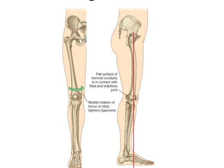
- The larger medial femoral condyle continue rolling as well as gliding posteriorly when the smaller lateral side stopped. These results in medial rotation of the femur on the tibia, in the last 5 degrees of the extension. A medial rotation of the femur at the final stage of extension is not voluntary or produced by muscular force, which is referred to as “Automatic” or even “Terminal Rotation”.
- A rotation within the knee joint brings a joint into the closed packed or locked position. The consequences of automatic rotation are also known as the “locking mechanism” or even the “screw home mechanism.”
- Open kinematic chain: Lateral rotation of the tibia on the femur.
True Knee Locking
- True locking at a knee is where a knee gets physically stuck & the patient physically cannot move a knee for a person times knee locking is caused by the mechanical block where something gets caught inside a knee joint, preventing movement.
- The truly locked knee is fairly rare & typically happens as the patient moves a knee into full extension, for instance towards being fully straight.
Pseudo Knee Locking
- Pseudo-knee locking is much more common than true locking, and knee motion is limited by temporary muscle spasming as a body tries to protect itself in response to the pain.
Unlocking the knee
- To initiate flexion, the knee should be unlocked.
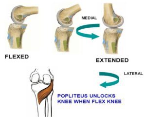
- The flexion force will automatically result in the lateral rotation of the femur.
- Due to the larger medial condyle will move before the shorter lateral condyle.
- Popliteus is the primary muscle to unlock the knee.
Characteristics of a locked knee
- The feeling of instability in the knees
- Pain while extending a knee
- Knee joint stiffness
- Swelling of the knee and around the affected knee
- The feeling of catching sensation when the knee is extended
- Difficulties in walking, climbing stairs, jumping, and running as well.
Causes behind the locked knee
- Damage to any of the structures with which a knee is made up (bone, ligaments, cartilage, tendons) might result in the locking of knee joints.
Musculoskeletal Causes: There are various musculoskeletal causes that may result in a locked knee. Wear as well as tear, tendon & muscle damage, damage to cartilage, ligament damage, inflammation as well as trauma are some of the main culprits. The infection or autoimmune disease may also lead to the locking of the knees. - Osteochondritis Dissecans: The joint condition, osteochondritis dissecans (OCD) happens when there is not ample flow of blood within the ends of the bone, beneath protective cartilage. Erosion of these layers of the bone embarks taking place making them separate from the main bone & also taking away cartilage with them. Children, as well as teenagers, are more susceptible to this condition.
- Injury to the Meniscus: The knees have two pieces of cartilage that serve as the shock absorbers known as the menisci. They are present in between the top of a shinbone & the lower end of a thighbone. The meniscus when torn is usually referred to as “torn cartilage” in the knees. Cartilage becomes worn out & thin with age in old people making the meniscus tear away.
- Patellar Subluxation: Repeated kneecap dislocation known as patellar subluxation refers to the continuous instability of a kneecap, which results in anterior knee pain, usually happening laterally.
- Knee Arthritis: Osteoarthritis results owing to the inflammation of one or more joints. The most common symptoms of arthritis are swelling, stiffness as well as pain. Though any joint in a body might be affected by this condition, osteoarthritis is specifically common in the knees.
- Loose Body
Another thing that may block a joint & cause true knee locking is when the small fragment of bone breaks off from a knee joint, known as the loose body, as well as floats around.
As with the meniscus tear, if it moves into the wrong place, it may get wedged in place & cause a joint to lock in the specific position. - Knee Injuries: Usually from the fall or even awkward twisting, for example, patella fractureKnee Arthritis: Bone spurs breaking off. The most common cause of knee locking is those over the age of 60.
- The most common causes of the pseudo locking at a knee include:
- Swelling: Excess fluid in the knee joint capsule may limit the motion owing to increased tension, preventing full flexion as well as extension.
- Inflammation: Inflammation of the structures in & around a knee can also limit motion. The most common causes of this are
rheumatoid arthritis as well as gout. - Patellar maltracking: The problem with a motion of a kneecap in a groove on the front of a knee may cause pseudo locking. This is usually very painful.
- Plica Syndrome: Irritation of a medial plica, the fold in a synovial tissue lining a knee joint may cause pseudo locking.
Clinical Significance
Restoration of the terminal extension is an important aim of a rehabilitation program. Terminal extension exercises, as well as terminal extension mobilizations, have important contributions to the restoration of the screw home movement /terminal extension. As does strengthening as well as stretching relevant muscles.
- Loss of the full knee extension range of motion (ROM) is a frequent finding in a population with knee osteoarthritis.
- Such loss of normal terminal knee extension may have important effects on knee mechanics while walking and standing as well.
- A primary aim of physical therapy is to have a positive effect on a patient’s ability to achieve terminal (end-range) knee extension while activities of daily living.
- A “screw-home” mechanism is considered to be a key element to knee stability for standing upright. A tibia rotates internally during a swing phase & externally during the stance phase.
- External rotation happens during the terminal degrees of knee extension & results in the tightening of both cruciate ligaments, which locks the knee.
- A tibia is then in the position of maximal stability with respect to the femur.
- In most knee issues generally, there is an insufficiency in terminal extension & vastus medialis obliquus function.

One Comment