
Cuboid Bone
Table of Contents
Introduction
The Cuboid bone is another small bone located in the foot. It is part of the midfoot and helps provide stability and support to the foot during movement. Common conditions or injuries associated with the cuboid bone include cuboid syndrome, cuboid subluxation, and cuboid fractures.
Cuboid syndrome occurs when the cuboid bone becomes misaligned, causing pain and discomfort. Treatment for cuboid syndrome usually involves rest, immobilization, and physical therapy to realign the bone. Cuboid fractures can occur due to trauma or repetitive stress, and treatment may involve immobilization, surgery, or a combination of both. It is important to seek medical attention if you suspect a cuboid fracture or any other foot injury to ensure proper diagnosis and treatment.
Anatomy of Cuboid Bone

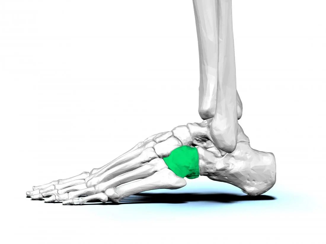
The cuboid bone is a small, cube-shaped bone located in the foot. It is one of the seven tarsal bones and is situated on the lateral side of the foot, between the calcaneus (heel bone) and the fourth and fifth metatarsals (bones of the foot). The cuboid bone forms joints with these adjacent bones, as well as with the navicular bone, forming the midfoot region.
The superior surface of the cuboid bone has a groove known as the peroneal sulcus. This groove serves as a passageway for tendons of the peroneus longus and brevis muscles. The peroneus longus tendon runs through this groove and helps in plantarflexion (pointing the foot downwards) and eversion (turning the foot outwards). The peroneus brevis tendon also passes through this groove and assists in the eversion of the foot.
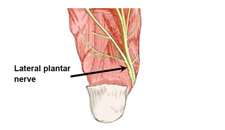
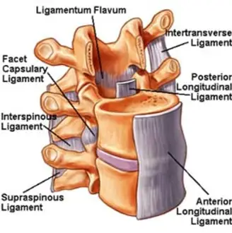
The lateral surface of the cuboid bone is rough and provides attachment sites for ligaments that help stabilize the foot during weight-bearing activities. These ligaments include the calcaneocuboid ligament, which connects the cuboid bone to the calcaneus, and the lateral plantar calcaneocuboid ligament, which connects the cuboid bone to the lateral plantar surface of the calcaneus.
On the plantar (bottom) surface of the cuboid bone, there is a tuberosity called the tuberosity of the cuboid. This prominence serves as an attachment point for muscles and ligaments involved in maintaining the arches of the foot. The plantar calcaneocuboid ligament attaches to this tuberosity, helping to support the longitudinal arch of the foot. Additionally, the tendon of the tibialis posterior muscle attaches to the tuberosity, contributing to arch support and inversion (turning the foot inwards).
The medial surface of the cuboid bone forms a joint with the navicular bone. This joint, known as the naviculocuboid joint, allows for movement and flexibility in the midfoot region. Ligaments, such as the plantar calcaneonavicular ligament (also known as the spring ligament), help stabilize this joint and maintain the arches of the foot.
Functions of the cuboid bone
The cuboid bone serves several important functions in the foot:
- Stability and weight-bearing: The cuboid bone helps to provide stability to the foot during weight-bearing activities such as walking, running, and jumping. It forms joints with the adjacent bones, including the calcaneus (heel bone), fourth and fifth metatarsals, and navicular bone. These joints allow for controlled movement while maintaining the structural integrity of the foot.
- Muscle attachment: The cuboid bone serves as an attachment point for several muscles and tendons involved in foot movement and arch support. The peroneus longus and brevis tendons pass through the peroneal sulcus on the superior surface of the cuboid bone. These tendons help in plantarflexion (pointing the foot downwards) and eversion (turning the foot outwards). The tibialis posterior tendon attaches to the tuberosity of the cuboid bone and contributes to arch support and inversion (turning the foot inwards).
- Arch support: The cuboid bone plays a role in maintaining the arches of the foot, specifically the longitudinal arch. The plantar calcaneocuboid ligament attaches to the tuberosity of the cuboid bone, providing support to the longitudinal arch. Ligaments such as the plantar calcaneonavicular ligament (spring ligament) also help stabilize the naviculocuboid joint and maintain the arches of the foot.
- Joint movement and flexibility: The cuboid bone forms joints with adjacent bones, including the calcaneus and navicular bone. These joints, such as the calcaneocuboid joint and naviculocuboid joint, allow for movement and flexibility in the midfoot region. This allows for adaptation to uneven surfaces and helps with shock absorption during activities.
- Blood supply and innervation: The cuboid bone receives a blood supply from branches of the posterior tibial artery. These blood vessels provide oxygen and nutrients necessary for the bone’s health and function. Nerves also innervate the cuboid bone, providing sensory information and motor control to the surrounding structures.
Overall, the cuboid bone plays a crucial role in providing stability, support, and movement to the foot. Its anatomical features and interactions with adjacent bones, muscles, ligaments, and blood vessels contribute to its various functions. Understanding the functions of the cuboid bone is important in diagnosing and treating conditions or injuries that may affect its role in foot mechanics.
Muscle attachment to the cuboid bone
The cuboid bone serves as an attachment point for several muscles and tendons involved in foot movement and arch support. These muscle attachments play a crucial role in the overall function and stability of the foot. Here are the main muscles and tendons that attach to the cuboid bone:
- Peroneus Longus and Brevis: The peroneus longus and brevis tendons pass through the peroneal sulcus on the superior surface of the cuboid bone. These tendons originate from the fibula (outer lower leg bone) and are responsible for plantar flexion, which is pointing the foot downwards, as well as eversion, which is turning the foot outwards. These movements are important for walking, running, and maintaining balance.
- Tibialis Posterior: The tibialis posterior tendon attaches to the tuberosity of the cuboid bone. This tendon originates from the posterior surface of the tibia (inner lower leg bone) and runs along the back of the leg and ankle. The tibialis posterior tendon plays a key role in arch support and inversion, which is turning the foot inwards. It helps maintain the medial longitudinal arch of the foot and provides stability during weight-bearing activities.
- Flexor Digitorum Longus: The flexor digitorum longus tendon also attaches to the tuberosity of the cuboid bone. This tendon originates from the posterior surface of the tibia and runs along the back of the leg and ankle. The flexor digitorum longus tendon is responsible for flexing (curling) the toes downwards and contributes to foot stability and grip during activities such as walking and running.
- Flexor Hallucis Longus: The flexor hallucis longus tendon runs adjacent to the cuboid bone but does not directly attach to it. This tendon originates from the posterior surface of the fibula and runs along the back of the leg and ankle. The flexor hallucis longus tendon is responsible for flexing (curling) the big toe downwards and contributes to foot stability and balance.
These muscle attachments to the cuboid bone are essential for proper foot mechanics and function. They allow for controlled movement, arch support, and stability during weight-bearing activities. Dysfunction or injury to these muscles and tendons can lead to foot pain, instability, and altered gait patterns.
Blood and lymph supply
The cuboid bone receives its blood supply from multiple sources, including branches of the anterior tibial artery, posterior tibial artery, and peroneal artery. These arteries provide oxygenated blood to the bone, allowing for its nourishment and maintenance.
The anterior tibial artery gives rise to the dorsal metatarsal arteries, which supply blood to the dorsal surface of the foot. These arteries also send branches to the cuboid bone, providing blood to its dorsal aspect.
The plantar surface of the foot is supplied blood via the posterior tibial artery. It gives rise to the lateral plantar artery, which sends branches to the cuboid bone, providing blood to its plantar aspect.
The peroneal artery also contributes to the blood supply of the cuboid bone. It gives off branches that supply blood to the lateral side of the foot, including the cuboid bone.
In addition to arterial supply, the cuboid bone also receives venous drainage through corresponding veins. These veins accompany the arteries and help remove deoxygenated blood from the bone.
Lymphatic vessels are also present in the cuboid bone. These vessels play a role in draining excess fluid and waste products from the bone tissue. The lymphatic vessels follow a similar course as the blood vessels, accompanying them throughout the bone.
Overall, the blood and lymphatic supply of the cuboid bone is crucial for its health and function. Adequate blood flow ensures proper nourishment and oxygenation of the bone tissue, while lymphatic drainage helps maintain a healthy environment by removing waste products and excess fluid.
Conditions that affect Cuboid Bone
There are several associated conditions that can affect the cuboid bone. These conditions can range from traumatic injuries to inflammatory conditions. Here are some of the most common associated conditions of the cuboid bone:
Cuboid syndrome: Cuboid syndrome, also known as cuboid subluxation or lateral plantar neuritis, is a condition characterized by the displacement or misalignment of the cuboid bone. This can occur due to trauma, overuse, or repetitive stress on the foot. Symptoms of cuboid syndrome include pain and tenderness on the lateral side of the foot, difficulty walking or bearing weight on the affected foot, and swelling.

Cuboid fracture: A cuboid fracture refers to a break or crack in the cuboid bone. This type of fracture usually occurs due to a direct impact or trauma to the foot, such as a fall or a sports-related injury. Symptoms of a cuboid fracture include pain, swelling, bruising, difficulty walking or bearing weight on the affected foot, and deformity.

Tarsal coalition: Tarsal coalition is a condition characterized by the abnormal fusion of two or more bones in the foot, including the cuboid bone. This condition can be present at birth or present later in life. Tarsal coalition can cause pain, stiffness, and limited range of motion in the affected foot. It may also lead to an increased risk of developing other foot conditions, such as flat feet or arthritis.
Arthritis: Arthritis refers to inflammation and degeneration of the joints. The cuboid joint can be affected by different types of arthritis, including osteoarthritis and rheumatoid arthritis. Arthritis in the cuboid joint can cause pain, stiffness, swelling, and difficulty moving the foot.
Tendinitis: Tendinitis is the type of inflammation of a tendon, which is the tissue that attaches muscles to bones. The tendons that surround the cuboid bone can become inflamed due to overuse or repetitive stress on the foot. This can lead to pain, swelling, and difficulty moving the foot.
Stress fractures: Stress fractures are small cracks or breaks in a bone that occur due to repetitive stress or overuse. The cuboid bone can develop stress fractures, especially in individuals who engage in activities that involve repetitive impact on the foot, such as running or jumping. Symptoms of a stress fracture in the cuboid bone include pain, swelling, and tenderness on the lateral side of the foot.
It is necessary to take medical attention if you experience any symptoms or suspect any associated conditions of the cuboid bone. A medical professional can provide an accurate diagnosis and recommend appropriate treatment options, which may include rest, immobilization, physical therapy, medication, or in severe cases, surgery.
Rehabilitation
Rehabilitation of the cuboid bone depends on the specific associated condition and the severity of the injury. Here is a general overview of the rehabilitation process for some common conditions:
- Cuboid syndrome: Rehabilitation for cuboid syndrome focuses on reducing pain, restoring normal alignment, and improving foot function. Treatment options may include rest, ice, compression, and elevation (RICE), as well as the use of orthotics or shoe inserts to support the foot. Physical therapy exercises are often prescribed to strengthen the muscles around the foot and improve stability. These exercises may include calf stretches, toe curls, ankle range of motion exercises, and balance training.
- Cuboid fracture: The rehabilitation process for a cuboid fracture typically involves immobilization of the foot with a cast, boot, or splint to allow the bone to heal. Once the fracture has healed, physical therapy may be recommended to restore range of motion, strength, and function. This may include gentle stretching exercises, strengthening exercises for the foot and ankle muscles, and gradual weight-bearing activities.
- Tarsal coalition: Treatment for tarsal coalition may involve conservative measures such as rest, immobilization with a cast or boot, and physical therapy to improve the range of motion and strengthen surrounding muscles. In some cases, surgical intervention may be necessary to separate the fused bones and restore normal foot function. Post-surgical rehabilitation will focus on pain management, wound healing, and a gradual return to weight-bearing activities.
- Arthritis: Rehabilitation for arthritis in the cuboid joint aims to reduce pain, improve joint mobility, and enhance overall foot function. Treatment options may include medications to manage pain and inflammation, physical therapy exercises to improve joint flexibility and strength, assistive devices such as braces or orthotics to support the foot, and lifestyle modifications to reduce stress on the joint.
- Tendinitis: Rehabilitation for tendinitis of the cuboid involves rest, ice, compression, and elevation to reduce inflammation and pain. Physical therapy exercises are often prescribed to improve flexibility, strength, and stability of the foot and ankle. This may include stretching exercises for the calf muscles and plantar fascia, strengthening exercises for the foot and ankle muscles, and a gradual return to activity or sports.
- Stress fractures: Rehabilitation for stress fractures in the cuboid bone typically involves rest and immobilization to allow the bone to heal. Once the fracture has healed, physical therapy may be recommended to restore strength, flexibility, and function. This may include gentle stretching exercises, progressive weight-bearing activities, and a gradual return to full activity or sports.
It is necessary to consult with a medical professional or physical therapist for an individualized rehabilitation plan tailored to your specific condition and needs. They can provide guidance on appropriate exercises, activity modifications, and progression of rehabilitation to ensure a safe and effective recovery.
FAQs
A cuboid bone fracture is typically diagnosed through a combination of physical examination, medical history, and imaging tests such as X-rays or CT scans. The doctor will assess the symptoms, conduct a thorough examination, and order appropriate tests to confirm the diagnosis.
In most cases, with proper treatment and rehabilitation, a cuboid bone fracture can heal without long-term complications. However, if the fracture is severe or not properly managed, it may result in chronic pain, instability, stiffness, or arthritis in the foot.
While it may not be possible to prevent all cuboid bone fractures, certain measures can reduce the risk. These include wearing appropriate footwear that provides support and stability, avoiding excessive stress or repetitive impact on the foot, and maintaining strong foot and ankle muscles through regular exercise and stretching.
5 Comments