
Cuboid Fracture
Table of Contents
INTRODUCTION
A cuboid fracture refers to a break or fracture of the cuboid bone, which is one of the seven tarsal bones located in the foot. The cuboid bone is located on the outer side of the foot, connecting the calcaneus and 4th-5th metatarsal bones. It plays a crucial role in maintaining the structure and stability of the foot.
Cuboid fractures are relatively rare and often occur due to traumatic injuries, such as a direct blow or forceful twisting of the foot. Athletes involved in sports that require sudden changes in direction or those that place repetitive stress on the foot, like soccer, basketball, or gymnastics, are more prone to these fractures. Additionally, cuboid fractures can also result from accidents, falls, or high-energy trauma, such as car accidents.
Clinical assessment to diagnose these fractures must be detailed and the differential diagnosis, particularly in the condition of vague symptoms, must have the exclusion of all lateral foot pain causes.
Traditional radiographs do not constantly reveal mysterious fractures, which can be underdiagnosed, particularly in children. In this case, additional analysis comprising magnetic resonance imaging or scintigraphy may be required. The treatment of these injuries is based on the individual fracture features. Non-displaced isolated fractures of the cuboid bone can be effectively treated conservatively method by immobilization and by preventing weight loading or unnecessary movement on the impacted limb. In the condition of contracting of the outer column greater than 3 mm or articular displacement greater than 1 mm, operative treatment of the fracture is compulsory in order to prevent negative biomechanical and functional outcomes for the foot and adverse effects, for example, arthritis and stiffness as well as painful walk. In this study, an update on the diagnosis and management of cuboid fractures is shown.
Core advice: The cuboid bone is a significant anatomic component of the midfoot that greatly affects foot biomechanics. Cube-shaped fractures Cuboid fractures are less common and commonly correlated with complex foot fractures and dislocations. Such fractures need a greater level of attention in demand to provide a convenient diagnosis. Excluding a thorough physical assessment, the additional radiological assessment will identify the presence and type of fracture. Although simple cuboid fractures are effectively treated conservatively, displaced fractures need operative treatment in order to prevent future devastating outcomes. Because of the lack of adequate scientific proof, the ideal operative approach is still not universally acknowledged.
Due to the unique bone architecture and the midfoot’s protective placement, cuboid single fractures are extremely uncommon. In the United Kingdom, their yearly frequency is 1.8 per 100000, and they commonly coexist with other midfoot fractures such as navicular or cuneiform fractures or are linked to Lisfranc and Chopart fractures and dislocations.
Cuboid single fractures are very uncommon because of the respective bone anatomy and the secure place of the midfoot. Their total commonness reaches 1.8 per 100000 in the UK and generally happens in complexes with other midfoot fractures, for example, navicular or cuneiform fractures, or are correlated with Lisfranc and Chopart fractures and dislocations.
Cuboid fractures can be the consequence of bone trauma because of the compression afterward to a road traffic accident or direct injury or crush injury of the outer aspect of the posterior of the foot as it may occur after a heavy thing drops on the foot. They may even be a consequence of cuboid ligamentous attachments being avulsed, which includes the calcaneocuboid ligament. For example, trauma is attributed to an ankle sprain as a cause of a bending injury of the foot with the hindfoot inverted and the forefoot adducted. A respective kind of isolated cuboid fracture was shown in the literature by Hermel and Gershon-Cohen in 1953 who coined the term “nutcracker fracture” in order to represent a cuboid fracture that results from compression between the calcaneus proximally and the bases of the fourth and fifth metatarsals distally. This fracture is the result of forced plantar flexion of the hindfoot and midfoot in front of the fixed and forced abduction forefoot. This injury is also demonstrated as an outcome of equestrian-related injuries in children and teenagers where reduction cuboid fractures are merged with other midfoot damages, for example, avulsion and compression navicular or cuneiform fractures.
Because the cuboid is not a weight-bearing bone, cuboid stress fractures are less prevalent than fractures in other tarsal bones like the calcaneus and navicular. These fractures can happen in both waddles and adults and can be a cause of overuse affecting sportsmen or military recruits. They can also follow osteoporosis and decreased bone strength. Although they are successfully managed non-operatively in the absence of having side effects because of vague symptoms, they may at first not come to attention.
Because of the unusual structure of the foot and the difficulty in understanding the radiologic information, cuboid fractures are not commonly quickly accepted. All the same, late identification and effective treatment of this trauma can have side effects on the biomechanics of the foot, for example, lack of length of the outer column causing forefoot abduction and also lesser metatarsals lateral subluxation, causing planus deformity correlated with compensatory hindfoot eversion and posterior tibial tendon insufficiency. Anatomical disorders of the bone articulations with tarsal bones can result in stiffness in the foot and painful arthritis as well as foot deformity.
This assessment examines the modern diagnostic and therapeutic method for these unique though difficult fractures whose unsuitable and late management, according to their typical feature can have significant negative effects on the mechanics and functionality of the foot, leading to pain and stiffness in the injured limb with a final negative impact on a patient’s quality of life.
This review analyzes the modern diagnostic and therapeutic approach to these rare though challenging fractures whose inappropriate and delayed management, depending on their unique traits, can have serious detrimental effects on the foot’s mechanics and functionality, resulting in discomfort and stiffness in the injured limb and a final negative impact on the patient’s quality of life.
CLINICAL ANATOMY
One of the seven bones located in the midfoot and hindfoot, the cuboid is situated most laterally in the distal tarsal row at the center of the lateral column of the foot. The single tarsus bone takes part in both the tarsometatarsal joint (Lisfranc complex) and the midtarsal joint (Chopart’s joint) because of its pyramidal shape and five articular surfaces that are articulated with the bases of the fourth and fifth metatarsal bones in front, the calcaneus in the back, the lateral cuneiform, and the navicular medially. It also links the lateral aspect to the transverse plantar arch]. Its articulation with the fourth and fifth metatarsals gives mobility three times more than the mobility of the first through third tarsometatarsal joints and has the biggest contribution to the dorsiflexion and plantar flexion of the midfoot. It also has a primary contribution to pronation and supination helping in this function by the calcaneocuboid joint
The cuboid is the primary supporting part of the rigid and static outer column and ensures that its length remains intact due to its uncommon architecture and location. Numerous ligamentous, capsuloligamentous, and tendinous constraints support its function.
Although it is not directly involved in weight bearing, the foot’s lateral column receives large stresses during walking and standing, and its role is crucial for the foot’s adaptability and capacity to adapt when walking on uneven ground. The posterior surface of the bone is uncovered, but on its plantar surface, there is a groove that runs diagonally distal and medially and from which the tendon of peroneus longus passes functioning as a fulcrum for the peroneus longus muscle contraction. Assembling forces in this region during activities for example running can lead to stress fractures.
Classification of Cuboid Fracture
Although there is not a universally acknowledged classification system, cuboid fractures are categorized according to their features concerning the displacement of the segments, the involvement of the articular surfaces, and the avulsion or comminuted kind of fracture.
The Orthopaedic Trauma Association separated these fractures into three primary types.
- Group A contains simple extraarticular fractures,
- Group B contains calcaneocuboid or metatarsocuboid joint fractures,
- and Group C contains fractures affecting both joints.
This type again separated the cuboid fractures from the least complicated ones up to the most complex in each type depending on the grade and the respective anatomical part of each fracture. The fractures per group are marked with numbers with the most complex fractures existing represented by greater numbers.
Fenton et al suggested the type of cuboid fractures into five groups.
- Type 1 fracture is the most common type of avulsion fracture concerning the capsule of the calcaneocuboid joint.
- Type 2 contains stable isolated additional-articular damages of the fracture that do not demand operative treatment due to the length of the foot lateral aspect being retained, and there is no intra-articular involvement.
- Type 3 injuries include calcaneocuboid, tarsometatarsal, or both of these joints affected solitary intra-articular fractures within the cuboid body.
- Type 4 fractures are correlated with disturbance of the midfoot as well as with tarsometatarsal trauma. These intra-articular fractures need anatomic reduction and stabilization.
- Ultimately, type 5 fractures are high-impact crushing injuries of the cuboid that may be attended by disturbance of the mid-tarsal joint and loss of length of the outer aspect alone or in a mixture with the inner aspect. These fractures are mainly operated surgically in conditions where the length of the foot lateral column is sustained.
Causes of Cuboid Fracture
Stress fractures are within a range of overuse syndrome to the bone resulting from changes in training regimens in professional sportsmen, highly competitive recreational sportsmen, and military recruits.
Common hazard aspects for stress fractures possess running, jumping, marching, reduced bone density, female sexes, and inadequate pre-activity training. Similarly, a precise triad has been created with the female sportsman affecting amenorrhea, low bone mineral density, and dietary restraint. This triad has been shown to enhance the threat of stress fractures by thirty to fifty percent
Advanced levels of fitness exercises in today’s people and advanced imaging technologies have resulted in an increase in reported cases of stress fractures, which now make up ten percent of cases in a specific sports medicine practice.
The thought about the underlying causes of stress injuries to the bone is that they are the cause of repeated mechanical stress, which can be either compressive or tensile.
These stresses, in precise for cuboid stress fractures, can be enhanced when there is overloading of the outer aspect because of structural anomalies or deficiencies in supporting soft tissue structures, for example weakening or rupturing the plantar fascia.
Taken separately, the single loading does not result in a failure of the bone cortex. Yet the combination of the various loading strains might cause the bone to mechanically break, resulting in a stress fracture. The earlier phase of bone failure is commonly known as a stress reaction. This diagnosis is made in a patient with symptoms who has a bone scan or MRI evidence of reactive alterations in the bone’s periosteum without a real fracture line. Several factors affect the threat of stress fractures, these being split into intrinsic (sexes, age, race), extrinsic (training regimen, footwear, surface, sport), biomechanics (bone geometry), hormonal (menses abnormalities, contraception, thyroid) and nutritional (eating disorders).
Can This Injury or Condition Be Prevented?
Risk Factor
There are various aspects that can participate in the result of this condition. These require to be considered and corrected with direction from the treating physiotherapist. A few of these aspects include:
- Foot bone instability.
- An ankle sprain.
- Obesity (overweight).
- Wearing inadequately-fitted or incorrectly-made shoes or orthotics.
- Improper or excessive training (particularly on hard or irregular surfaces)
- Allowing inadequate recovery period later to exercise.
- Performing physical training on irregular surfaces.
- Inadequate foot posture (mainly flat feet or high arches)
- Weakness of the muscle (majorly of the gluteals, quadriceps, calf, and core stabilizers)
- Insufficient flexibility (mainly of the calf or personal)stiffness joint (mainly of the ankle or foot)
- Inadequate running method
- Insufficient diet
- Improper or excessive training (particularly on hard or irregular surfaces)
- Wearing high heels or rigid heels.
- Stomping the foot.
- Landing hard on the ground from a jump.
Prevention
To prevent cuboid fracture people should:
- Prevent violent stomping on hard surfaces, like concrete or rock, particularly when wearing rigid heels or high-heeled shoes.
- Restrict violent stomping activities in all-around.
- Perform cool-down and mild stretching exercises next to high-level sports or dance exercises.
- When running, dancing, taking long walks, or simply being active generally, try to avoid wearing high or difficult heels.
- Seek guidance on appropriate footwear.
- Consult your workplace with a physiotherapist, who can provide an estimation of your job tasks and offer recommendations for lessening your chance of injury again.
- Maintain your muscles strong and flexible. Participate in a constant protocol of physiotherapy to keep a healthy weight and wellness level.
To prevent the repetition of cuboid fracture, obey the above direction, and:
- Resume the new foot exercises that you learned from your physiotherapist to keep your improvements.
- Resume to do any other house exercises your physiotherapist instructed you to assist keep your improvements.
- Resume to be physically active and remain health
CLINICAL EVALUATION
The diagnosis of cuboid fractures, especially of those that are not attended by other foot trauma, may be hard. The earlier detection of these fractures needs a high degree of prognostication. Concerning children, regional swelling and antalgic limp with rejection to load the weight on the lateral aspect of the foot may indicate a fracture of the cuboid. Cuboid fractures can be correlated with lateral foot pain, particularly with push-off when walking. Generally, there is tenderness to direct palpation of the cuboid around the outer side of the midfoot while the fracture can be accompanied by deformity, ecchymosis, or fracture blisters. In the possibility that the calcaneocuboid joint is not stable, diagnostic maneuvers that control the integrity of this joint can lead to pain.
Stress fractures of the cuboid can have no pronounced clinical signs. There may be slight soreness on the outside of the foot and discomfort when walking without noticeable swelling or hematomas. Pain can be mild with a progressive nature and accompany a recent increase in athletic or other daily activity levels of a person. A palpable mass may be found in the location of greatest discomfort if there is a large periosteal response or sclerosis in the fracture area. The placement of a vibrating tuning fork just above the region of maximum tenderness can lead to an increase in pain intensity and has been suggested as a diagnostic procedure for stress fractures.
Imaging method
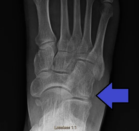
Plain X-ray radiography commonly interprets uncomplicated fractures of the cuboid and comprises the anterior, posterior, lateral, and traditional diagonal views of the foot. Imaging assessment must comprise the contralateral foot for the comparison and determination of the length of the foot’s outer aspect as well as for the appropriate preoperative planning. The integrity of the foot’s outer aspect can be evaluated by the anteroposterior view, which also permits the detection of any transverse plane deformity. The lateral radiograph examined the congruity of the calcaneocuboid joint and can also reveal avulsion fractures. The traditional medial diagonal view is especially helpful for looking at cuboid fractures due to it permits the view of the cuboid and its articulation with the metatarsals calcaneus, which has an open cuboid joint without any superimposed bones. It likewise participates in the evaluation of the size of the outer aspect.
Although traditional radiography can deliver a ton of facts related to the character of cuboid bone trauma, the significant overlapping of bony structures in the foot outcome in a failure in the depiction of invisible cuboid fracture. Only seven of the seventeen patients with pain on the outside of the midfoot who underwent X-ray radiography for diagnosis had cuboid fractures, said Miller.
Computed tomography
Computed tomography (CT) can give extra information related to the size, exact place, and type of fractures of the cuboid, as well as corealted trauma for example other fractures and dislocations of other bones of the foot. It clearly represents tarsometatarsal joints and the Lisfranc joint and even gives information on the fracture curative advancement.
Magnetic resonance imaging
In the condition of non-diagnostic radiography findings utilizing plain film radiographs, the taking part of magnetic resonance imaging (MRI) is a precious, delicate examination for the diagnosis of cuboid fractures in both children and elders. Μiller showed that in seventeen elders with isolated invisible cuboid fractures, the existence of a fracture was confirmed by MRI in eight of them. In their latest analysis, O’Dell utilized MRI to ensure cuboid fractures in nineteen minors aged eighteen months to seventeen years, nine of whom the 1st radiography was -ve for fracture. This radiology examination discovered fractures that were the consequence of recent trauma or were stress fractures linear in pattern and most usually side by side to the tarsometatarsal joint.
In the condition of cuboid fractures in the T1 weighted sequence, constant hypointense signals are discovered in the cuboid bone extending from the cuneiform joint surface toward the lateral side as well as fat development, while in the T2 sequence, a hyperintense signal in the bone, as well as the lack of the bone marrow signal, is noticed.
Sonography
Sonography, although not the method of preference for the linking of cuboid fractures, is a dedicated and immediate way when painful swelling is present side by side with the bone exists in the condition of subtle cuboid fractures with non-diagnostic plain radiography. Bilateral ultrasonography that considers longitudinal and transverse scan planes brings out a cortical separation and cortical step in the region of the fracture as well as soft tissue edema. Wang reported an examination of 268 patients with post-traumatic pain in the region of the foot and ankle with negative findings in the earlier radiology examination, where ultrasound examination discovered a fracture in 24 of them. Two of the patients examined experienced cuboid fractures.
Scintigraphy
Scintigraphy can show focal uptake in the cuboid and participate in the initial diagnosis of cuboid fractures, especially in stress fractures in children in the condition of a negative earlier physical assessment and non-diagnostic radiographs. Regardless, it must be taken into account that because of the complicated nature of the anatomy of the midfoot, it is hard to establish the precise information site of the radiopharmaceutical, which decreases the diagnostic precision of the method.
DIFFERENTIAL DIAGNOSIS
Differential diagnoses exclude bony results of lateral foot pain for example ankle sprain, stress fractures of the foot, fracture of the fifth metatarsal distal to tuberosity, avulsion of the anterior calcaneus process, injuries of the theLisfranc, tarsal coalition, cuboid syndrome, and injury to the os perineum. Likewise, soft tissue pathogens, for example, peroneus, longus tendon tenosynovitis or partial sprain, peroneal tendons subluxated, extensordigitorum, tendonitis, sinus tarsi syndrome, and entrapment of lateral plantar nervous can also possess an equivalent clinical image to cuboid fractures and must be excluded during the initial diagnosis in the condition of negative radiology evaluation with a clinical view equivalent to a cuboid fracture.
Differential diagnosis of outer foot pain
- Ankle sprain
- Peroneal tendonitis
- Peroneus longus tendon tear
- Subluxing peroneal tendons
- Stress fracture
- Fracture at the shaft of the 5th metatarsal
- Avulsion at the fifth metatarsal’s base
- Apophysitis of the fifth metatarsal
- Jones fracture
- Avulsion of the anterior calcaneus process
- Lisfranc injuries
- Tarsal coalition
- Cuboid syndrome
- Os perineum fracture
- Sinus tarsi syndrome
- Lateral plantar nerve entrapment
Cuboid syndrome, which typically affects athletes or dancers, is brought on by the calcaneocuboid joint’s anatomical congruency and cuboid subluxation. It is characterized by pain in the region of the plantar aspect of the cuboid or in the foot. Two clinical maneuvers have been suggested for the clinical diagnosis of the syndrome, viz. the midtarsal adduction and the midtarsal supination examination test.
The Os perineum is a supportive bone placed within the peroneus longus tendon side by side with the lateral plantar side of the cuboid, which can result in foot pain. It is established in four percent to thirty percent of normal feet. In several conditions, it is symptomless but can be the result of acute and chronic foot pain as in conditions of stress fracture or diastasis. For the diagnosis of the syndrome, plain radiography films can be a diagnostic, or further examination by computed tomography (CT), magnetic resonance imaging (MRI), or sonography can be needed.
Treatment of Cuboid Fracture
Avulsion fractures and stress fractures, which are non-displaced low-impact fractures, can be successfully treated with conservative methods of care, but complex high-energy injuries involving fractures with significant intra-articular involvement and displacement call for anatomic reduction and either external or internal osteosynthesis, depending on the specific fracture characteristics.
Conservative treatment
With cuboid fractures with minimal pain and edema, walking with some weight on your feet while using a fracture boot or an elastic bandage may be an adequate treatment. In the condition of extreme initial pain, a short walking cast for four to six weeks is suggested.
Stress fractures need to limit the movement and use of the extremity while it is instructed to have plantar arch support. Avulsion fractures are managed with protected weight bearing as bearable and a fracture boot for four to six weeks. These fractures need a standard radiographic evaluation each month as they can be complicated with fibrous non-union. In this condition as well as in the existence of resistant pain, an excisional operation can be crucial.
Surgical approach
According to Borrelli, open cuboid fractures are the best wholly used for urgent operative management. Operation is also suggested when an articular displacement of more than one mm or a contraction of the foot’s outer aspect length. According to Holbein, this contracting demands the operative treatment of the fracture if it is higher than three mm. The operative intervention of cuboid fractures includes external fixators, open reduction, internal fixation may or may not requirement of bone grafting, and midtarsal arthrodesis which is a fusion of the bone. The main objective of this operative intervention is to repair the length of the outer aspect as well as the nature of the anatomy and mobility of the nearby joints and specifically for the calcaneocuboid joint as well as the 4th and 5th tarsometatarsal joints.
In the condition of extreme comminution and displacement of fracture segments and inadequate skin quality that does not permit open fixation, the application of spanning external fixation provides that the length of the outer aspect is restored.
With lumbar or epidural anesthesia, cuboid fracture internal osteosynthesis can be carried out. The patient is put in the position in which the patient lying on their back with a thigh tourniquet and a bump below the same side hip to prevent excessive external rotation of the foot and to make sure adequate access to the cuboid bone. A six cm lateral longitudinal incision over the cuboid from the tip of the fibula to the tip of the 5th metatarsal exposes the calcaneocuboid and tarsometatarsal joints and permits the anatomic reduction of the fracture or fractures.
The extension of this section, five mm centrally of the calcaneocuboid joint and above the level of the lateral base of the 5th metatarsal, permits a view of the joints and reduction of the fractures. The incision is kept at the outer border of the extensor digitorum brevis muscle and proximal to the sural nerve, which should be recognized and rescued. Additional dissection is conducted to imagine the extensor digitorum brevis and then the muscle belly is somewhat elevated off the periosteum to permit assessment of the cuboid and the calcaneocuboid and tarsometatarsal joints.
After fixing the length of the cuboid, the dorsolateral wall is rebuilt. The articular surfaces must be reconstructed beginning from the media segments utilizing the intact aspect of the joint as a model. To support the reduction provisional fixation with little Kirschner wires can be needed. Internal osteosynthesis is achieved utilizing locking or unlocking screws or mini fragment plates under fluoroscopy. Any hole is sealed with autografts, allogenic, or cancellous bone chips, and an anatomic plate is seated over the cuboid to keep the length.
Postoperatively, the patient ambulates in the absence of weight loading for four to six weeks and for another six weeks with partial weight loading and with a walking cast. Maximum weight-loading walking is indicated after twelve-week
Midtarsal primary arthrodesis is an operative intervention approach suggested for extreme crush trauma with extensive comminution of the cuboid bone and articular involvement. Regardless, this approach presents the drawback of significant loss in outer aspect movement and is mainly indicated to less active patients. In the condition of failed internal fixation and continued pain, secondary fusion can be an alternative treatment method.
PHYSIOTHERAPY TREATMENT
The Cuboid fracture frequently responds to treatment quickly.
Your physiotherapist will work with you to design a specific treatment protocol that will quicken your recovery, comprising exercises and treatments that you can do at-house. The physiotherapist will assist you to return to your everyday lifestyle and activities. The time it carries to recover the condition differs, but noticeable progress can be achieved in one or two physiotherapy sessions, with complete recovery within some weeks or less when a proper treatment protocol is implemented.
During the 1st 24 to 48 hours next to your diagnosis of cuboid fracture, your physiotherapist can instruct you to:
- Prevent all weight-loading activities like jumping, hopping, and running.
- Prevent as much as prolonged walking.
- Wear flat, stiff-soled shoes.
- Be active around the house, and go on short walks many times per day. The movement will reduce the pain and stiffness, and help you feel good.
- Use ice packs on the painful region for fifteen to twenty minutes every two hours.
Your physiotherapist will work with you to:
- Reposition the cuboid and stabilize it. The cuboid bone can be moved back to its native anatomical position by your physiotherapist’s hands (manual therapy) so that it can move with greater mobility. This can ease most of the pain and discomfort and enhance the ability to stand and walk. The physiotherapist then can use multiple exercises and techniques to help the cuboid bones in the proper position. These can passing exercises of the foot, taping of the foot, and instructing on footwear and when to return to AdLs.
- Decrease pain and other symptoms. Your physiotherapist will work with you to identify the actions that helped to the injury along with ways to stop or alter them so that healing can start. Your physiotherapist may use various kinds of therapies and techniques to control and relieve your pain and other discomfort. The concentration will be placed on physiotherapy, cryotherapy, and soft movement to decrease pain without the required medicine for the pain.
- Enhance movement. Your physiotherapist will select characteristic activities and treatments to assist restore regular movement in the foot or in any stiff joints. These might initiate with “passive” motions that the physiotherapist passively performs for you to move a joint, and advancement to active exercises and stretches that you do yourself. You can do these movements at the house and in your job place to assist in quick recovery and pain relief.
- Enhance flexibility. Your physiotherapist will determine whether any muscles in the area are tight, begin to expand them out for you, and show you at-home stretching techniques.
- Enhance power. If your physiotherapist finds any weak muscles, the therapist will determine, and guide you, through the proper exercises to steadily restore your power and agility.
Exercises for the foot, ankle, and cuboid are typically introduced when treating cuboid fractures in order to strengthen the muscles and tendons surrounding the cuboid bone, foot arch, and ankle. - Enhance endurance. Restoring muscular endurance is significant after a trauma. Your physical therapist makes a treatment plan of activities that helps you recover the endurance you had prior to injury, and enhance it.
- Learn a home protocol. the physiotherapist will guide you through strengthening, stretching, and pain-reduction exercises to do at-house. These exercises will be specific to your demands; if you do them as prescribed by your physiotherapist, you can quicken up your rehab.
- Return to ADLs. Your physiotherapist will examine your activity levels with you and utilize them to develop your work, sport, and house-life rehab objectives. Your treatment protocol will help you to fulfill your goals in the safest, quickest, and most effective way achievable. Your physiotherapist can advise you on the best kind of shoes to wear to prevent exacerbating your cuboid bone condition and to add specialized support such as orthotics.
Once your pain is run away, it will be crucial for you to restart your foot exercises at house, to help maintain your foot healthy and pain-free.
In all but the many severe conditions, physiotherapy treatment provides exceptional outcomes. Operation intervention and pain medication (for example opioid medication) are not commonly required for this condition.
FAQ
It may be sufficient to manage cuboid fractures with minimal pain and edema with an elastic bandage or a fracture boot and to walk with some weight on one foot until the symptoms are sufficiently relieved. When there is severe initial pain, a four- to six-week walking cast is recommended.
For four to six weeks in a well-molded below-knee cast, the non-weight bearing can be used to treat cuboid fractures that are not dislocated. It is possible to get stress or weight-loading X-rays as a follow-up after two weeks to rule out the possibility of an undetectable fracture, ligamentous damage, or subluxation.
Acceptable first-aid care for fractures is essential. Moving the broken bones can enhance the pain and bleeding and harm tissues near the wound. This can result in difficulties in the restoration and recovery of the injury subsequently on. First aid for fractures is all about immobilizing (limiting movement of) the injured region.
One test I do is to have the patient stand up and roll a small Super Ball beneath the feet, straight under the cuboid. This will produce intense sharp pain on the involved limb while resulting in minimal or no pain on the contralateral foot. This test also becomes a treatment for the patient to do at the house daily.
On average, a broken bone can take anywhere from six to eight weeks to heal before it can be used too. For young youths, the recovery procedure can happen faster. For more senior adults or those who have an underlying health pathology, for example, diabetes, recovery can take longer.
Many individuals make the association between dairy items, calcium, and bone health, but multiple don’t know that eggs play a role, too! Eggs are one of the best sources of vitamin D, which works well with calcium to help develop strong bones.


2 Comments