
Proximal Radial Head Fracture
Table of Contents
What is a Proximal Radial Head Fracture?
A proximal radial head fracture is a type of elbow fracture that occurs when the upper part of the radius bone in the forearm breaks near the elbow joint. This type of injury is commonly caused by falls onto an outstretched arm or direct impact to the elbow. Treatment typically involves immobilization, pain management, and physical therapy, although more severe cases may require surgery. Recovery depends on the severity of the fracture and the individual’s overall health.
Introduction
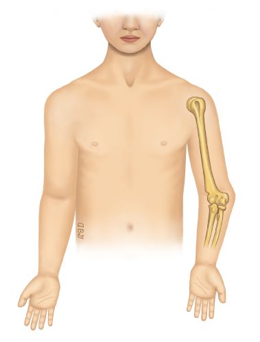
The radius, situated in the lateral forearm, is the shorter of the two bones that form the forearm. The radius communicates with the ulna, the 2nd bone in the forearm. both the two bones and their articulations constitute the radioulnar and radiocarpal joints at the elbow and wrist, and vice versa.
Radius fractures possess the proximal part of the radius, the neck, and the head. This kind of fracture is very expected in adults. Proximal radial fractures are common when falling on an outstretched hand (FOOSH), which pushes the radius into the humerus, or direct hit to the elbow. Fractures at the proximal radius position the radial head at a more raised threat for avascular necrosis (AVN).
Clinical Anatomy
The elbow is a synovial hinge joint constructed up of 3 articulations– the humeroulnar, humeroradial, & radioulnar. The arm’s humerus completes the forearm’s ulna and radius to make the hinge, while the radius and ulna articulate to form a pivot joint to
permit forearm pronation and supination movement.
Significant milestones of the proximal radius possess the radial head, neck, and tuberosity. The head is rounded with a flat though little concave consistency. The flat surface articulates with the humerus. The rim of the head is held within the annular ligament and in front of the radial notch of the ulna where it rotates and glides during pronation and supination. The radial tuberosity acts as an attachment area for the biceps brachii and supinator brevis muscles.
Epidemiology
Proximal radial head fractures are most familiar in years 20-64. Nevertheless, radial head fractures are noticed more prone in younger males than females. This distinction is linked with males participating more in falls related to sports or heights whereas females tend to undergo fractures after in life because of falls and the fragility of the bone.
Cause of Proximal Radial Head Fracture
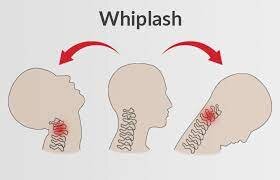
Falling on an extended hand or falling with the elbow extended and the forearm pronated, which distributes the trauma force to the radius head through the wrist and forearm, are the two most common ways to injure the radial head. Few typically typical damages with this kind of fracture can be ligamentous for example lateral collateral ligament (LCL) or medial collateral ligament (MCL) injuries. Dislocation of the elbow can also have what is understood as the “terrible triad” which possesses a dislocation of the elbow, a radial head fracture, and a coronoid fracture.
Symptoms of Proximal Radial Head Fracture
The most common symptoms are:
- Elbow Pain on the outside
- Elbow joint Swelling
- Difficulty in moving elbow
- Difficulty in rotating the forearm
Types of Proximal Radial Head Fracture
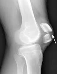
Proximal radial head fractures, typically represented by using the Mason classification, have four grades. The orthopedic surgeon selects the diagnosis of different type of Mason fractures, and they decide which interventions are required. The surgeon’s judgment is driven by diagnostic imaging, X-ray, MRI, or CT scans. Likely interventions possess immobilization which may apply splinting, slings, and or surgery.
Mason Type 1
A fissure or margin sector fracture with a non-displaced or lightly substituted radius and a disparity of more than 2 mm is known as Mason Type 1 fracture. These fractures can be questioned to identify on an X-ray when the fracture is non-displaced. A CT or MRI scan is needed for more distant research. In a Mason type 1 proximal radius fracture, there is no mechanical restriction of movement in the supination and pronation direction.
A five percent (5%) chance of a non-union happens with a Mason Type 1 fracture of the proximal radius. A proximal radius
non-union can lead the radial head to subluxate. Nevertheless, the reduction of associated segments of the fracture does not enhance the possibility of subluxation.
Mason Type 2
A Mason Type 2 radial head fracture is clear when the radial head is partly fractured with a >2mm displacement
Mason Type 3
Mason Type 3 fractures are entire breaks of the continuity or comminuted, broken into numerous parts or segments. Mason Type 3 fractures can be further defined by subclasses. Type 3a is the total displacement of the radial head from the shaft, with the fracture
via the radial neck. Type 3b is a joint fracture where the head breaks into two or more pieces. Type 3c applies to an articular fracture that is rotated and affected.
Differential Diagnosis
The increased likelihood of other damages happening with a fall on an outstretched hand (FOOSH) offers X-ray, MRI, and
sometimes CT scans are required to verify the diagnosis and to certify the integrity of all nearby structures and tissues.
The potential injuries that must be assessed and eliminated with a radial fracture of the head are capitellar fractures, distal ray fractures, injuries to the medial collateral ligament (MCL), the biceps and triceps tendons, the distal radio-ulnar joint, and dislocation of the elbow
Diagnostic Procedures
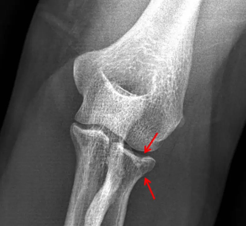
The preliminary diagnostic tool used for determining radial head fractures is a radiograph. Mason Type 1 fracture do not ever show
on X-ray. In such circumstances, a ‘sail sign’ might mean a fracture. A ‘sail sign’ is a silhouette on a radiograph driven by an enlarged fat pad at the elbow. Recent investigations are glancing at the use of sonography in seeing occult fractures more fast. Existing diagnostic processes can take upward of three weeks before the determination of a fracture.
Outcome Measures
The Impairment of the Arm, Shoulder, and Hand is a result of measures used to regulate the capabilities of a patient’s upper limb. The survey lists AdLs for example opening a container, holding shopping bags, donning and doffing clothes, etc. The patient scales the difficulty of individual activities in this questionnaire from 1 to 5, with 1 presence of ‘No Difficulty’ and 5 presence of ‘Unable.’ Note that few queries are expressed contrarily so the 1 to 5 range is relabeled as required, for example, none to ‘Extreme’ or ‘Strongly Disagree’ to ‘Strongly Agree.’ The score is considered utilizing the formula ([sum of n responses/n]-1)(25). n = completed items. The final score and level of disability have a great association. An extra module is provided for patients using workman recompense or athletes and musicians.
The Quick DASH is a modified form of the DASH outcome measure that is briefer but with an indication of being as precise as the DASH. The Quick DASH comprises only 11 questions and employs a similar rating scale and scoring formula.
These two result measures can be utilized with Mason Type 1, 2, and 3 fractures irrespective of the mode of injury.
Examination
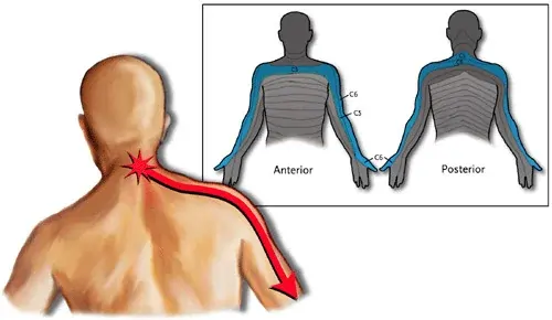
The mode of injury is frequently falling on an outstretched hand or a direct hit to the elbow. Radial head fractures are communal together with elbow dislocation. Swelling and bruising of the posterior elbow may be noticeable. Swelling and heat are palpable. Type III fractures may result in noticeable deformity. The patient has incomplete active elbow extension or flexion and forearm pronation or supination movement. Few may experience inadequate wrist movement as well. Passive range of motion (PROM) is limited because of pain. The empty end feels of muscle guarding can be likely. Palpation of the radial head is painful. Few patients experience the feeling of numbness in the region of the forearm, hands, and digits.
Medical Treatment of Proximal Radial Head Fracture
Non-Surgical Treatment
There should be a brief period of immobilization of the arm, which mainly applies to Mason Type 1 fracture. This can be accomplished by the patient using a sling that is recommended for no more than 7 days.
Surgical Treatment
- An open reduction internal fixation (ORIF) of the radial head has been discovered to be useful for Type 2 and 3 fractures of Mason classification.
- A radial head resection is a choice for the inactive patient or when there is sustained pain because of a lonely radial head fracture.
- For comminuted Mason Type, 3 fractures radial head arthroplasty is indicated that possessing higher than 25% of the radial head is added effective choice.
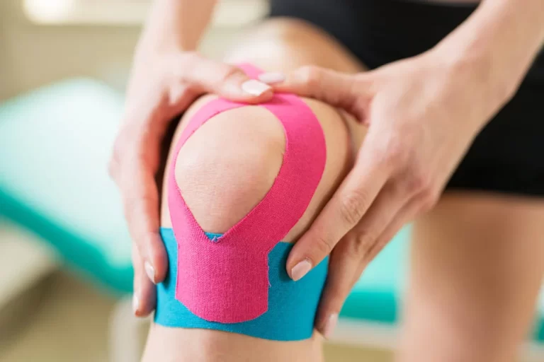
Physical Therapy Management
Mason Type 1 Fracture
Non-operatively management is indicated in type 1 non-displaced proximal radial fractures, and the patient is advised to have a sling or splint for some days. Cryotherapy techniques can be used as required to aid minimized the presence of swelling. As pain permits, the patient must achieve an active range of motion (AROM) in the initial stages of rehabilitation, possessing movement of the forearm in supination and pronation. A vital part of this type of fracture is to mobilize the joint initiative to minimize any presence of complications of post-traumatic stiffness in the joint. ROM must be considered watchfully when the fracture possesses one-third of the articular surface. If the fracture does include one-third of the articular surface, a sling or splinting must be implemented for at least a two-week duration.
Mason Type 2 and 3 Fractures
An ORIF is indicated with Mason Type 2 and 3 fractures, which has established the greatest recovery outcomes. After fixation, there are three phases of physical therapy. In the initial first phase, which comprised 0-14 days, the patient starts with gentle movements like elbow flexion and extension AROM. At the last of the 2nd week, the elbow ROM must be 15-105 degrees. After these range of motion supplies are met, the patient initiates gripping strengthening exercises with clay-putty and isometric strengthening exercises for the elbow and wrist. In phase two, which is normally around from 15 days to 6 weeks, the physiotherapist must measure the shoulder and wrist strength along with ROM.
The patient persists with elbow AROM and an active-assisted range of motion (AAROM). Elbow flexion and extension ROM must be at full at the end of six weeks. Later this time, the patient starts AROM and AAROM supination and pronation. The patient will continue the isometric strengthening exercises from the initial phase working precisely on flexion and extension movement. In stage three, from week 7 to week 12 duration, the patient persists in working on active ROM and actively assisted ROM with supination and pronation. In the eighth week, complete pronation and supination must be attained. The patient must further advance isometric strengthening exercises of elbow flexion and extension, along with wrist pronation and supination. The physiotherapist also wants to measure and focus on any extra deficits caused by the fracture and or the surgery.
Education
If surgery happens, the splint must stay in position until the patient’s first postoperative visit, which commonly happens 1-2 weeks later to the surgery. Showering is allowable on the 2nd day, but care and precaution must be taken to preserve the splint clean and dry. Submersion of the elbow region is restricted for at least four weeks later to surgery.
Once at home, the patient can walk as much as wanted. The patient can drive a car on one occasion approved by the surgeon, which is commonly four to six weeks later to surgery. Returning to occupation is based on the patient’s duties required to complete their job and should be cleared by the surgeon to return to employment duties.
The patient must be supposed to see some swelling present in the region nearby the arm after the surgery. The patient must interact with the surgeon directly if skin changes with or without discharge or bleeding are noted nearby the incision site, or if a fever happens >101 degrees.
What the patient must not be doing for the first six weeks is:
- using the arm to push themselves up on a couch or a chair.
- utilizing forceful contraction of muscles vital to push off.
- overuse of the elbow and arm can result in problems with the curing procedure.
- lifting weight that is weightier than a glass of water.
- putting the arm in a risky position, possessing straight out to the side or back of the patient’s body.
When required, the patient must ask for help with activities.
At the first postoperative visit with the surgeon (1-2 weeks), the patient’s staples or stitches are removed, the wound is inspected, and X-rays are got to guarantee appropriate recovery. The patient is given instructions on the next steps and strategies for the future weeks after everything is estimated.
Rehabilitation & Pain Management
Non-Surgical Rehabilitation
The initial range of motion for non-operated simple and complex radial head fractures and initial active range of motion & active assisted range of motion of the elbow aids to evade the complication like the presence of edema, stiffness in the joint, and the development of adhesions in the capsule and annular ligament. It is also overbearing for the patient to focus on the adjacent joints for example the shoulder, wrist, hand, and scapulothoracic joint to ensure a range of motion and utilization of the arm has been sustained.
Surgical Rehabilitation
Simple and complex are the two types of fractures: A simple type radial head fracture references as a fracture only in the head of the radius. On the Mason classification, Type 2 and Type 3 radial head fractures necessity of surgical interference to fix the radius. A complex type radial head fracture is defined as added instability because of another element out from the radial head fracture. In both cases, it is suggested that immobilizing the arm is valuable to defend and caring for the arm after surgery. Still, immobilization of the arm for only up to one week later surgery for simple types of fractures and on the other hand up to three-six weeks of the period with a long-arm splint for the complex types of fractures.
Recovery
Mason Type 1 Fracture
The non-surgical method comprises donning a splint or sling for some days, next step is a gradual gentle improvement in elbow and wrist movement based on how painful it feels. Lack of active elbow extension, mild loss of forearm pronation and supination movement, and rare fatigue and pain with overdoing in the forearm are the most typical complications with Mason type 1 proximal radius fractures may embrace. If the unnecessary motion is encouraged too initial, it is likely to move and displace the bones.
Mason Type 2 Fractures
If the displacement is negligible the management comprises the patient wearing a sling or a splint for 1 to 2 weeks and must be full with a range of motion exercises.
Mason Type 3 Fractures
Initial stretching exercises and elbow flexion are essential to evade contractures of the elbow or stiffness in the elbow range of motion.
Nearly Type 3 fractures need the patient to be located in a splint or sling for a brief period of time. The surgeon will mention the patient not weight-bear through the arm or wrist or lift weights that are weightier than a couple of pounds for 6 to 12 weeks. Fractures that occur in the proximal part of the radial head will cause in lack of elbow ROM. The patient must execute exercises to restore range of motion and strength to return to functional activities. A second surgery can be essential to eliminate any scar tissue that develops and limits the elbow’s range of motion.
FAQ
This category of fracture is very typical in adults. Proximal radial fractures occur when falling on an outstretched hand (FOOSH), which forces the radius into the humerus, or direct hit to the elbow. Fractures at the proximal radius position the radial head at a more raised threat for avascular necrosis (AVN).
Radius fractures comprise the proximal part of the radius, the radius fractures of the neck, and radius fractures of the head. This kind of fracture is very typical in adults. Proximal radial fractures happen when falling on an outstretched hand (FOOSH), which forces the radius into the humerus, or direct hit to the elbow. Fractures at the proximal radius position the radial head at a more raised threat for avascular necrosis (AVN).
For instance, if you fall off monkey bars, skateboards, or a vehicle or if you take a solid impact in a contact game for example lacrosse, hockey, or football you can get a proximal radius fracture. The designation for this fracture arises from the portion of bone near the elbow where it typically ensues: the radial head.
Surgical procedure is constantly obligatory to moreover fix or remove the broken fragments of bone and repair the soft-tissue impairment. If the damage is very extreme, the total radial head may necessity to be detached. In this circumstance, an artificial radial head can be positioned to progress long-term function. Initial movement to stretch and flexion the elbow is essential to evade the stiffness in the joint.
The radial head fuses with the radial shaft for about 16 years (Tibone, 1981). The procedure for a proximal radial fracture must follow a stepwise strategy from closed reduction, to percutaneous helped in reduction, to open treatment. (Basmajian, 2014) The choice is completed based on the quantity of angulation and displacement.
The most typical clinical symptoms of a radial head fracture comprise Pain on the outer aspect of the elbow. Presence of swelling around the elbow joint. Difficulty in flexing or straightening the elbow accompanied by pain. Inability to bear weight or difficulty in twisting the forearm (palm up to palm down or respectively)
One Comment