
Cuneiform Bone
Table of Contents
Introduction
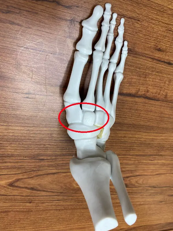
The Cuneiform bones are a group of three small tarsal bones located in the foot, specifically in the midfoot region. They are named based on their shape, with the names referring to their resemblance to a wedge or triangular shape.
There are three cuneiform bones:
- the medial cuneiform
- the intermediate cuneiform
- the lateral cuneiform
The medial cuneiform is the largest and is located on the inner side of the foot, closest to the big toe. The intermediate cuneiform is located in the middle, between the other two cuneiform bones. The lateral cuneiform is located on the outer side of the foot, adjacent to the cuboid bone.
The cuneiform bones play an important role in maintaining the arches of the foot and providing stability during weight-bearing activities. They help support the body’s weight and distribute it evenly across the foot, allowing for efficient movement and shock absorption.
Anatomy of Cuneiform Bone
The cuneiform bones are a group of three small tarsal bones located in the midfoot region of the foot. They are named based on their shape, resembling a wedge or triangular shape. Each cuneiform bone has distinct anatomical features.
- Medial Cuneiform: The medial cuneiform bone is the largest and is located on the inner side of the foot, closest to the big toe. It articulates with several other bones, including the first metatarsal bone (base of the big toe) and the navicular bone (a bone in the midfoot). It has six surfaces:
- Dorsal surface: This is the top surface of the bone, which can be felt just below the skin.
- Plantar surface: This is the bottom surface of the bone that contacts the ground during weight-bearing activities.
- Medial surface: This surface faces inward towards the midline of the body.
- Lateral surface: This surface faces outward away from the midline of the body.
- Proximal surface: This surface articulates with the navicular bone.
- Distal surface: This surface articulates with the first metatarsal bone.

- Intermediate Cuneiform: The intermediate cuneiform bone is located in the middle, between the other two cuneiform bones. It articulates with several bones, including the second metatarsal bone (base of the second toe) and the navicular bone. It also has six surfaces similar to the medial cuneiform.
- Lateral Cuneiform: The lateral cuneiform bone is located on the outer side of the foot, adjacent to the cuboid bone. It articulates with several bones, including the third metatarsal bone (base of the third toe) and the navicular bone. It also has six surfaces similar to the other cuneiform bones.
The cuneiform bones are connected to each other and to neighboring bones by ligaments, which provide stability and allow for proper movement of the foot. They also have important attachments for tendons, which are responsible for transmitting forces from muscles to bones, allowing for movement.
Overall, the cuneiform bones play a crucial role in maintaining the arches of the foot and providing stability during weight-bearing activities. They work together with other bones, ligaments, and muscles in the foot to support the body’s weight and distribute it evenly across the foot, allowing for efficient movement and shock absorption.
Functions of Cuneiform Bone
The cuneiform bones play several important functions in the foot:
- Arch Support: The cuneiform bones, along with other bones in the midfoot region, help maintain the arches of the foot. They form a stable base for the arches, which are essential for weight-bearing activities such as walking, running, and standing. The medial cuneiform bone, in particular, contributes significantly to the support of the medial longitudinal arch, which runs along the inner side of the foot.
- Weight Distribution: The cuneiform bones help distribute the body’s weight evenly across the foot. As weight is transferred from the hindfoot (heel) to the forefoot during activities like walking or running, the cuneiform bones act as a bridge between the rearfoot bones (talus and calcaneus) and the metatarsals (long bones in the forefoot). This distribution of weight prevents excessive pressure on specific areas of the foot and reduces the risk of injuries such as stress fractures.
- Stability: The cuneiform bones, along with their ligamentous attachments, provide stability to the midfoot region. They help maintain proper alignment between the hindfoot and forefoot, ensuring that the foot remains stable during weight-bearing activities. This stability is crucial for maintaining balance and preventing excessive movement or rolling of the foot, which can lead to ankle sprains or other injuries.
- Joint Articulation: The cuneiform bones articulate with several other bones in the foot, including the metatarsals and navicular bone. These articulations allow for smooth movement and flexibility in the midfoot region. The cuneiform bones, along with other tarsal bones, contribute to the complex movements involved in walking, running, and other weight-bearing activities.
- Tendon Attachments: The cuneiform bones have important attachments for tendons, which are responsible for transmitting forces from muscles to bones. Tendons from various muscles in the foot and lower leg attach to the cuneiform bones, allowing for coordinated movement and control of the foot. These tendon attachments help generate power and provide stability during activities such as pushing off during walking or running.
In summary, the cuneiform bones are crucial for maintaining the arches of the foot, distributing weight evenly, providing stability, facilitating joint articulation, and allowing for proper tendon function. They work in conjunction with other bones, ligaments, and muscles in the foot to support the body’s weight, absorb shock, and enable efficient movement.
Articulation of Cuneiform Bone
The cuneiform bones are a group of three small bones located in the midfoot region of the foot. They are named based on their position relative to the other tarsal bones: the first cuneiform bone is closest to the inner side of the foot, the second cuneiform bone is in the middle, and the third cuneiform bone is closest to the outer side of the foot. These bones articulate with several other bones in the foot, including the navicular bone, the metatarsal bones, and the cuboid bone.
The articulations between the cuneiform bones and the other bones in the foot are classified as synovial joints, specifically gliding joints. Gliding joints allow for limited movement in multiple directions, such as sliding or gliding motion. The specific type of gliding joint involved in the articulation of the cuneiform bones is called an intertarsal joint.
- First Cuneiform Bone:
- Naviculo-cuneiform joint: This joint is formed between the first cuneiform bone and the navicular bone. It is a saddle joint, which means that it allows for movement in multiple directions. The articular surfaces of these bones have a convex-concave shape, allowing for rocking and sliding motions. This joint is important for maintaining the medial longitudinal arch of the foot and facilitating movements such as inversion and eversion.
- First metatarsal-cuneiform joint: This joint is formed between the first cuneiform bone and the first metatarsal bone. It is a gliding joint that allows for limited movement in multiple directions. This joint contributes to the flexibility and stability of the midfoot region during weight-bearing activities.
- Second Cuneiform Bone:
- Naviculo-cuneiform joint: The second cuneiform bone also articulates with the navicular bone at this joint, similar to the first cuneiform bone. The articular surfaces allow for gliding and rocking motions, contributing to the overall mobility and stability of the foot.
- Second metatarsal-cuneiform joint: This joint is formed between the second cuneiform bone and the second metatarsal bone. It is also a gliding joint that allows for limited movement. This joint plays a role in weight distribution and shock absorption during walking and running.
- Third Cuneiform Bone:
- Third metatarsal-cuneiform joint: The third cuneiform bone articulates with the third metatarsal bone at this joint. It is a gliding joint that allows for slight movement in multiple directions. This joint contributes to the overall flexibility and adaptability of the foot during various activities.
The articulations between the cuneiform bones and the other bones in the foot are supported by ligaments, which provide stability and prevent excessive movement. These ligaments include the dorsal and plantar cuneonavicular ligaments, as well as the interosseous and plantar cuneocuboid ligaments.
Overall, the articulation of the cuneiform bones with other bones in the foot allows for coordinated movement, shock absorption, and weight distribution. These joints play a crucial role in maintaining foot function and stability during various activities.
Muscle Attachment of Cuneiform Bone
The cuneiform bones have important attachments for tendons, which are responsible for transmitting forces from muscles to bones. These tendon attachments play a crucial role in the movement and stability of the foot. Here are the specific muscle attachments for each cuneiform bone:
First Cuneiform Bone:
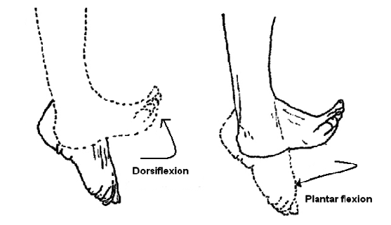
- Tibialis anterior tendon: This tendon originates from the tibialis anterior muscle in the front of the leg. It passes over the top of the first cuneiform bone and attaches to its medial surface. The tibialis anterior muscle is responsible for dorsiflexion (lifting the foot upwards) and inversion (turning the foot inward).
Second Cuneiform Bone:
- Tibialis posterior tendon: This tendon originates from the tibialis posterior muscle, which is located deep in the back of the leg. It runs behind the medial malleolus (bump on the inner side of the ankle) and attaches to the plantar surface of the second cuneiform bone. The tibialis posterior muscle is involved in inversion and plantarflexion (pointing the foot downward).
Third Cuneiform Bone:
- Extensor hallucis longus tendon: This tendon originates from the extensor hallucis longus muscle, which is located in the front of the leg. It passes over the top of the third cuneiform bone and attaches to its dorsal surface. The extensor hallucis longus muscle is responsible for extending (straightening) the big toe.
Navicular Bone:
- Tibialis posterior tendon: In addition to attaching to the second cuneiform bone, the tibialis posterior tendon also attaches to the navicular bone. This attachment helps stabilize and support the medial longitudinal arch of the foot.
These tendon attachments allow for coordinated movement and control of the foot. When muscles contract, they pull on these tendons, which in turn exert forces on the cuneiform bones. This generates power and provides stability during activities such as walking, running, and jumping. The proper functioning of these muscle attachments is essential for maintaining the arches of the foot, distributing weight evenly, and facilitating efficient movement.
Blood and lymph supply
The blood supply to the cuneiform bones is primarily provided by branches of the dorsalis pedis artery and the medial plantar artery. These arteries arise from the anterior tibial artery and the posterior tibial artery.
The dorsalis pedis artery gives off branches that supply the first and second cuneiform bones. These branches include the first dorsal metatarsal artery and the deep plantar arch, which give rise to smaller arteries that reach the cuneiform bones.
The medial plantar artery supplies the third cuneiform bone. It gives off branches that include the deep plantar arch and the lateral tarsal artery. These branches provide blood flow to the third cuneiform bone.
In terms of lymph supply, the cuneiform bones are drained by lymphatic vessels that accompany the blood vessels. The lymphatic vessels collect interstitial fluid from the cuneiform bones and transport it toward regional lymph nodes for filtration and immune response.
Overall, the blood and lymph supply to the cuneiform bones is essential for maintaining their health and supporting their function in the foot. Adequate blood flow ensures the delivery of oxygen and nutrients, while lymphatic drainage helps remove waste products and maintain tissue homeostasis.
Conditions that Affect Cuneiform Bone
Associated conditions of the cuneiform bones can vary depending on the specific bone affected and the underlying cause. Here are some commonly associated conditions:

Cuneiform fractures: Fractures of the cuneiform bones can occur due to trauma or excessive stress on the foot. These fractures can be classified as stable or displaced, and their severity can vary. Symptoms may include pain, swelling, bruising, difficulty walking, and deformity. Treatment options range from conservative measures such as immobilization and rest to surgical intervention, depending on the extent of the fracture.
Tarsal coalition: Tarsal coalition refers to an abnormal connection between two or more tarsal bones, including the cuneiform bones. This condition is usually congenital, meaning it is present at birth, but it may also develop as a result of trauma or infection. Tarsal coalition can cause foot pain, stiffness, limited range of motion, and an abnormal gait. Treatment options include physical therapy, orthotic devices, and in some cases, surgical intervention.
Arthritis: Arthritis can affect the cuneiform bones, leading to inflammation, pain, stiffness, and limited mobility. Osteoarthritis, which occurs due to wear and tear of the joints over time, and rheumatoid arthritis, an autoimmune condition that causes joint inflammation, are the most common types of arthritis that can affect the cuneiform bones. Treatment options may include medication, physical therapy, joint injections, and in severe cases, surgical intervention.
Tendonitis: Tendonitis refers to inflammation of the tendons that attach to the cuneiform bones. Overuse or repetitive stress on these tendons can lead to tendonitis. Symptoms may include pain, swelling, tenderness, and difficulty with activities that involve the affected tendon. Treatment typically involves rest, ice, compression, elevation (RICE), physical therapy, and anti-inflammatory medications.
Sesamoiditis: The sesamoid bones, small bones located under the first metatarsal bone, can also be associated with the cuneiform bones. Sesamoiditis refers to inflammation of the sesamoid bones or the tendons surrounding them. It is often caused by repetitive pressure or injury to the sesamoids. Symptoms involve swelling, pain, and hard-to-bear weight on the affected foot. Treatment may include rest, immobilization, orthotic devices, and in some cases, surgical intervention.
It is mandatory to notice that these are just some examples of associated conditions, and there may be other less common conditions that can affect the cuneiform bones. Proper diagnosis and treatment by a medical professional are essential for managing these conditions effectively.
Rehabilitation
The rehabilitation of a cuneiform bone typically refers to the process of restoring its function and strength after an injury or surgery. The cuneiform bones are located in the midfoot region of the foot, specifically in the arch area.
- Initial Assessment: The rehabilitation process begins with a thorough assessment by a healthcare professional, such as a physical therapist or orthopedic specialist. They will evaluate the extent of the injury or surgical procedure, assess the patient’s overall health, and determine the appropriate rehabilitation plan.
- Immobilization: In some cases, immobilization may be necessary to allow the cuneiform bone to heal properly. This can involve the use of a cast, splint, or walking boot to restrict movement and protect the bone during the initial healing phase. The total time of immobilization depends on the severity of the injury or surgical procedure.
- Pain and Inflammation Management: Pain and inflammation are common after cuneiform bone injuries or surgeries. Rehabilitation often involves techniques to manage these symptoms, such as ice or cold therapy, non-steroidal anti-inflammatory drugs (NSAIDs), or other pain management modalities prescribed by a healthcare professional.
- Range of Motion Exercises: Once the initial healing phase is complete, range of motion exercises are introduced to improve joint mobility and flexibility. These exercises may include toe curls, ankle circles, and gentle stretching of the foot and ankle to gradually restore normal movement.
- Strengthening Exercises: Strengthening exercises play a crucial role in rehabilitating the cuneiform bone. These exercises target the muscles surrounding the foot and ankle to improve stability and support. Examples of strengthening exercises include calf raises, toe raises, resistance band exercises, and balance training.
- Proprioception and Balance Training: Proprioception refers to the body’s ability to sense its position in space. After a cuneiform bone injury or surgery, proprioception and balance may be affected. Rehabilitation often includes exercises that challenge balance and coordination, such as standing on one leg, using a wobble board, or performing specific proprioceptive drills.
- Gait Training: Restoring normal walking or gait pattern is an important aspect of cuneiform bone rehabilitation. A physical therapist may analyze the patient’s walking pattern and provide guidance on correcting any abnormalities or compensatory movements.
- Gradual Return to Activities: As the cuneiform bone continues to heal and the patient’s strength and function improve, a gradual return to activities is initiated. This may involve a gradual increase in weight-bearing activities, such as walking, jogging, running, or specific sports-related movements.
Throughout the rehabilitation process, regular follow-up appointments with a healthcare professional are essential to monitor progress, make necessary modifications to the treatment plan, and ensure a safe and effective recovery.
FAQs
Cuneiform bone injuries can occur due to trauma, such as a direct blow to the foot or a fall. Surgeries may be performed to repair fractures, correct deformities, or address other foot and ankle conditions.
The total time of immobilization depends on the severity of the injury or surgical procedure. It can vary from some weeks to several months.
Pain and inflammation management techniques may include ice or cold therapy, NSAIDs, and other pain management modalities prescribed by a medical professional.
The duration of the rehabilitation process varies depending on the individual and the extent of the injury or surgical procedure. It can vary from some weeks to several months.
The timing of returning to activities depends on the individual’s progress and the guidance of a medical professional. A gradual return to activities is typically initiated once the cuneiform bone has healed and the patient’s strength and function have improved.
Regular follow-up appointments with a medical professional are essential during cuneiform bone rehabilitation to monitor progress, make necessary modifications to the treatment plan, and ensure a safe and effective recovery.
One Comment