
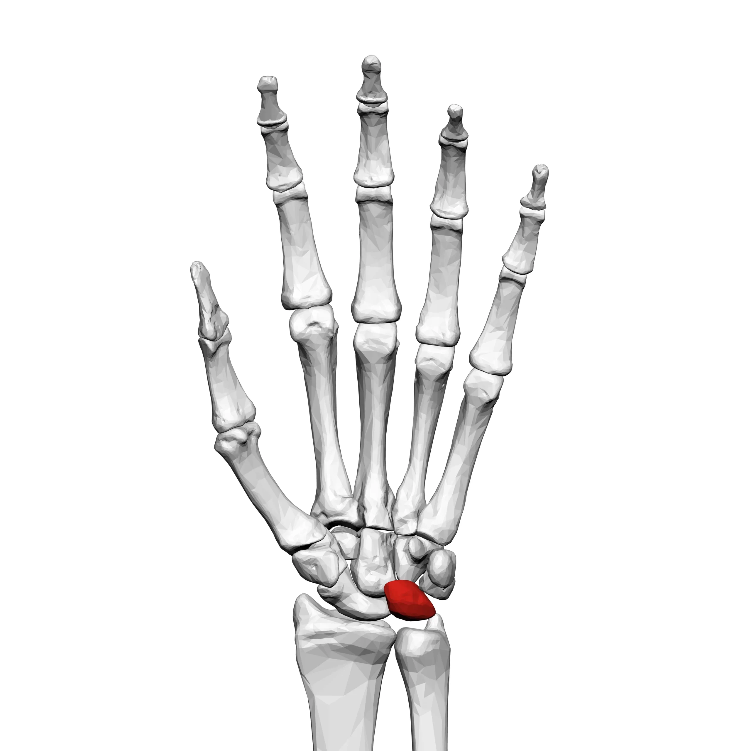
Lunate Bone
Table of Contents
Introduction
The Lunate bone is another one of the eight carpal bones in the wrist. It is located between the capitate bone and the triquetrum bone. The lunate bone is roughly moon-shaped, which is where its name originates (luna means moon in Latin).
The lunate bone plays a crucial role in wrist movement and stability. It articulates with the radius bone of the forearm, forming the radiocarpal joint, which allows for flexion, extension, and rotation of the wrist. It also connects with other carpal bones, contributing to the overall stability and mobility of the wrist.
Anatomy of the Lunate Bone
The anatomy of the lunate bone includes several key features:
- Articular surfaces: The lunate bone has multiple articular surfaces that allow it to connect with other bones in the wrist joint. On its superior (upper) surface, it articulates with the radius bone of the forearm, forming the radiocarpal joint. This joint is responsible for wrist flexion, extension, and rotation. On its inferior (lower) surface, it connects with the capitate bone, contributing to wrist stability and movement.
- Proximal and distal surfaces: The lunate bone has proximal and distal surfaces that interact with neighboring carpal bones. The proximal surface faces toward the forearm and forms a joint with the radius bone. The distal surface faces toward the hand and connects with the capitate bone.
- Medial and lateral surfaces: The lunate bone also has medial and lateral surfaces that allow for connections with adjacent carpal bones. The medial surface faces toward the midline of the wrist and forms a joint with the triquetrum bone. The lateral surface faces away from the midline and articulates with the scaphoid bone.
- Dorsal and volar surfaces: The dorsal surface of the lunate bone faces toward the back of the hand, while the volar surface faces toward the palm. These surfaces provide attachment sites for ligaments and tendons involved in wrist movement and stability.
Understanding the proper anatomy of the lunate bone is essential for diagnosing and treating conditions or injuries that may affect its function. Medical professionals specializing in orthopedics or hand surgery can evaluate and provide appropriate treatment options for any issues involving the lunate bone.
Articulation of the Lunate Bone
The lunate bone articulates with several other bones in the wrist joint, allowing for movement and stability. The main articulations of the lunate bone include:
- Radiocarpal joint: The superior surface of the lunate bone forms a joint with the radius bone of the forearm. This joint, known as the radiocarpal joint, is one of the main joints in the wrist and allows for flexion, extension, and rotation of the wrist. The lunate bone acts as an intermediate bone between the radius and other carpal bones, helping to distribute forces and facilitate smooth movement.
- Capitate-lunate joint: The inferior surface of the lunate bone connects with the capitate bone. This joint, known as the capitate-lunate joint, is important for wrist stability and movement. It allows for gliding and rotational movements between the lunate and capitate bones, contributing to the overall range of motion in the wrist.
- Triquetrum-lunate joint: The medial surface of the lunate bone forms a joint with the triquetrum bone. This joint, known as the triquetrum-lunate joint, provides additional stability to the wrist and allows for limited gliding movements between the two bones.
- Scaphoid-lunate joint: The lateral surface of the lunate bone articulates with the scaphoid bone. This joint, known as the scaphoid-lunate joint, contributes to the overall mobility and stability of the wrist.
These articulations are facilitated by the presence of articular surfaces on the lunate bone, which are smooth and covered with cartilage. The articular surfaces allow for smooth gliding movements between the lunate bone and its neighboring bones, reducing friction and enabling efficient wrist function.
It is important to note that any disruption or damage to these articulations can lead to wrist pain, instability, and limited range of motion. Injuries such as fractures, dislocations, or ligament tears can affect the articulation of the lunate bone and may require medical intervention to restore proper function and stability to the wrist joint.
Functions of the Lunate Bone
The lunate bone plays several important functions in the wrist joint:
- Support and stability: The lunate bone is one of the key bones that help support and stabilize the wrist joint. It acts as a central bone between the radius bone of the forearm and the other carpal bones, distributing forces and providing a stable foundation for wrist movements.
- Load transmission: The lunate bone helps to transmit forces from the forearm to the hand and fingers. When we grip or grasp objects, the forces generated by the muscles of the forearm are transmitted through the radius bone to the lunate bone, which then distributes these forces to the other carpal bones and ultimately to the hand and fingers.
- Range of motion: The lunate bone is involved in various movements of the wrist joint, including flexion, extension, and rotation. Its articulations with other bones allow for smooth gliding and rotational movements, contributing to the overall range of motion in the wrist.
- Shock absorption: The lunate bone, along with the other carpal bones, helps absorb shock during activities such as landing from a jump or when the hand hits a surface. It acts as a cushion, reducing the impact forces transmitted to the rest of the hand and forearm.
- Force distribution: The lunate bone helps distribute forces evenly across the wrist joint during activities that involve weight-bearing or repetitive motions. By distributing forces, it helps prevent excessive stress on any one area of the wrist joint, reducing the risk of injury or overuse conditions.
- Joint stability: The lunate bone’s articulations with other bones, such as the radius, capitate, triquetrum, and scaphoid bones, contribute to the overall stability of the wrist joint. These articulations provide support and limit excessive movements that could lead to instability or dislocation.
The lunate bone plays an important role in wrist function by providing stability, transmitting forces, facilitating smooth movements, absorbing shock, distributing forces, and contributing to joint stability. Its proper functioning is essential for normal wrist function and a wide range of activities involving the hand and forearm.
Blood and lymph supply
The lunate bone is one of the eight carpal bones located in the wrist. It is situated between the scaphoid bone and the triquetrum bone. The blood and lymph supply to the lunate bone is crucial for its nourishment and overall health.
Blood Supply:
The arterial blood supply to the lunate bone primarily comes from two main sources: the dorsal branch of the anterior interosseous artery and the palmar branch of the anterior interosseous artery.
- Dorsal Branch of the Anterior Interosseous Artery: This branch arises from the ulnar artery, which is a major artery in the forearm. The dorsal branch of the anterior interosseous artery enters the wrist joint through the extensor retinaculum, a thick band of connective tissue that holds the tendons in place. It then runs along the dorsal (back) surface of the wrist joint and supplies blood to the lunate bone.
- Palmar Branch of the Anterior Interosseous Artery: This branch also arises from the ulnar artery. It passes through the carpal tunnel, a narrow passageway in the wrist formed by the carpal bones, and a thick ligament called the flexor retinaculum. The palmar branch of the anterior interosseous artery supplies blood to the palmar (front) surface of the wrist joint, including the lunate bone.
Lymphatic Supply:
The lymphatic drainage of the lunate bone follows a similar pattern to its blood supply. Lymphatic vessels drain excess fluid, waste products, and immune cells from tissues and return them to the bloodstream. The lymphatic vessels associated with the lunate bone drain into regional lymph nodes located in the wrist and hand.
The lymphatic vessels accompanying the arterial branches mentioned above collect lymph fluid from the lunate bone. They then transport this fluid through a network of lymphatic vessels that eventually drain into the lymph nodes in the wrist region. From there, the lymph fluid continues its journey through larger lymphatic vessels and eventually returns to the bloodstream.
In summary, the lunate bone receives its blood supply primarily from the dorsal and palmar branches of the anterior interosseous artery. The lymphatic drainage of the lunate bone follows a similar pattern to its blood supply, with lymphatic vessels accompanying the arterial branches and eventually draining into regional lymph nodes in the wrist and hand.
Conditions that affect Lunate Bone
The lunate bone is one of the eight carpal bones in the wrist. It is located between the capitate bone and the triquetrum bone. The lunate bone plays a crucial role in the movement and stability of the wrist joint. However, it can be affected by several associated conditions, including:

Avascular necrosis of the lunate bone is a characteristic of Kienböck’s disease. It occurs when the blood supply to the bone is compromised, leading to the death of bone tissue. Although the precise origin of Kienböck’s disease is unknown, a genetic and environmental cocktail is thought to be responsible. Symptoms include pain, swelling, limited range of motion, and eventually, wrist stiffness.

Lunate dislocation: This occurs when the lunate bone becomes displaced from its normal position within the wrist joint. It is often caused by a traumatic injury, such as a fall onto an outstretched hand. Symptoms include severe pain, swelling, deformity, and difficulty moving the wrist. Immediate medical attention is required to reset the bone and stabilize the wrist.
Lunate fracture: A fracture of the lunate bone can occur due to a direct impact or as a result of a force transmitted through the wrist joint. Fractures can vary from hairline breaks to complete breaks. Symptoms include pain, swelling, tenderness, and difficulty moving the wrist. Treatment options depend on the severity of the fracture and may include immobilization with a cast or surgical intervention.
Lunate cysts: These are fluid-filled sacs that can develop within the lunate bone. The exact cause of lunate cysts is unknown, but they are often associated with degenerative conditions such as osteoarthritis. Symptoms may involve pain, swelling, and limited range of movement. Treatment options include rest, immobilization, medication for pain management, and in some cases, surgical removal of the cyst.
Lunate malalignment: This refers to an abnormal positioning or alignment of the lunate bone within the wrist joint. It can occur due to congenital abnormalities, trauma, or degenerative conditions. Lunate malalignment can lead to wrist pain, instability, and limited range of motion. Treatment options depend on the underlying cause and may include conservative measures such as splinting, physical therapy, or surgical intervention to realign the bone.
It is necessary to note that these associated conditions of the lunate bone can differ in severity and require proper diagnosis and treatment by a medical person. Getting medical attention is crucial to prevent further complications and restore function to the wrist joint.
Rehabilitation
The rehabilitation of the lunate bone depends on the specific condition or injury affecting it. Here is a general overview of the rehabilitation process for common lunate bone conditions:
- Kienböck’s disease: The goal of rehabilitation for Kienböck’s disease is to relieve pain, improve range of motion, and restore function to the wrist joint. Treatment may involve immobilization with a splint or cast to reduce stress on the lunate bone. Physical therapy exercises are typically prescribed to strengthen the muscles around the wrist and improve flexibility. In some cases, surgical interventions such as lunate bone grafting or joint fusion may be necessary.
- Lunate dislocation: Rehabilitation for lunate dislocation focuses on reducing pain and swelling, restoring normal alignment of the bone, and regaining wrist function. Immediate medical attention is required to reset the bone and stabilize the wrist. Following reduction, a period of immobilization with a cast or splint may be necessary to allow the ligaments and tissues to heal. Physical therapy exercises are then introduced to gradually restore the range of motion, strength, and stability of the wrist.
- Lunate fracture: The rehabilitation process for a lunate fracture depends on the severity and stability of the fracture. Treatment may involve immobilization with a cast or splint to allow the bone to heal. Physical therapy is typically initiated once the fracture has healed to restore the range of motion, strength, and function of the wrist joint. The specific exercises and progression of rehabilitation will depend on the individual’s condition and the guidance of a healthcare professional.
- Lunate cysts: Rehabilitation for lunate cysts aims to reduce pain, and swelling, and improve wrist function. Treatment options may include rest, immobilization with a splint or cast, and medication for pain management. Physical therapy exercises can help improve the range of motion, strength, and stability of the wrist joint. In some cases, surgical removal of the cyst may be necessary, followed by a period of rehabilitation to restore function.
- Lunate malalignment: The rehabilitation approach for lunate malalignment depends on the underlying cause and severity of the condition. Treatment options may involve conservative measures such as splinting, physical therapy exercises, and activity modification. In cases where conservative measures are not effective, surgical intervention may be needed to realign the bone. Rehabilitation following surgery will involve a gradual progression of exercises and activities to restore wrist function.
In all conditions, it is important to work closely with a medical person, such as a physical therapist or orthopedic specialist, to develop an individualized rehabilitation plan. The specific exercises, duration of immobilization, and progression of rehabilitation will vary depending on the condition and individual factors. Compliance with the prescribed rehabilitation program is crucial for optimal recovery and restoration of function to the wrist joint.
FAQs
It contributes to wrist movement and stability.
Kienböck’s disease, lunate dislocation, lunate fracture, ligament tears.
Physical examination and imaging diagnoses such as X-rays or MRI.
Immobilization, medications, physical therapy, and surgery if necessary.


3 Comments