
Back muscles Anatomy, Function, Exercise
Table of Contents
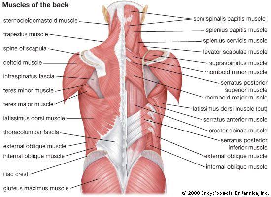
What are the back muscles?
- The back muscles are the main structural support for the trunk (torso). These muscles help you move the body, including the head, neck, shoulders, arms & legs. The back muscles work together to allow you to bend over, twist, turn the head & extend the back. These muscles also help you sit & stand up straight. They play an essential role in supporting the spine & helping you breathe.
- The back muscles start just under the skull, extend across the shoulders & down to the lower back just above the hips. These muscles attach to the ribs, vertebrae (bones in the spine), shoulder blades & neck.
- The back muscles are divided into three-layer – The superficial layer, the intermediate layer, and the Deepest layer the muscles.
- Superficial back muscles :
The superficial back muscles are the muscles situated just beneath the skin. Within this group of back muscles, you will find the latissimus dorsi, the trapezius, levator scapulae, & the rhomboids. - Intermediate back muscles :
The intermediate muscles are the erector Spinae. They contain the longissimus, iliocostalis, & Spinalis muscles. Their attachments further divide these muscles, & they all have a commonest tendinous origin. They play an important role in the movement of the thoracic cage & flexion of the upper vertebral column & head. - Deep back muscles :
The deep back muscles included the semispinalis, multifidus, & rotatores. These muscles help to stabilize the vertebral column & also have a role in proprioception & balance. Moreover, these muscles help with the movements of the vertebral column & also help to maintain posture.
- Superficial layer: splenius (splenius Capitis, splenius Cervices, latissimus dorsi, trapezius, levator scapulae, & the rhomboid minor & rhomboid major)
- Intermediate layer: erector spinae (iliocostalis, longissimus, spinalis)
- Deep layer: transversospinales (semispinalis, multifidus, rotatores)
- The deeper layer of Muscles: segmental muscles (Levatores Costarum, Interspinales, & Intertransversarii)
Superficial back muscles:
- Superficial back muscles include:
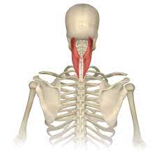
- Splenius Capitis Muscle (Musculus splenius Capitis)
- Splenius cervices muscle (Musculus splenius Cervices)
Splenius Capitis muscle
Origin:
The spinous processes of vertebrae C7 to T3 & the lower half of the nuchal ligament.
Insertion :
Inserted over the mastoid process & the lateral third of the superior nuchal line of the occipital bone.
Blood supply :
Blood supplies are the vertebral, occipital, superior intercostal, deep cervical, & transverse cervical arteries.
Nerve supply :
Innervation of splenius Capitis are posterior rami of the middle cervical spinal nerves & lower cervical spinal nerves
Action :
Lateral flexion of the neck & rotation of the neck at the same side.
Splenius Cervices muscle
Origin:
The spinous processes of vertebrae T3-T6
Insertion :
The transverse processes of vertebrae C1-C3 or C4.
Blood supply :
The blood supply of splenius Cervicis vertebral, occipital, superior intercostal, deep cervical, & transverse cervical arteries.
Nerve supply :
Splenius cervicis innervated by posterior rami of the middle cervical spinal nerves & lower cervical spinal nerves
Action :
Lateral flexion of the neck & rotation of the neck at the same side.
Latissimus dorsi muscle
Origin :
Spinous processes of thoracic T7 to T12, thoracolumbar fascia, iliac crest, & inferior 3 or 4 ribs, inferior angle of the scapula
Insertion :
As the latissimus dorsi inserts into the floor of the intertubercular groove of the humerus, it is surrounded by 2 major muscles. The teres major inserts medially on the medial lip of the intertubercular groove & the pectoralis major inserts laterally onto the lateral lip of the intertubercular groove.
Blood supply :
The latissimus dorsi muscles receive vascular supply from dorsal perforating branches of the lower 3 posterior intercostal arteries and the upper three lumbar arteries.
Nerve supply :
The thoracodorsal nerve, a branch of the posterior cord of the brachial plexus (C6-C8 with C7 predominant) provides innervation to the latissimus dorsi muscles.
Action :
Adduction, medially rotation, & extension of the upper limb at the glenohumeral joint.
Trapezius muscle
Origin :
The muscle attaches to the medial 3 of the superior nuchal line; external occipital protuberance, nuchal ligament, & spinous processes of C7 to T12 vertebrae.
Insertion :
Trapezius muscle inserts on the lateral 3 of the clavicle, acromion, and spine of the scapula.
Nerve Supply :
The spinal root of the accessory cranial nerve XI (motor)
C3 & C4 Cervical nerves (pain & proprioception)
Blood Supply :
Transverse cervical artery (cervicodorsal trunk)
Action :
The function of the trapezius muscles is to move & stabilize the scapula. The upper fibers of the trapezius elevate & upwardly rotate the scapula & extend the neck. The middle fibers of the trapezius do retract (adduct) the scapula. The lower fibers of the trapezius depress & aid the upper fibers in upwardly rotating the scapula.
Levator scapulae muscle
Origin :
The levator scapulae originate from the posterior tubercle of the transverse process of cervical vertebrae C1 to C4.
Insertion :
The muscle is inserted into the medial border of the scapula extending from the superior angle to the junction of the spine & medial border of the scapula.
Nerve Supply :
cervical nerve C3 & C4 & dorsal scapular nerve C5.
Blood Supply :
Dorsal scapular artery
Action :
The action of the Levator scapulae is to an elevation the scapula & tilt the glenoid cavity inferiorly by rotating the scapula downward.
Rhomboid minor muscle
Origin :
The rhomboid minor is a cylindrical muscle that originates from the Ligamentum nuchae & C7 & T1 vertebra.
Insertion :
Rhomboid minor inserts into the scapula’s medial border near the base of the spine of the scapula. The rhomboid major is a quadra-angular muscle in shape which is located inferior to the rhomboid minor muscles.
Nerve Supply :
Rhomboid minor is supplied by the dorsal scapular nerve from the brachial plexus of the C5 root.
Blood Supply :
Dorsal scapular artery.
Action :
Retraction & rotation of the scapula fixes the scapula to the thoracic wall.
Rhomboid major muscle
Origin :
The origin of the rhomboid muscles is from the spinous processes of the T2-T5 thoracic vertebra.
Insertion :
The rhomboid major is inserted into the medial border of the scapula, just inferior to the rhomboid minor.
Nerve Supply :
Rhomboid major is supplied by the ventral primary ramus through the dorsal scapular nerve C5.
Blood Supply :
Deep branch of the transverse cervical artery & dorsal scapular artery.
Action :
Retraction, elevation, & rotation of the scapula. The medial border of the scapula also protracts by the rhomboid major, keeping it in position at the posterior thoracic wall.
Intermediate back muscles:
- This layer contains the large erector spinal muscles which are sometimes known as the long muscles of the back. This muscle group is the largest of the deep muscles of the back & lies on either side of the vertebral column between the spinous process of the vertebrae & the ribs angle.
- The Intermediate layer muscles contain 3 vertical columns of muscle that lie side to side. From the lateral to the medial side, these are the iliocostalis, longissimus, & Spinalis muscles. Each column of muscle is further divided into different regions (like cervicis, capitis, lumborum, thoracic) based on which region of the axial skeleton it attaches to superiorly.
- Erector Spinae muscles,
- Iliocostalis cervicis
- Iliocostalis thoracic
- Iliocostalis lumborum
- Iliocostalis muscle (Musculus iliocostalis):
- The lateral column of the erector spinae muscle group forms by the iliocostalis muscles.
- The muscle is divided into 3 different regions according to its attachments:
- Iliocostalis cervicis
- Iliocostalis thoracic
- Iliocostalis lumborum
Iliocostalis cervicis muscle
Origin:
The angle of the ribs is 3-6.
Insertion:
Transverse processes of vertebrae C4-C6.
Nerve supply :
The nerve supply of iliocostalis cervicis is lateral branches of the posterior rami of cervical, thoracic & lumbar spinal nerves.
Blood supply :
The iliocostalis cervicis is supplied by the occipital, deep cervical, & vertebral arteries.
Action:
Laterally flexion & Extention of the lower cervical region.
Iliocostalis thoracic muscle
Origin:
The angle of the ribs is 7-12.
Insertion:
Angles of ribs 1- 6, transverse process of the C7 vertebra.
Nerve supply :
The iliocostalis thoracic muscles are innervated by lateral branches of the posterior rami of cervical, thoracic & lumbar spinal nerves.
Blood supply :
The vascular supply of iliocostalis thoracic muscles is supplied by the dorsal branches of posterior intercostal & subcostal arteries.
Action:
Extension & laterally flexion of the thoracic spine.
Iliocostalis lumborum muscle
Origin:
The origin of the Iliocostalis lumborum is the lateral crest of the sacrum, thoracolumbar fascia, and medial end of the iliac crest.
Insertion:
The angle of ribs 5 to 12, transverse processes of vertebrae L1 – L4.
Blood supply :
The iliocostalis lumborum muscles are supplied by the dorsal branches of the lumbar & lateral sacral arteries.
Nerve supply :
The nerve supply of iliocostalis lumborum muscles is lateral branches of the posterior rami of cervical, thoracic & lumbar spinal nerves.
Action :
Extension of the spine- action bilaterally, extension & hyperextension of the spine.
Laterally flexion of the spine when acting unilaterally.
Longissimus muscle (Musculus longissimus):
- The longissimus muscle forms the central column of the erector spinae muscle group & is the longest & thickest muscle in this group. It is divided into 3 regions according to their attachments:
- Longissimus Capitis
Longissimus Cervicis
Longissimus Thoracis
Longissimus thoracis further divided into thoracic & lumbar parts.
Attachments of the Longissimus muscle,
Longissimus capitis muscle
Origin:
Transverse processes of vertebrae C4 to T5.
Insertion:
Mastoid process of the temporal bone.
Nerve supply:
Longissimus capitis is innervated by the lateral branches of posterior or dorsal rami of cervical spinal nerves.
Blood supply :
Longissimus capitis receive blood supply from the vertebral artery, deep cervical artery, superficial & deep descending branches of the occipital artery, & deeper branch of the transverse cervical artery.
Action :
Extension of the neck vertebrae. Neck lateral flexion of the same side.
Longissimus cervicis muscle
Origin:
Transverse processes of vertebrae T1-T5.
Insertion:
Transverse processes of vertebrae C2-C6.
Nerve supply :
Longissimus cervicis is innervated by the lateral branches of posterior or dorsal rami of cervical spinal nerves.
Blood supply :
Longissimus cervicis receive blood supply from the vertebral artery, deep cervical artery, superficial & deep descending branches of the occipital artery, & deeper branch of the transverse cervical artery.
Action :
Extension of the neck vertebrae. Neck lateral flexion of the same side.
Longissimus Thoracis muscle,
Lumbar part-
Origin:
It originates from the lumbar intermuscular aponeurosis, the medial part of the sacropelvic surface of the ilium & posterior sacroiliac ligament.
Insertion:
Accessory & transverse processes of vertebrae L1-L5.
Nerve supply :
The longissimus thoracic muscles are innervated by the intermediate & lateral branches of the posterior rami of the lumbar spinal nerves.
Blood Supply :
The posterior branches of superior intercostal, posterior intercostal, lateral sacral, & median sacral arteries.
Action :
Spine extension & lateral flexion.
Thoracic part-
Origin:
Transverse & spinous process of vertebrae L1-L5, median sacral crest, posterior surface of the sacrum, & posterior iliac crest.
Insertion:
Transverse process of vertebrae T1-T12 ribs 7-12 angle.
Nerve supply :
The lateral & medial branches of the posterior rami of thoracic spinal nerves.
Blood Supply :
The dorsal branches of superior intercostal, posterior intercostal, lateral sacral, & median sacral arteries.
Action :
Spine extension & lateral flexion.
Spinalis muscle (Musculus spinalis);
- Synonyms: Musculi Spinalis
- The Spinalis muscle is the small & most medial of the Erector Spinae group of muscles.
- Like the longissimus, the Spinalis muscle is also divided into 3 parts:
- Spinalis Capitis
- Spinalis Cervicis
- Spinalis Thoracic
Attachments of the Spinalis muscle,
Spinalis Capitis muscle
Origin:
Spinous processes of vertebrae C7-T1.
Insertion:
Occipital bone (midline).
Nerve supply :
The innervation of the spinal capitis muscle comes from the lateral branches of the posterior or dorsal rami of adjacent spinal nerves (cervical, thoracic & lumbar).
Blood supply :
Muscular branches of the vertebral, deep cervical, & occipital arteries supplied spinalis capitis.
Action :
Produced cervical extension as well as lateral flexion & rotation of the spine & head.
Spinalis cervicis muscle
Origin:
Spinous processes of vertebrae C7-T1, nuchal ligament.
Insertion:
The spinous process of vertebrae C2 to C4.
Nerve supply :
The innervation of the spinal cervicis muscle arrives from the lateral branches of the posterior or dorsal rami of adjacent spinal nerves (cervical, thoracic & lumbar).
Blood supply :
Muscular branches of the vertebral, deep cervical, & occipital arteries supplied spinalis cervicis.
Action :
Produced lateral flexion, extension & rotation at the cervical spine level.
Spinalis Thoracic muscle
Origin:
The spinous process of vertebrae T11 to L2.
Insertion:
The spinous process of vertebrae T2 to T8.
Nerve supply :
The innervation of the spinal thoracic muscle arrives from the lateral branches of the posterior or dorsal rami of adjacent spinal nerves (cervical, thoracic & lumbar).
Blood supply :
The Spinalis Thoracic muscle arterial is supplied by dorsal branches of the superior & posterior intercostal arteries, & branches of the lumbar arteries.
Action :
Produced thoracic extension as well as lateral flexion & rotation of the spine.
Multifidus muscle (Musculus multifidus);
- This relates to the intermediate layer of the transversospinalis muscles group. The multifidus muscle is composed of many short, triangular muscles that span the entire length of the vertebral column but are the thickest & most developed in the lumbar region. The multifidus is divided regionally into three parts:
- Multifidus Cervicis
- Multifidus Thoracic
- Multifidus Lumborum
Multifidus cervicis muscle
Origin :
That arises from the superior articular processes of vertebrae C4-C7.
Insertion :
Lateral aspect & the tips of the spinous processes of C2-C5 vertebrae.
Multifidus thoracic muscle
Origin :
That arises from the transverse process of the thoracic vertebra.
Insertion :
Lateral aspect & tips of the spinous processes of vertebrae 2-5 levels above the origin.
Multifidus Lumborum muscle
Origin :
It originates from the mammillary processes of lumbar vertebrae, posterior aspect of sacrum, posterior superior iliac spine (PSIS) of the ilium, & posterior sacroiliac ligament.
Insertion :
lateral aspect & tips of the spinous processes of vertebrae 2 to 5 levels above the origin.
Nerve supply :
The multifidus muscle is innervated from the medial branches of the posterior rami of spinal nerves in the corresponding cervical, thoracic & lumbar regions.
Blood Supply :
Multifidus muscle’s arterial supply comes from the vertebral, deep cervical, occipital, posterior intercostal, subcostal, lumbar, & lateral sacral arteries according to the areas the muscle parts occupy.
Function :
The multifidus muscles’ important function is to stabilize the vertebrae during movements of the spine. Bilateral contraction of the muscle results in extension of the vertebral column at all levels (cervical, thoracic & lumbar regions), while unilateral contraction produces ipsilateral( same side) lateral flexion & contralateral rotation of the vertebral column.
Rotatores muscles:
- Rotatores breves & Rotatores Longi muscles (Musculi rotatores breves et Longi),
- Synonyms: Thoracic rotatores muscles, Long rotatores muscles, & short rotatores muscles.
- Deep to the multifidus is the small rotator muscles, which are the deeper of this muscle group. the rotatores muscles are also present along the entire length of the vertebral column, but are more prominent & best developed in the thoracic area. They consist of short rotatores (rotatores breves) which attach to the spinous processes of adjacent superior vertebrae & long rotatores (rotatores Longi) which attach to 2 levels up vertebrae.
Attachments of the rotatores muscles,
Rotatores breves muscle
Origin:
Transverse processes of T2-T12 vertebrae.
Insertion:
Laminae or spinous process of the vertebra (1 level above the origin).
Rotatores Longi muscle
Origin:
Transverse processes of the thoracic vertebrae.
Insertion:
Laminae or spinous process of the vertebra (2 levels above the origin).
Nerve supply :
The rotatores muscle is innervated by the medial branches of the posterior rami of spinal nerves.
Blood supply :
Vascular supply through dorsal branches of posterior intercostal & lumbar arteries.
Function :
The main function of the rotator muscles is to stabilize the spine. Bilateral contraction of these muscles causes extension of the vertebral column, while unilateral contraction causes rotation of the trunk to the contralateral side.
Deep muscles of the back:
- The levatores costarum, interspinal, & Intertransversarii muscles make up the deepest layer of the deep back muscles & are sometimes known as the segmental muscles or the minor deep muscles of the back.
Levatores Costarum muscles (musculi levatores costarum)
Synonyms: Levator costae muscles
The Levatores Costarum muscles are situated in the thoracic part of the vertebral column.
Origin :
From the transverse processes of C7 to T11 vertebrae & travel inferolateral to,
Insertion :
Between the tubercle & the angle of the corresponding rib below.
Nerve supply :
The levatores costarum are innervated from the lateral branches of the posterior rami of thoracic spinal nerves (T1-T12).
Blood supply :
Levatores costarum blood is supplied by the dorsal branch of the posterior intercostal artery.
Function :
The main function of levatores costarum muscles is to elevate the ribs & facilitate inspiration during breathing.
Interspinales muscles (Musculi interspinales)
- The Interspinales muscles are short & paired muscles that connect adjacent spinous processes of the vertebral column.
- These muscles are divided regionally into 3 parts :
- Interspinales cervicis
- Interspinal Thoracic
- interspinales lumborum
- They are well developed in the cervical & lumbar parts of the spine but may be entirely absent in the thoracic area.
Attachments of the Interspinales muscles
Interspinal cervicis muscle
Origin:
The superior aspect of the spinous processes of vertebrae C3-T1.
Insertion:
The inferior aspect of spinous processes of vertebrae C2-C7.
Interspinal Thoracic muscle
Origin:
The superior aspect of the spinous process of vertebrae T2, T11-T12 (variable).
Insertion:
The inferior aspect of spinous processes of vertebrae T1, T10-T11.
Interspinal lumborum muscle
Origin:
Superior aspects of spinous processes of vertebrae L2-L5.
Insertion:
inferior aspects of spinous processes of vertebrae L1-L4.
Nerve supply :
The Interspinales muscles are supplied by the posterior rami of the respective spinal nerves.
Blood supply :
The Interspinales muscles receive vascular supply from dorsal branches of respective regional arteries, namely the vertebral, deep cervical, occipital, transverse cervical, superior & posterior intercostal, subcostal, & lumbar arteries.
Function :
The main function of the Interspinales is to stabilize the adjoining vertebrae of the vertebral column. They also assist in the extension of the cervical & lumbar spine.
Intertransversarii muscles (Musculi Intertransversarii)
The Intertransversarii muscles are small muscles that pass between the transverse processes of adjacent vertebrae & are most developed in the cervical & lumbar area of the spine.
Intertransversarii Colli is found In the cervical region & is composed of anterior & posterior sets.
Intertransversarii Lumborum is found in the lumbar region. They contain 4 pairs of muscles that extend between adjacent transverse processes of lumbar vertebrae.
Nerve supply :
The Intertransversarii Colli is innervated by the anterior & posterior rami of cervical spinal nerves, while the lumbar intertransversarii is innervated by the posterior or anterior rami of lumbar spinal nerves.
Blood supply :
The Intertransversarii Colli receive blood supply from the occipital, deep cervical, ascending cervical & vertebral arteries, while lumbar Intertransversarii is supplied by the dorsal branches of lumbar arteries.
Function :
The function of the intertransversarii Colli muscles is to assist in lateral flexion & stabilization of the cervical spine. Similarly, the function of the lumbar Intertransversarii is to assist in the same side lateral flexion of the lumbar spine when acting unilaterally & to stabilize the lumbar spine when acting bilaterally.
Clinical significance of back muscles:
- Back pain in the lower or upper back region is the commonest significance of back muscles. It can be occurring due to numerous reasons depending upon different causes & symptoms.
- Muscle strain or ligament sprain: Repeated heavy lifting or a sudden jerky movement can strain back muscles & sprain spinal ligaments.
- Back Muscle spasm.
- Bulging or ruptured disks.
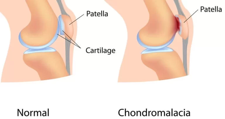
- Arthritis: Osteoarthritis can affect the lower back. In many cases, arthritis of the spine will cause narrowing of the space around the spinal cord, a condition called spinal stenosis.
- Osteoporosis: The vertebrae in the spine can develop painful fractures if the bones become porous & brittle.
- Injuries, fractures, or falls.
- Sciatica: A sharp & shooting type of pain travels through the buttock & down the back of the leg, caused by a bulging or herniated disk pressing on a nerve.
- Abnormal curvature of the spine: If the spinal curves are in an unusual form, back pain can result. An example is a scoliosis, in which the spine curves shift to the side in an s shape.
- Cancer of the spine: A tumor on the spine might be the reason for nerve root compression, resulting in back pain.
- Cauda equina syndrome: The cauda equine is a bunch of spinal nerve roots that arise from the lower end of the spinal cord. Symptoms include a dull pain in the lower back & upper buttocks area, as well as numbness in the buttocks, genitalia, & thighs. There are sometimes bowel & bladder functions get disturbed.
- Spina bifida.
Back muscles exercise:
Back muscles exercise mainly of two types:
- Back muscles strengthening exercise
- Back muscles stretching exercise
Strengthening exercise of Back muscles:

Transverse Abdominal Contraction:
- Lie down on the back with both knees bent, the feet flat. contract your core muscles by pulling the belly button towards the back.
- Hold for 10 secs & relax.
- Repeat 10 times. Perform 2 times per day.

Bridging:
- Lie down on the back with both knees bent. Squeeze the buttocks together then slowly lift the buttocks off the floor, keep the stomach tight & buttocks contracted.
- Slowly lower & release to starting position.
- Hold the Bridging position for 5 secs & repeat 10 times.
- Perform 2 times per day.
Gluteal Squeeze:
- Lie down on the back with both knees bent & squeeze the buttocks together.
- Hold for 10 secs & relax. Repeat 10 times.
- Perform 2 times per day.
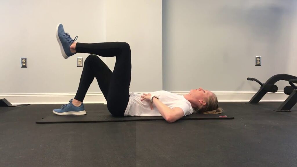
Transverse Abdominal March:
- Lying down on the back with both the knees bent. Contract the abdominal muscles by bringing the belly button toward the spine.
- Slowly march by lifting one leg off the ground, then change to the other.
- Continue pulling the belly button toward the spine during the transverse abdominal march exercise.
- Repeat 10 times on each side.
- Perform 2 times per day.

Wall Squats:
- Stand with the back against a wall with feet approximately 1-2 feet away from the wall.
- With feet, shoulder-width apart, squat approximately ½ of the way down, and make sure that the knees do not go past the toes.
- Hold Wall Squats position for 10 secs & repeat 10 times.
- Perform 2 times per day.
Stretching Exercise :

Child’s Pose:
- This traditional yoga pose is used to stretch the gluteus maximus, thigh muscles, & spinal extensors. It helps reduced pain & tension all along the spine, neck, & shoulders.
- To do Child’s Pose, follow these steps:
- With the hands & knees on the floor, sink back through the hips to rest them on heels.
- Hinge the hips as you fold forward, walking the hands out in front of you.
- Rest the belly on the thighs.
- Extend the arms in front of or alongside the body with the palms facing upward.
- Focus on breathing deeply & relax any areas of tension or tightness.
- Hold Childs Pose position for up to one minute.
- You can do this position several times during the stretching routine.

Cat-Cow:
- To do Cat-Cow, follow these steps:
- Come onto all 4 in a tabletop position (hands & knees on the floor).
- Press into the hands & feet as you breathe in to look up, allowing the belly to fill with air.
- Breath out, tucking the chin towards your chest & arching the spine toward the ceiling.
- This pattern of motion continues, moving with each breath.
- Do this for 1 to 2 minutes.

Supine Hamstring Stretch:
- Lie on the back, starting with both knees bent. Wrap a rope or towel around one foot of the self. While holding both ends of the towel, slowly lift 1 leg off the floor until a stretch is felt in the back of the leg.
- Hol this position for 20 secs.
- Return to the starting position.
- Repeat 3 times on each side.
- Perform 2 times per day.

Knee to Chest:
- In supine lying with both knees bent. Grab behind one knee & gently pull the knee towards the chest until a comfortable stretch is felt in the lower back.
- Hold Knee to Chest: position for 20 secs then return to starting position.
- Repeat 3 times on each side.
- Perform 2 times per day.
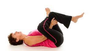
Piriformis Stretch:
- In supine lying with both knees bent. Pull one side of the knee to the opposite shoulder. Keep the back flat, do not twist.
- Hold this position approx for 20 secs then return to the starting position.
- Repeat 3 times on each side.
- Perform 2 times per day.

Prone Quadriceps Stretch:
- Lie down on your stomach.
- Put a rope, sheet, or belt around one of the feet & pull the heel toward the buttock until you can feel a stretch in the front of the thigh.
- Hold this position approx for 20 secs then return to the starting position.
- Repeat 3 times on each side.
- Perform 2 times per day.
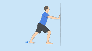
Calf stretch:
- Stand facing a wall. Keep back leg straight with heel on surface & foot facing forward.
- Bend the front knee slightly & lean towards the wall until a stretch is felt in the calf. It is necessary to keep the back heel on the surface throughout the entire stretch.
- Hold this position approx for 20 secs. Return to starting position.
- Repeat 3 times on each side
- Perform 2 times per day.

22 Comments