
Hamstring muscles: Anatomy, Origin, Insertion, Function, Exercise
Table of Contents
What are Hamstring muscles?
- Hamstring muscles are part of the skeletal muscles.
- These muscles are voluntary means a person controls the muscle move & work.
- The human body has three hamstring muscles that lie at the back of the thigh.
- These muscles are used to walk, climb stairs, squatting & perform many other leg movements.
- The function of these hamstring muscles is to flex the knee, extend the thigh at the hip joint & rotate the lower leg from side to side when the knee joint is bent.
How are the hamstring muscles structured?
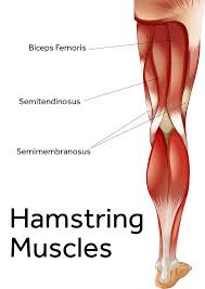
The three hamstring muscles are:
Biceps femoris, closest to the outside of body. The function of this hamstring is to flex the knee, extend the thigh at hip and rotate lower leg from side-to-side when knee is bent.
Semimembranosus, closest to the middle of body. This hamstring flexes knee joint, extends thigh at hip and offers medial rotation for hip and lower leg.
Semitendinosus, between the semi-membranous and the biceps femoris. The function of this hamstring muscle is same as semimembranosus.
What is the Function of the hamstring muscles?
The hamstrings cross and act upon 2 joints – the hip and the knee – and as such they are known as bi-articular muscles.
Semitendinosus & semimembranosus extend the hip when the trunk is fixed, they also flex the knee and medially rotate the lower leg when the knee is bent.
The long head of the biceps femoris extends the hip, as when starting to walk, both short and long heads flex the knee and laterally rotate the lower leg when the knee is bent.
The hamstrings play a important role in many daily activities such as walking, running, jumping, and controlling some motion in the gluteus. In the walking, they are most important as an antagonist to the quadriceps in the deceleration of knee extension.
Semitendinosus muscle
Semitendinosus is a fusiform muscle of the posterior compartment of thigh. Along with semimembranosus & long head of biceps femoris it comprises a group known as the hamstring muscles with which it shares these 3 common features:
- They all attach between the ischial tuberosity of pelvis & bones of the leg
- They cross both hip & knee joints on their course & act on them
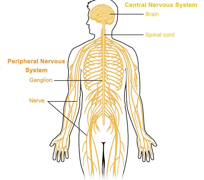
- They all are innervated by the tibial division of sciatic nerve
As a prime mover, semitendinosus extends and internally rotates the thigh, flexes and internally rotates the leg. It also has a postural role to stabilising the pelvic girdle.
Origin:
It arises, by a common tendon origin with the long head of the biceps femoris, from the lower medial facet of the lateral section of the ischial tuberosity. The semitendinosus muscle mainly originates from the medial surface of the tendon of the long head of the biceps femoris, and also originates from the ischial tuberosity with a thin tendon & a muscular part.
Insertion:
The semitendinosus tendon inserts at the upper part of the medial surface of the tibia, behind the attachment of sartorius & infero-anterior to the attachment of gracilis.
Function:
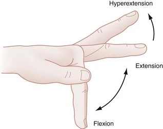
Flexion of the knee joint, Extension of hip joint, & Medial rotation of the tibia (with knee flexion)
Nerve supply:
Tibial nerve
Structure:
The semitendinosus, remarkable for the great length of its tendon of insertion, is situated at the postrior & medial aspect of the thigh.
It arises from the lower & medial impression on the upper part of the tuberosity of the ischium, by a tendon common to it & the long head of the biceps femoris; it also arises from an aponeurosis which is connect to the adjacent surfaces of the 2 muscles to the extent of about 7.5 cm. from their origin.
The muscle is fusiform & ends a little below the middle of the thigh in a long round tendon which is situated along the medial side of the popliteal fossa; it then curves around the medial condyle of the tibia & passes over the medial collateral ligament of the knee-joint, from which it is separated by a bursa, & is inserted into the upper part of the medial surface of the body of the tibia, nearly as far forward as its anterior crest.
The semitendinosus is more superficial than the semimembranosus (with which it shares very close to the insertion & attachment points). However, because the semimembranosus is wider & flatter than the semitendinosus, it is until possible to palpate the semimembranosus directly.
At its insertion it gives off from its lower border a prolongation to the deep fascia of the leg and situated behind the tendon of the sartorius, & below that of the gracilis, to which it is united. These 3 tendons form what is known as the pes anserinus, so named because it looks like the foot of a goose.
Relations:
Together with semimembranosus, semitendinosus contain the superomedial border of popliteal fossa. Its tendon can be palpated when the knee is flexed against resistance (for example while squatting), as a most lateral & posterior tendon of the superomedial border.
The ischial attachment of the semitendinosus situated at deep to gluteus maximus muscle. Being in the same plane & located medial to biceps femoris, the muscle belly & its tendon course posteriorly to adductor magnus & semimembranosus in the thigh.
Tibial attachment have a couple of interesting relations;
- Within pes anserinus, semitendinosus inserts along a vertical line posteriorly to sartorius & posteroinferiorly to gracilis.
- All 3 muscles in pes anserinus are separated from each other with a bursa. In addition, pes anserinus as a whole is separated from the medial collateral ligament of the knee by the anserine bursa.
Blood supply:
This muscle is supplied by branches from 3 large arterial sources;
- Femoral arteries are supplied via deep femoral artery & its first perforating branch, & medial femoral circumflex artery
- Internal iliac artery supplied through inferior gluteal artery
- Popliteal artery supplied via inferior medial geniculate artery.
Semimembranous muscle
- The semimembranosus muscle is the most medial of the 3 hamstring muscles in the thigh. It is so called because it has a flat tendon of origin. It is located at posteromedially in the thigh, deep to the semitendinosus muscle. It extends the hip joint & flexes the knee joint.
- The semimembranosus muscle, so called from its membranous tendon of origin, is situated at the posterior & medial side of the thigh. It is wider, flatter, & deeper than the semitendinosus (with which it shares very close insertion and attachment points). The muscle overlying the upper part of the popliteal vessels.
Origin:
The semimembranosus muscle attaches by a thick tendon from the superolateral aspect of the ischial tuberosity. It arises above & medial to the biceps femoris muscle & semitendinosus muscle. The tendon of origin enlarge into an aponeurosis, which covers the upper part of the anterior surface of the muscle; from this aponeurosis, muscular fibers arise, & converge to another aponeurosis which covers the lower part of the posterior surface of the muscle & contracts into the tendon of insertion.
Insertion:
The semimembranosus muscle inserts on the:
- medial condyle of the tibia
- medial margin of the tibia
- intercondylar fossa of femur
- lateral condyle of femur
- fascia of the popliteus muscle
The tendon of insertion gives off certain fibrous expansions. one, of considerable size, passes upward & laterally to be inserted into the posterior lateral condyle of the femur, forming part of the oblique popliteal ligament of the knee-joint; a 2nd is continued downward to the fascia which covers the popliteus muscle; while a few fibers join the medial collateral ligament of the joint & the fascia of the leg.
Function:
- Semimembranosus extends across both the hip & knee joints & is consequently responsible for motion about the joints. However, semimembranosus works in co-occurrence with the other hamstring muscles to carry out its function. When the feet are firmly planted on the floor, semimembranosus causes extension at the hip, which is pulls the upper torso to go into an erect position.
- Semimembranosus along with semitendinosus can also source internal rotation of the thigh when the hip is fully extended. When the legs are suspended off the floor, it causes flexion of the knee & internal rotation of the leg on the thigh.
- Semimembranosus & the other posterior thigh muscles are inactive whenever an individual is standing symmetrically. However, once the individual tilts too far forward, semimembranosus is activated & counteracts the forward movement.
Nerve supply:
The semimembranosus is supplied by the tibial part of the sciatic nerve. The sciatic nerve include of the anterior divisions of ventral nerve roots from L4 through S3. These nerve roots are the part of the wider nerve network–the sacral plexus. The Tibial part of the sciatic nerve is too responsible for innervation of semitendinosus & the long head of biceps femoris.
Variation:
The semimembranosus muscle may be reduced or absent, or double, arising mainly from the sacrotuberous ligament & giving a slip to the femur or adductor magnus.
Structure:
The muscle begins as a flat & membranous structure that develops a fleshy belly about midway down the thigh. The fleshy component is medially related to its tendon & the fibers are oriented inferomedially. The tendon is actually trifurcates distally to give:
- the main important part that inserts on the medial tibial condyle,
- a 2nd part that fuses with the popliteal fascia
- and a 3rd part that becomes the oblique popliteal ligament.
Another main feature of semimembranosus is that its lateral border forms the superomedial wall of the popliteal fossa.
Relations:
- The semimembranosus muscle has numerous adjacent muscular & neurovascular structures along its course. Semimembranosus is deep to semitendinosus, superficial to adductor magnus & medial to biceps femoris along its entirety. The proximal part of the muscle is covered by gluteus maximus & medial to adductor minimus. Distally, semimembranosus crosses over & becomes medially related to the medial head of gastrocnemius before inserting on the medial tibial condyle. The distal portion of the semimembranosus is also medial to the adductor canal, which accommodates the vessels of the lower limb.
- There is a U-shaped bursa that surrounded the semimembranosus tendon. It separates the tendon from the medial tibial plateau, medial head of gastrocnemius, semitendinosus & the medial cruciate ligament.
- The largest nerve in the human body is the sciatic nerve its travels lateral to semimembranosus until it reaches the apex of the popliteal fossa. At this point, the popliteal artery & vein are lateral to & partially covered by semimembranosus.
Blood supply:
The femoral popliteal arteries give off deep perforating branches that supply the semimembranosus muscle. Occasionally, the inferior gluteal artery supplies the proximal portion of the muscle.
Biceps femoris muscle
Biceps femoris is the long muscle of the posterior aspect of the thigh. Together with the semitendinosus & semimembranosus muscles, it makes the group of muscles commonly known as the hamstrings.
Biceps femoris muscle runs from the ischial tuberosity, all the way to the proximal portion of the fibula. In doing so the muscle crosses 2 joints; the hip joint & the knee joint. Acting simultaneously on the these joints, biceps femoris has many important functions; flexion & external rotation at the knee joint, extension & external rotation in the hip joint.
As its name suggests, this muscle consists of 2 heads, one lying deep to the other. Each head has a different origin & innervation but they share the same insertion.
Origin:
Long head: ischial tuberosity
Short head: linea aspera & lateral supracondylar line of the femur
Insertion:
Lateral aspect of fibular head
Function:
In general, the biceps femoris muscle acts on both the knee & hip joints. Although, due to this attachments, the short head of this muscle acts only on the knee joint while the long head acts on both.
When acting on the hip joint, biceps femoris produces the motion of hip extension. This action is the strongest when the trunk is bent forward & is to be brought in an upright position. The biceps femoris is also for sometimes described as assisting with external rotation (when the hip joint is in an extended position). When acting on the knee joint, the most prominent action of the biceps femoris muscle is flexion of the leg. This happen when the lower limb is in an anatomical position. In contrast, when the knee is semiflexed, biceps femoris acts to produce external rotation of the leg at the knee joint.
Together with other hamstring muscles the biceps femoris stabilizes pelvis, especially during the forward flexion of the trunk occurs. Therefore it has a major role in the gait cycle. Its most important antagonist is the quadriceps femoris muscle which is nearly 3 times stronger than the hamstrings.
Nerve supply:
- It is a composite muscle as the short head of the biceps femoris develops in the flexor compartment of the thigh and is thus supplied by common fibular branch of the sciatic nerve (L5, S1), while the long head is supplied by the tibial branch of the sciatic nerve (L5, S1).
Variations:
- The short head may be absent; additional heads may attaches from the ischial tuberosity, the linea aspera, the medial supracondylar ridge of the femur, or from various other portion. The tendon of insertion may be attached to the Iliotibial band & to retinacular fibers of the lateral joint capsule.
- A slip may be pass to the gastrocnemius.
Structure:
- It has 2 heads of origin:
- The long head arises from the lower & inner impression on the posterior part of the tuberosity of the ischium. This is a common tendon origin with the semitendinosus muscle,& from the lower part of the sacrotuberous ligament.
- The short head, arises from the lateral lip of the linea aspera, between the adductor magnus & vastus lateralis extending up almost as high as the insertion of the gluteus maximus, from the lateral prolongation of the linea aspera to within five cm of the lateral condyle;& from the lateral inter-muscular septum.
- The 2 muscle heads joint together distally & unite in an intricate fashion. The fibers of the long head form a fusiform belly, which passes obliquely downward & lateral-ward across the sciatic nerve to end in an aponeurosis which is covers the posterior surface of the muscle & receives the fibers of the short head. Inferiorly, the aponeurosis condenses to form a tendon which is predominantly inserts onto the lateral side of the head of the fibula. There is a 2nd small insertional attachment by a small tendon slip into the lateral condyle of the tibia.
- At its insertion the tendon divides into 2 portions, which embrace the fibular collateral ligament of the knee joint. Together, this joining of tendons is generally referred to as the conjoined tendon of the knee.
- The posterior border of the tendon is a thin expansion is given off to the fascia of the leg. The tendon of insertion of this muscle forms the lateral hamstring, the common fibular (peroneal) nerve is descends along its medial border.
Relations:
- For its largest part, the biceps femoris muscle runs superficially in the posterolateral thigh and sitting deep only to skin, fat & fascial layers. The exception to this is at its superior side, where it is covered by the gluteus maximus muscle. While the descending from the pelvis into the posterior thigh region, biceps femoris passes on the top of semimembranosus muscle, adductor magnus muscle & the lateral head of gastrocnemius muscle.
- Along the way, it is also located superficial to the sciatic nerve and providing protection for it. The sciatic nerve is gives its terminal branch (common fibular nerve) near the insertion of the biceps femoris. The nerve travels to briefly along with the medial border of biceps femoris, adhering to the tendon. This is an important clinical relation when considering injury and performing surgery procedures in this region.
Blood supply:
The muscles vascular supply is derived from the anastomoses of several arteries: the perforating branches of the profunda femoris artery, the inferior gluteal artery, & the popliteal artery.
Clinical Importance of Hamstring muscles:
Hamstring Injuries:
Overuse injuries to the hamstrings are more common, particularly in sports like soccer, football, basketball, & tennis, where running is combined with rapid starts & stops. The long head of the biceps femoris is a particularly prone to injury in sports such as these, likely because it exerts the most of force compared to the others muscles in the hamstring group.
Hamstring sprains & tears are also relatively common. They can become more serious when there is remarkable bruising behind the thigh. Repetitive stress injuries from running or walking are also a common causes of hamstring pain & injury.
Strains & Contusions
The onset of injury to the hamstring muscle group is often sudden & is usually identified as a strain (sprain or tear) or contusion (bruising). Strains range from mild to severe and the include the following traits.
Mild strains involve minimal damage to the muscle & heal quickly. They can be treated with rest & over the counter pain medication.
Moderate strains cause a partial rupture of the muscle & result in a loss of function.
Severe strains result in a total rupture of tissue & lead to short or long term functional disability.
Contusions are caused by external force and making contact with the hamstring muscles, such as with many contact sports.
Symptoms of contusions include:
- Hamstring Muscle pain
- Swelling
- Bruising & discoloration
- Limited range of motion
- Stiffness
If pain resulting from a hamstring injury does not resolve in a few days or is inhibiting your ability to walk normally & perform everyday activities, see your doctor for a diagnosis & treatment. In addition, studies show that 12% to 33% of hamstring injuries are reoccurring. If you play a sport & sustain a hamstring injury, you will likely need to fully rehabilitate prior returning to normal activity. This will allow the muscle group sufficient time to recover & repair, which can help prevent a relapse.
Regularly doing stretching & strengthening exercises, & warming up prior to exercise, may help to reduce the risk of injuring hamstring.
Stretching exercise of hamstring muscles
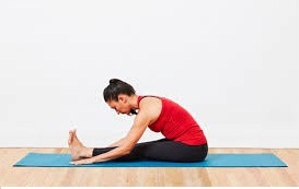
1. Hamstring muscle stretch:
- Sit on the ground with both legs are out straight.
- Extend the arms & reach forward by bending at the waist as far as possible while keeping the knees straight.
- Hold this position for 15-30 seconds.
- Relax back into the starting position.
- Repeat three times.
Be sure to stretch until a moderate pull is felt in the back of the thighs. If you are feel any excessive pain, you might be stop the exercise.
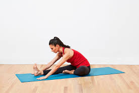
2. Hurdler hamstring muscle stretch:
- Sit on the ground with one leg are out straight.
- Bend the other leg at the knee & position the sole of that foot against the opposite inner thigh.
- Extend the arms & reach forward over the one straight leg by bending at the waist as far as possible.
- Hold this position for 15 seconds.
- Relax and than Repeat with the other leg.

3. Standing hamstring muscle stretch( Both legs are together):
This hamstring stretch is a simple one to do anywhere at the all. It is done in the standing position & stretches both legs at once. Here is how you to do the standing hamstring stretch:
- Stand & cross the right foot in front of the left.
- Slowly lower the forehead to the right knee joint by bending at the waist.
- Keep the both knees straight.
- Hold this position for 15-30 seconds.
- Relax and then Repeat for the other side by crossing the left foot in front of the right.
4. Standing hamstring muscle stretch (one leg at a time)
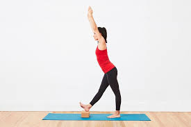
The one-legged standing hamstring stretch is completely possibly the easiest hamstring stretch to do. It can be done anywhere home, office, or outdoors & it requires no special tools.
- Stand up the straight with one heel resting on a small stool. if it is outside, one can use the curb, but be sure to watch for cars.
- Keep the knee straight.
- Reach both arms up toward the place where the wall & ceiling meet. If outside where there is no wall and ceiling, when simply reach up into the air so that the arms are about even with the ears. Reaching the arms up and as opposed to reaching down toward the foot, will keep the back straight.
- Keep the back straight & One should be bending forward slightly from the hips.
- Reach the forward & feel a stretch in the hamstring behind the thigh.
- Hold this position for 15-30 seconds, & repeat 3 times.
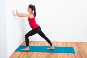
5. Runner’s hamstring stretch
The runner’s stretch is a common flexibility exercise for hamstrings
- Stand one foot from a wall & place the hands on the wall at shoulder height, shoulder width apart.
- Take a step back with 1 leg while pushing into the wall
- Keep the back straight & press the heels into the floor.
- Hold for 15-30 seconds.
- Step forward and the repeat with the other leg.
- Repeat the exercise 3 times on each side.
Strengthening exercise of the hamstrings muscle:
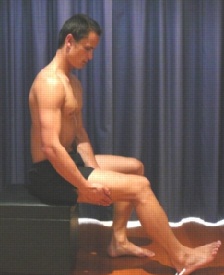
1. Static Hamstring contraction
- Start this exercise in sitting with the knee bent to about 45 degrees.
- Press the heel into the ground tightening the back of thigh.
- Hold for 5 seconds & repeat 10 times as hard as possible pain free.

2. Bridging exercise
- Start this exercise lying on the back in the supine position.
- Gradually, lift the bottom pushing through feet, until the knees, hips & shoulders are in a straight line.
- Tighten the back of the thigh while performing this exercise.
- Hold for 3-5 seconds then slowly lower the bottom of the back down.
- Perform this exercise 3 sets of 10 repetitions provided the exercise is pain free.
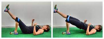
3. Single leg bridging
- Lie flat on the back with the knees bent & feet flat on the floor. The feet & knees might be hip distance apart.
- Tighten the abdominal muscles slightly to engage the core and stabilize the lower back.
- Lift up into simple bridge position but as one hold the bridge position, lift 1 foot off the floor & extend the knee as shown.
- Hold for 3-5 seconds, bring the foot back down, & then lower the buttocks to the floor.
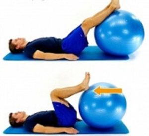
4. Hamstring curls with gym ball:
- Lies on the back with the feet & lower calves resting on a gym ball with knees straight & arms crossed over the chest.
- Slowly curl the ball towards the body by bending the knees & then slowly roll the ball away.
- Repeat this exercise for 10 times.
34 Comments