
Radial Artery
The radial artery, a major blood vessel in the human body, plays a crucial role in the circulatory system. Arising from the brachial artery in the forearm, it runs along the radius bone, hence its name, and is responsible for supplying oxygenated blood to the forearm and hand.
Table of Contents
Introduction
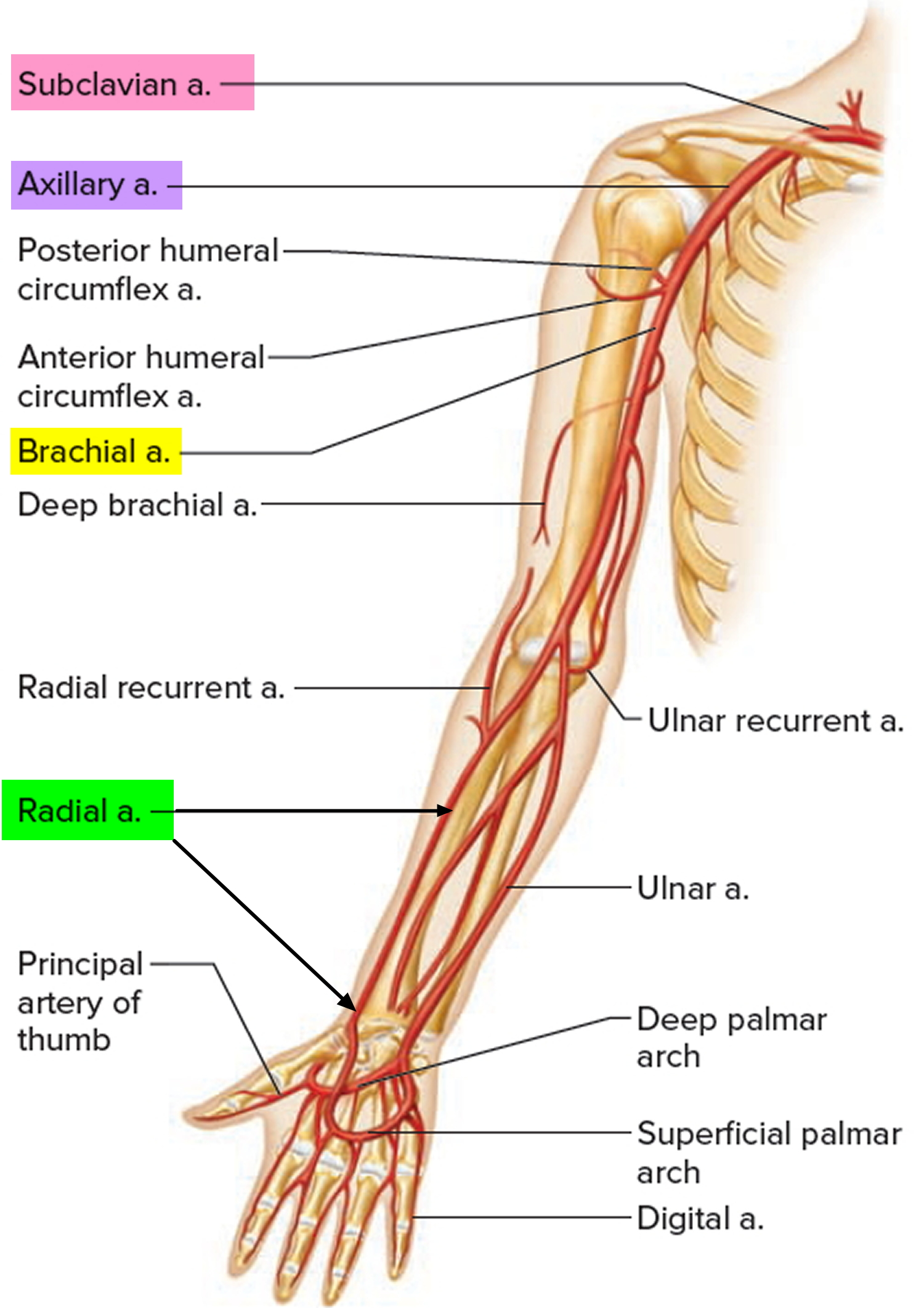
One of the brachial artery’s two branches is the radial artery; the other is the ulnar artery. It provides blood to the forearm’s anterior compartment. The radial and ulnar arteries are the forearm’s main perforators and result from the axillary artery bifurcating in the cubital fossa.
The radial artery passes between the brachioradialis and flexor carpi radialis muscles on the lateral portion of the forearm after bifurcating. It divides into superficial and deep palmar branches only anterior to the wrist, where it forms an anastomosis with the hand’s distal ulnar artery branches.
In the human forearm, the radial artery is a significant artery. The radial artery is located at the surface of the forearm’s underside; it points upward as the hand’s palm does. The radial artery transports oxygen-rich blood from the lungs to the hand and arm.
The radial artery is the most frequently used artery to monitor a patient’s pulse because of its size and closeness to the arm’s surface. Since the radial artery is closest to the surface of the wrist, this is the best place to examine the pulse. The radial artery is also frequently employed when taking arterial blood to measure “arterial blood gas” (ABG).
Three factors underlie this: first, the arm receives blood from other sources. The ulnar artery will take over in the event of injury to the radial artery. Second, getting to it is simple. Thirdly, because the radial artery is superficial, damage can be readily fixed, and the patient is rarely at risk.
What is a Radial Artery?
The radial artery supplies blood to the lower arm’s hand and forearm. The blood arteries in the body carry the blood throughout. Your lungs produce the oxygen in this blood, which is then distributed throughout your body.
A system of blood vessels, including the radial artery, transports blood to the heart and other parts of the body. The radial artery is used by medical professionals to do cardiac treatments and examinations.
Anatomy
The radial artery extends from the elbow to the thumb on the inside of the forearm. The artery is situated just beneath the epidermis. The blue or purple vein that supplies blood to your thumb may be visible inside your wrist.
Running parallel to one another down the forearm and into the hand are the radial and ulnar arteries. The hands, fingers, and forearms receive blood supply from them.
The radial artery’s first segment descends the pre-axial border of the forearm in conjunction with two radial veins, known as venae comitantes.
All along the forearm, the radial artery is covered by superficial and deep fasciae, except the upper portion, where the brachioradialis muscle’s belly overlaps it.
The radial pulse is felt against the bone on the anterior surface of the distal end of the radius, which is reached by passing inferolaterally between the brachioradialis and flexor carpi radialis after running inferiorly between the brachioradialis laterally and pronator teres medially.
While selecting the puncture site, the presence of bone posteriorly should be confirmed sonographically, as it greatly facilitates both manual compression and radial artery puncture to produce patent hemostasis during closure.
Just below the styloid process and into the first interosseous space, the lower portion of the radial artery’s middle segment curves across the wrist joint’s radial aspect. It travels between the thumb’s long and short extensors (extensor policies longus and extensor policies brevis) in the anatomic snuffbox after passing beneath the long abductor tendon (abductor policies longus).
There, it is crossed superficially by the beginning of the cephalic vein (which should be avoided during distal radial artery access) and superficial branches of the radial nerve. It is abutted by the distal scaphoid and trapezium, where the distal radial pulse can be palpated for anatomic sniffbox access.
The radial artery’s final segment enters the palm via the two heads of the first dorsal interosseous muscle.
Next, it turns medially, travels over the base of the metacarpals, and joins the deep branch of the ulnar artery to form the deep palmar arch. It passes between the transverse and oblique heads of the adductor pollicis.
Path/Course
In the antecubital fossa, the brachial artery bifurcates to become the radial artery. It runs distally, acting as a marker for the separation of the forearm’s anterior band and posterior compartments. The radial and ulnar arteries are the forearm’s main perforators and result from the axillary artery bifurcating in the cubital fossa.
The radial artery passes between the brachioradialis and flexor carpi radialis muscles on the lateral portion of the forearm after bifurcating. It divides into the superficial and deep palmar branches only anterior to the wrist, forming an anastomosis with the hand’s distal ulnar artery branches.
The anatomical snuff box, the first dorsal interosseous muscle heads, and the posterior compartment all weave around each other as they start immediately lateral to the artery and wrap laterally around the wrist. It moves between the adductor pollicis heads anteriorly.
Clinical Importance
Radial-arterial pulse
The radial artery is simply covered by skin and fascia and is located on the anterior aspect of the forearm at its distal end, which is before the wrist, as was previously mentioned. This is a typical location to take a patient’s pulse in order to evaluate heart rate, cardiac rhythm, and pulse strength.
Each forearm’s radial pulse can be taken simultaneously to evaluate radio-radial delays in the event of an aortic dissection, or it can be compared to the femoral pulse to evaluate radio-femoral delays in the event of a potential aortic coarctation.
Supplied Structure
Along with providing blood to the elbow joint through the radial recurrent artery, the radial artery supplies the brachioradialis, supinator, brachialis, and extensor carpi radialis longus and brevis.
The muscular branches of the radial artery feed the lateral aspect of the forearm with blood. These muscles are the extensor carpi radialis longus, extensor carpi radialis brevis, flexor pollicis longus, flexor digitorum superficialis, and pronator teres.
In addition to contributing to the palmar and dorsal carpal arches, which supply the wrist joint and hand, it gives tiny branches to the superficial radial nerve.
The hand, wrist, and forearm are the radial artery’s outflow points.
Branches
There are several branches of the radial artery at the wrist, hand, and forearm.
- Muscular branches
- Radial recurrent artery
- Palmar carpal branch
- Dorsal carpal branch
- Superficial palmar branch
- Deep palmar branch
At the forearm
The radial artery is divided into:
Muscular branches
The extensor muscles of the posterior compartment of the forearm, in particular, are supplied by these tiny branches on the radial aspect of the forearm. But only a subset of the forearm extensors is supplied by those muscular branches.
The anterior and posterior interosseous arteries are branches of the common interosseous artery that supply certain specific muscles of the posterior compartment, or only portions of the corresponding muscles. The ulnar artery is the source of the latter.
Radial recurrent artery
This branch lies directly distal to the point where the brachial artery and radial artery split. It is a major blood supply to the elbow joint and anastomoses with the radial collateral artery, which is derived from the deep brachial artery.
Palmar carpal branch
This branch travels along the anterior side of the carpal bones and emerges close to the distal edge of the pronator quadratus muscle. It anastomoses with the anterior interosseous arteries and the palmar carpal branch of the ulnar artery. To supply the carpal bones and their joints, this creates the palmar carpal arch.
At the wrist
Dorsal carpal branch
At the proximal end of the anatomical snuffbox, the dorsal carpal branch splits off from the radial artery and travels medially over the wrist. To produce the dorsal carpal arch, it anesthesizes in conjunction with the posterior interosseous arteries and the dorsal carpal branch of the ulnar artery.
Superficial palmar branch
The superficial palmar arch’s lateral portion, which is primarily supplied by the ulnar artery’s direct continuation, is completed by this branch. Between the palmar aponeurosis and the long flexor tendons of the fingers is the superficial palmar arch.
Deep palmar branch
This branch generates the hand’s deep palmar arch and is a direct extension of the radial artery. The deep palmar branch of the ulnar artery completes the medial portion of the deep palmar arch. Between the long flexor tendons of the fingers and the bases of the metacarpal bones, the palm is crossed by the deep palmar arch.
On the other hand,
Princeps pollicis artery: As the radial artery turns medially towards the deep part of the hand, the Princeps pollicis artery, the principal artery of the thumb, emerges. It passes beneath the tendon of the flexor pollicis longus muscle, descends between the oblique head of the adductor pollicis and the first dorsal interosseous muscle, goes down the medial side of the first metacarpal bone to the base of the proximal phalanx, and divides into two branches.
They emerge between the adductor’s medial and lateral insertions, running the length of the thumb and forming an arch on the palmar surface of the distal phalanx, from which branches extend to the thumb’s subcutaneous tissue and integument.
Radial indices branch: The branch of the radial artery that supplies blood to the index finger is known as the radial indices. It originates near the princeps pollicis artery, descends between the transverse head of the adductor pollicis and the first dorsal interosseous muscle, and travels down the side of the index finger to its extremity, where it anastomoses with the appropriate digital artery.
This vessel anastomoses with the princeps pollicis near the lower border of the transverse head of the adductor pollicis, and it provides a connected branch to the superficial palmar arch. The first palmar metacarpal artery may serve as the shared source for the Princeps pollicis and radialis indicis.
Deep volar arch or deep palmer arch: An artery network located in the palm is called the deep volar arch or deep palmer arch. Usually, the radial artery’s terminal segment is primarily responsible for its creation, with the ulnar artery’s deep palmar branch serving as an anastomosis. On the other hand, the ulnar artery forms the superficial palmar arch, which is different. The oblique head of the adductor pollicis muscle, the flexor tendons of the fingers, and the lumbrical of the hand cover the deep palmar arch, which rests on the bases of the metacarpal bones and the hand’s interossei.
The deep branch of the ulnar nerve runs along it, but in the opposite direction, towards the radial side of the hand. Compared to the deep palmar arch, the superficial palmar arch is placed farther away. This is the level of the superficial palmar arch, or Boeckel’s line if one fully extends the thumb and draws a line from the thumb’s distal border across the palm. This is approximately one finger’s width away from the deep palmar arch.
Allen’s test can determine whether there is an anastomosis, which is demonstrated by the link between the deep and superficial palmar arterial arches. The palmar metacarpal arteries come from the deep palmar arch.
Purpose of the Radial Artery
The radial artery allows blood to travel from the heart to the forearm as part of the circulatory system. The branches of the radial artery are many. They give blood that is oxygenated to:
- elbow joint.
- muscles of the forearm.
- Thumbs and index fingers.
- Radial nerve: regulates feelings and movement of the hands and arms.
- wrist joints and bones (carpal).
Summary
The radial artery supplies the hand and lower arm with oxygen-rich blood. Radial artery catheterizations and other noninvasive (nonsurgical) cardiac exams and treatments are performed by healthcare providers.
Heart bypass surgery is another procedure that providers do using the radial artery. A damaged or absent radial artery can be replaced if necessary by the ulnar artery in the forearm.
FAQs
What is the function of the radial artery?
The elbow joint, lateral forearm muscles, radial nerve, carpal bones and joints, thumb, and lateral side of the index finger 2 are all supplied with blood by the radial artery.
What are the radial artery branches?
Numerous branches of the radial artery can be found in the hand, wrist, and forearm. These little branches supply the extensor muscles of the posterior compartment of the forearm, which are located on the radial aspect of the forearm. But only a subset of the forearm extensors are supplied by those muscular branches.
Why is the radial artery important?
The elbow joint, lateral forearm muscles, radial nerve, carpal bones and joints, thumb, and lateral side of the index finger are all supplied with blood by the radial artery.
Can you live without a radial artery?
In carefully chosen individuals, radial artery harvesting is thought to be safe [2-4]. However, nothing is known about the state of digital flow following radial artery harvesting.
Where is the radial artery pulse?
On either wrist, you can take your radial pulse. Feel the pulse in your radial artery between the tendon on the thumb side of your wrist and your wrist bone using the tips of your other hand’s index and third fingers.
Reference:
- Radial Artery. (n.d.). Physiopedia. https://www.physio-pedia.com/Radial_Artery
- Professional, C. C. M. (n.d.). Radial Artery. Cleveland Clinic. https://my.clevelandclinic.org/health/body/21856-radial-artery#function
- Radial Artery | Complete Anatomy. (n.d.). www.elsevier.com. https://www.elsevier.com/resources/anatomy/cardiovascular-system/arteries/radial-artery/20303
2 Comments