
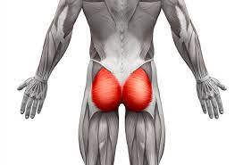
Gluteal muscles
Table of Contents
What is Gluteal muscle?
- The gluteal muscles are also called the glutes which are a group of the three muscles which is made by to gluteal region & commonly known as the buttock.
- This gluteal region is an anatomical area that is located posteriorly to the pelvic girdle.
- These three muscles of gluteal region are the gluteus maximus, gluteus medius & gluteus minimus.
- These three muscles originate from the ilium&sacrum & insert on the femur bone.
- The functions of these muscles include abduction, extension, external rotation -ER, & internal rotation – IR of the hip joint.
Where is the gluteal located?
- Gluteus Maximus muscle is the largest & heaviest muscle in the body. It is a most superficial of all gluteal muscles that are located at the back side of the hip joint.
- It arises from the posterior gluteal line of the inner upper ilium, a bone of the pelvis, as well as above it to the iliac crest & slightly below it; from the lower part of the sacrum & the side of the coccyx, the tailbone; from the aponeurosis of the erector spinae (lumbodorsal fascia), the sacrotuberous ligament, & the fascia covering the gluteus medius (gluteal aponeurosis).
Gluteus maximus
Introduction
- Gluteus Maximus muscle is the largest & heaviest muscle in the body. It is a most superficial of all gluteal muscles that are located at the back side of the hip joint. It is a largest muscle at the hip representing 16% of the total cross-sectional area.
- Gluteus Maximus’s size allows it to bring about the large amount of force (the muscle evolved from an adductor of the hip which is a still seen in lower primates today). The development of the muscle’s function is the associated with the erect posture & changes to the pelvis, now functioning to maintain the erect posture, as a hip extensor.
- Gluteus maximus muscle covers the all of the gluteal muscles except for the antero_superior third of the Glutes medius.
- The ischial tuberosity can be felt deep to the lower site of the Glutes maximus. When the thigh is flexed the lower border of Glutes Maximus moves to the superiorly , exposing the ischial tuberosity subcutaneously (in sitting you sit on the ishial tuberosity , ischial bursae , subcutaneous fat & skin).
Origin:
- Outer slope of dorsal segment of the iliac crest
- Gluteal surface of the ilium
- Dorsal surface of the lower part of sacrum
- Side of coccyx
- Sacrotuberous ligament
- Gluteal aponeurosis
- Attaches to thoracolumbar fascia and its associated raphe. By this attachment Glutes maximus is a coupled to the ipsilateral multifidus & contralateral Latissimus dorsi forming posterior oblique & deep longitudinal myofascial slings.
Insertion
- Gluteal tuberosity
- Iliotibial tract
Structure:
- The gluteus maximus muscle is the outermost muscle of the buttocks. It arises from connections to nearby structures in this site. It arises from the posterior gluteal line of the inner upper ilium, a bone of the pelvis, as well as above it to the iliac crest & slightly below it; from the lower part of the sacrum & the side of the coccyx, the tailbone; from the aponeurosis of the erector spinae (lumbodorsal fascia), the sacrotuberous ligament, & the fascia covering the gluteus medius (gluteal aponeurosis).
- The fibers are directed obliquely inferiorly & laterally;
- The gluteus maximus ends in 2 important sites:
- Those forming the upper & larger portion of the muscle, together with the superficial fibers of the lower portion, end in a thick tendinous lamina, which passes across the greater trochanter, & inserts into the iliotibial band of the fascia lata;
- The deeper fibers of the lower portion are inserted into the gluteal tuberosity of the linea aspera, between the vastus lateralis & adductor magnus. If present, the third trochanter also to be serves as an attachment.
Bursae:
- 3 bursae are usually found in relation with the deep surface of this muscle:
- One of these, of large size, and separates it from the greater trochanter;
- A second (often missing) is a situated on the tuberosity of the ischium;
- A third is the found between the tendon of the muscle & that of the vastus lateralis.
Function
- Chief extensor of the thigh
- Essential for maintaining an erect posture
- Lateral rotation of the thigh
- Abduction of the thigh
Role in Activity of daily living (ADLs) eg:
- As the powerful extensor of the hip joint, the gluteus maximus is suited to powerful lower limb motion eg stepping onto a step, climbing, or running but is not used greatly during normal walking.
- The Gluteus maximus & the hamstrings work together to extend the trunk from a flexed position by pulling the pelvis backward, eg standing up from a bent forward position. Eccentric control is the also provided when the bending forward.
- If the gluteus maximus muscle is paralyzed climbing stairs & running will become a very difficult however, other muscles can extend the hip. The Gluteus maximus can be trained to produce functional knee extension when the quadriceps femoris is a weak or paralyzed.
- Research has indicated that contraction of the deep abdominal muscles may be assist with the contraction of the gluteus maximus to assist with the control of anterior pelvic rotation. Gluteal muscle weakness has been proposed to be associated with the number of the lower limb injuries.
Relations
- The gluteus maximus is the most superficial & the largest of all 3 gluteal muscles. It is surrounded with a thin fascia that separates the muscle from the adjacent subcutaneous tissue.
- The deep surface of the gluteus maximus muscle are covers a number of structures; gluteus medius muscle, pelvic bones, the proximal attachments of the hamstring muscles & several lateral rotators of the hip (piriformis, inferior gemellus, superior gemellus & obturator internus muscles).
- The deep surface of the muscle is also in relation to the 3 bursae:
- The trochanteric bursa is a separates the muscle from the greater trochanter.
- The ischiofemoral bursa, when present, is located on the tuberosity of the ischium.
- The gluteofemoral bursa is found between the tendon of the gluteus maximus & that of the vastus lateralis.
Nerve supply
- The gluteus maximus muscle is the supplied by the inferior gluteal nerve (root L5, S1 and S2). Cutaneous supply is mainly provided by L2 and L3.
Blood supply
- The gluteus maximus muscle is a vascularized by the muscular branches of the inferior gluteal & superior gluteal arteries, the branches of the internal iliac artery.
Gluteus medius muscle
Introduction
- Gluteus medius muscle is the large fan-shaped muscle lies in the posterior hip, extending from the ilium to the proximal femur. Together with the gluteus maximus, gluteus minimus & tensor fascia latae muscles, it belongs to the muscles of the gluteal region.
- The gluteus medius muscle acts on the hip joint producing 2 movements; its anterior part internally rotates the thigh, while the contraction of the whole muscle abducts the thigh. Additionally, the gluteus medius muscle stabilizes the pelvis during standing & walking.
- Gluteus maximus muscle covers the all of the gluteal muscles except for the anterior-superior third of the Gluteus medius. This uncovered part of Gluteus medius is the safe area at which we might be apply buttocks dorso gluteal intramuscular injections.
Origin
Gluteal, or lateral surface of the ilium between the posterior & anterior gluteal lines & gluteal aponorosis . This is the large area, extended from the iliac crest above to the almost the sciatic notch below.
Insertion
Glutes medius is divided into 3 portions similar to deltoid muscle of the shoulder joint.
- Fibers of the posterior part pass forwards & downwards.
- Fibers of the middle portion pass downwards.
- Fibers of the anterior portion pass backwards & downward. All Fibers combine to form a flattened tendon which attaches to the posterior & lateral part of the superior portion of the greater trochanter of the femur.
Structure
- The gluteus medius muscle starts, or “originates”, on the outer surface of the ilium between the iliac crest & the posterior gluteal line above, & the anterior gluteal line below; the gluteus medius also arise from the gluteal aponeurosis that covers its outer surface.
- The fibers of the muscle converge into the strong flattened tendon that inserts on to lateral surface of the greater trochanter. More specifically, the muscle’s tendon inserts into an oblique ridge that runs downward & forward on the lateral surface of the greater trochanter.
Function
- Gluteus medius is a prime mover of abduction at hip joint.
- Anterior portion of Gluteus medius abduct, and assist in flexion & medial rotation of hip.
- Posterior portion of Gluteus medius abduct, and assist in ext & lateral rotation of hip.
- In the hip flexion all portions internally rotate the hip & it has shown that at 90` of hip flexion the leverage of medial rotation of Gluteus medius is increased eight folds.
- All portions of Gluteus medius will produce the hip abduction regrades the position of the hip.
- Glutes medius is an extremely important muscle in maintaining the frontal plane stability of the pelvis it forms with the ipsilateral tensor fascia latae & contralateral quadratus lumborum a lateral fascial sling whose main role is the provide frontal plane stability.
- Gluteus medius is an important muscle in walking, running & single-leg weight-bearing because it prevents the opposite side of the pelvis from dropping during walking, running & single leg weight-bearing.
- When a limb is taken off the floor the pelvis on the that side will tend to drop through loss of support from below.
- Gluteus medius function is maintain the side of the pelvis that drops therefore allowing the other limb to swing forward for the next step.
- Gluteus medius also to be supports the pelvis during gait by producing rotation of hip with assistance from gluteus minimus & tensor fascia lata. Conversely, the hip is the supported during the stance phase by acting on the same side.
Relations
- Gluteus medius is a middle gluteal muscle, located deep to the gluteus maximus & superficial to the gluteus minimus. Only the posterior third of the muscle is the covered by gluteus maximus, whereas the larger anterior portion is a covered by the deep fascia of the hip. These fascia is sometimes referred to as the gluteal aponeurosis. The posterior margin of the muscle located anterior to the piriformis muscle, & sometimes it may be blended with it.
- Gluteus medius is related to the branches of the superior gluteal artery & nerve, which run between the adjacent surfaces of the gluteus medius & minimus muscles. This is an important relation, as these vessels & nerves may be damaged during surgical procedures that involve the incision of gluteus medius.
Variations
- The posterior border is more or less closely united to the piriformis, or some of the fibers end on its tendon.
- The posterior fibers of gluteus medius contract to produce hip extension, lateral rotation & abduction. During the gait, posterior fibers help to decelerate internal rotation of the femur at the end of swing phase.
Nerve Supply
- The gluteus medius is supplied by the superior gluteal nerve (root L4,L5 & S1). Cutaneous supply is mainly provided by L1 & L2.
Blood supply
- The blood supply to the gluteus medius muscle comes from the deep branch of superior gluteal artery. The tendon, however, is mainly supplied by the trochanteric anastomosis. This anastomosis is formed between the ascending branch of the medial circumflex femoral artery & descending branches of the superior gluteal & inferior gluteal arteries.
Gluteus minimus
Introduction
- The gluteus minimus muscle is the small triangular muscle located deep in the posterior region of the hip, spanning from the gluteal surface of the ilium to the proximal end of the femur. It belongs to the group of gluteal muscles, along with the gluteus maximus, gluteus medius & tensor fascia latae.
- Gluteus minimus acts in synergy with the gluteus medius to produce the motion of the hip joint; the internal rotation & abduction of the thigh. Moreover, this muscle is an important stabilizer of pelvis in the gait cycle.
Origin
- Outer surface of ilium between middle and inferior gluteal lines .
Insertion
- Anterior surface of greater trochanter of femur .
Function
- The gluteus medius & gluteus minimus abduct the thigh, when the limb is extended, & are principally called into action in supporting the body on one limb, in conjunction with the tensor fascia lata.
- Their anterior fibers also flex the hip, and by drawing the greater trochanter forward, rotate the thigh inward, in which action they are also assisted by the Tensor fascia lata.
- Additionally, with the hip flexed, the gluteus medius & minimus internally rotate the thigh. With the hip extended, the gluteus medius & gluteus minimus externally rotate the thigh.(contradictory)
- The attachment to the superior capsule of the hip may also serve to retract the capsule away from the joint during movement.
- This mechanism may prevent the capsular impingement similar to the role of the articularis genus in the knee.
Nerve supply
- Superior gluteal nerve (L4, 5, S1) .
Variations
- The muscle may be divided into an anterior & a posterior portion, or it may send slips to the piriformis, the superior gemellus or the outer part of the origin of the vastus laterali.
Blood supply
- The blood supply to the gluteus minimus muscle comes from the deep branch of the superior gluteal artery. The distal part of the muscle & its tendon also to be receives the contribution from the trochanteric anastomosis; an arterial network formed by the ascending branch of the medial circumflex femoral artery & descending branches of the superior & inferior gluteal arteries.
Relations
- The gluteus minimus is the smallest & the deepest of all 3 gluteal muscles. Its deep surface is entirely related to the ilium, while superficially it is covered by the gluteus medius muscle. The branches of the superior gluteal artery & nerve course between these 2 muscles.
- The anterior margin of muscle is the related to tensor fascia latae, while the posterior one is the related to piriformis muscle. At the level of the insertion, the tendon of the gluteus minimus is lies superficial to the reflected tendon of the rectus femoris & the capsule of the hip joint.
Clinical Importance of Gluteal muscles:
- Sitting for long periods can be lead to the gluteal muscles atrophying through constant pressure & disuse. This may be associated with lower back pain, difficulty with some movements that naturally require the gluteal muscles, such as the rising from the seated position, & climbing stairs.
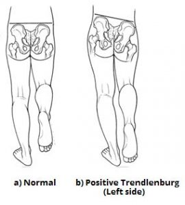
- The Trendelenburg sign is produced when the patient is asked to stand unassisted on both legs in turn. As a positive sign, the pelvic drop will happen on the unsupported leg. Pelvic drop can be recognized by observing the level of the iliac crests on each side.
- For example, if the left gluteal muscles are weak, the right side of the pelvis will drop when the patient stands on their left leg (& the right leg is unsupported).
Gluteus minimus tendinopathy results in a Greater Trochanteric Pain Syndrome (GTPS). Which is characterized by lateral hip pain, tenderness at the greater trochanter & Trendelenburg gait, it might be differentiated from trochanteric bursitis which is very rare.
Anteroposterior radio graph of the pelvis is the done to rule out hip osteoarthritis. Gluteus minimus trigger points referred pain starts at the end of the lumbar spine & ends at the ankle, following a similar pain pathway of sciatic nerve but without the neurological symptoms of the sciatic nerve such as weakness & numbness.
A supplemental injury of the superior gluteal whim-whams may lead to loss of motor function. The classical sign is the pelvis dropping to the healthy side when standing on one leg( Trendelenburg’s sign). In order to maintain the balance, the cases compensatorily bend their upper body to the side of the station leg. likewise, they walk with conspicuous side ward movements( Duchenne gait, also “ lurching gait ”).
Physiotherapy Relevance
Weak gluteal muscles have been associated with the variety of lower limb issues. It may be lead to other muscles or tissues to become overloaded through a cascade of events
- Patellofemoral pain syndrome
- Gluteal Tendinopathy
- Iliotibial Band Syndrome
- Trochanteric Bursitis
- Greater Trochanteric Pain Syndrome
- Differentiating Buttock Pain – Gluteal Tendinopathy
- Exercise to strengthen the muscles of the gluteus plus exercises to improve core strength results in a greater decrease in low back pain & disability than core strengthening alone.
- Patients with impaired hip abduction may be present with an abnormal gait. Impaired hip abduction is commonly due to damage to the superior gluteal nerve. This may occur secondary to pelvic fractures, or space-occupying lesions & as a complication of hip surgery.
Gluteus muscle exercise:
- Exercises that strengthen the anterior part: resisted hip abduction-extension exercise.
- Exercises that strengthen the Posterior part: single leg bridge, side lie abduction, the resisted hip abduction-extension exercise, and single-leg squat.
- Exercises that build low activity in both segments: side lie clam.
- Low activity was produced in the anterior segment of the single-leg bridge.
Stretching for Gluteus muscle
- Gluteus stretch
- Lying Deep Gluteus Stretch
- Groin and long adductor muscle stretch
- Seated figure-four stretch
- Seated glute stretch
- Supine Hip Twist Stretch
- Wall Glute Stretch:
- Supine Leg Extension Stretch
- Gluteus stretch:
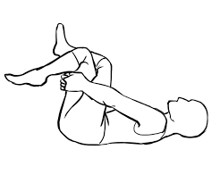
- The starting position for the exercise is supine position.
- The patient is Lying down onto the back after that lift to the knee joint & cross to the right ankle joint over to the left knee joint.
- Then Grip the left thigh gently.
- Pull the hip joint in towards the chest.
- Hold this stretching position for 30 seconds.
- Then repeat this stretching one the other leg.
- Do this stretching 3 times in 1 session.
- Then do the 3 sessions per day.
2.Lying Deep Gluteus Stretch:

- The starting position for the exercise is supine position.
- the patient is lying on the back with the legs being too bent.
- then Raise to the right ankle joint & rest the right ankle onto the left knee joint.
- then Using both hands lace with the fingers behind the left thigh & gently pull it toward the patient,
- keep the head & back on to the floor.
- Hold this stretching position for 20 to 30 seconds.
- then Repeat this exercise with to the other leg.
- do this stretching 3 times in 1 session.
- then do the 3 sessions per day.
3. Groin and long adductor muscle stretch

- The starting position for the exercise is the sitting position.
- The patient is Sitting down on the floor with the legs are spread out too far as the patient so that is straight in front of the body.
- Place the hands onto the floor in front of the body onto the floor & angle to the torso toward the floor.
- Lean-to the forward, leaving the elbows on to the floor.
- Hold to the position for 30 seconds.
- Stop this stretch if they feel any pain.
- Do this stretching 3 times in 1 session.
- Then do the 3 sessions per day.
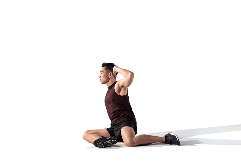
4. Seated figure-four stretch:

- This is also known as the Seated Pigeon Pose.
- Starting position of the exercise is the sitting position.
- The patient is sitting upright in a sturdy chair.
- Place the right ankle on the left thigh, just above the knee joint.
- Then Place the hands on the shins.
- After that Keep the spine straight & then lean slightly forward to deepen the stretch.
- Hold for the stretching position for 30 seconds & do the 3 times in 1 session.
- Then Return to the starting position of the stretching.
- After that Repeat with the other leg.

This stretching is also done in sitting on the floor & standing position.
5. Seated glute stretch:
- The starting position of the exercise is the sitting position.
- Sit on the floor & extend your legs in front of you.
- Keeping the back straight, lift the left leg.
- Place the left ankle joint on the right knee joint.
- Then Lean-to slightly forward &feel the patient deepen the stretch.
- Hold this stretching position for 30 seconds & do the 3 times in 1 session.
- Repeat this stretching on the other side.
- Do the 3 times in 1 session.
6. Z – Sit exercise
- Start by sitting comfortably on the ground.
- Bring the left knee to a 90-degree position in front of the body (as much as your body allows).
- Do the same with your right leg, toward the back of your body.
- You can sit upright in this pose or lean the torso forward toward your front leg.
- Hold the pose for 30 seconds, & then repeat on the other side.
- Supine Hip Twist Stretch
This exercise stretches the outer hip, where the gluteus minimus muscle is located, without placing stress on your spine. Lie on the floor on the back with arms out to your sides & with your hands on the floor. Put the feet flat on the floor about hip-width apart. Cross left foot over right thigh near your knee. Exhale & turn your pelvis toward your right so that your outer right knee & your left foot move toward the floor. Do not lift your left shoulder off the ground or twist your upper back. Hold this stretch for 5 to 6 deep breaths. Perform 2 sets of stretches on each side of the body.
7. Wall Glute Stretch:
This exercise stretches the entire hip region, which also takes the pressure off the sciatic nerves, which run through either side of the buttocks. Lie on the ground on your back, & put your feet on the wall about hip-width apart. Cross the left foot on your right thigh near the knee. Do not change the position of your pelvis as you cross your foot. Push your left knee toward the wall by using your hip. Hold this stretch for 5 to 6 deep breaths. Switch leg position, & repeat the stretch on the opposite hip.
8. Supine Leg Extension Stretch

This exercise stretches the entire back of your hip & leg from your buttocks & into your calves. Lie on the ground on your back, & lift your left knee to your ribs. Grab the back of your knee, & point your knee so that it faces toward your right shoulder or the right side of your chin. Slowly extend your leg straight up, & flex your left foot toward your right shoulder. Hold the stretch for 5 to 6 deep breaths. Repeat the stretch on the opposite leg.
Strengthening exercise of the gluteus muscle
- Buttocks
- Knee to the opposite shoulder
- Side Leg Raises
- Prone Straight Leg Raises [ hip extension ]
- Glute bridge with band
- Seated hip abduction with resistance band
- Spiky ball roll on glutes
- Bridges
- Knee to Chest
- Downward-Facing Dog
- Pigeon Pose
- Clam shell exercise
- Leg bridge exercise

1. Buttocks:
- The starting position for the exercise is supine.
- Patient Lay on to the back.
- Then bring the affected leg up to the right angle.
- Grasping to the with the help of both hands behind of to the thigh.
- Then locking to the fingers of the head.
- Do the take unaffected leg & place to the ankle joint against to the knee joint.
- This exercise Repeat to the opposite sides.
- Do this exercise 3 times in 1 session.
- Then do the 3 sessions per day.

2. Knee to the opposite shoulder:
- The starting position for the exercise is the supine position.
- The patient is lying on the back with to both legs are extended & feet flexed to the upward.
- Then Bend to the right leg & clasp both hands around to the knee joint.
- After that Gently pull the leg across to the body toward the left shoulder.
- Hold this exercise position for 30 seconds.
- Push the knee joint so that the leg is returned to the starting position.
- Do this exercise 3 times in 1 session.
- Then do this exercise 3 sessions per day.
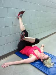
3.SIDE Leg Raises (hip abduction) exercise

- The starting position for the exercise is the side-lying position.
- The patient is lying on one side with the legs stacked.
- The patient is Bend to the bottom leg for support.
- Then Straighten to the top of the leg and maybe upraise to 45 degrees.
- Hold this exercise position for 10 seconds & then relax.
- Then Repeat this exercise 10 times in the 1 session.
- Do this exercise in 3 sessions per day.
- Woman doing Side Leg Raise Exercise with lying in 2 Step on blue mat. Illustration about Thighs Workout.
4.Prone Straight Leg Raises (hip extension) exercise :

- The starting position for the exercise is the prone position.
- Patient Lie to flat on to the stomach.
- Must be Keep the legs straight.
- Then Tighten to the muscles in the bottom & then the hamstring muscle of the one leg.
- One leg is lifted toward the ceiling.
- Hold this exercise position for 10 seconds.
- Repeat this exercise 10-15 times in 1 session.
- Do this exercise in 3 sessions per day.

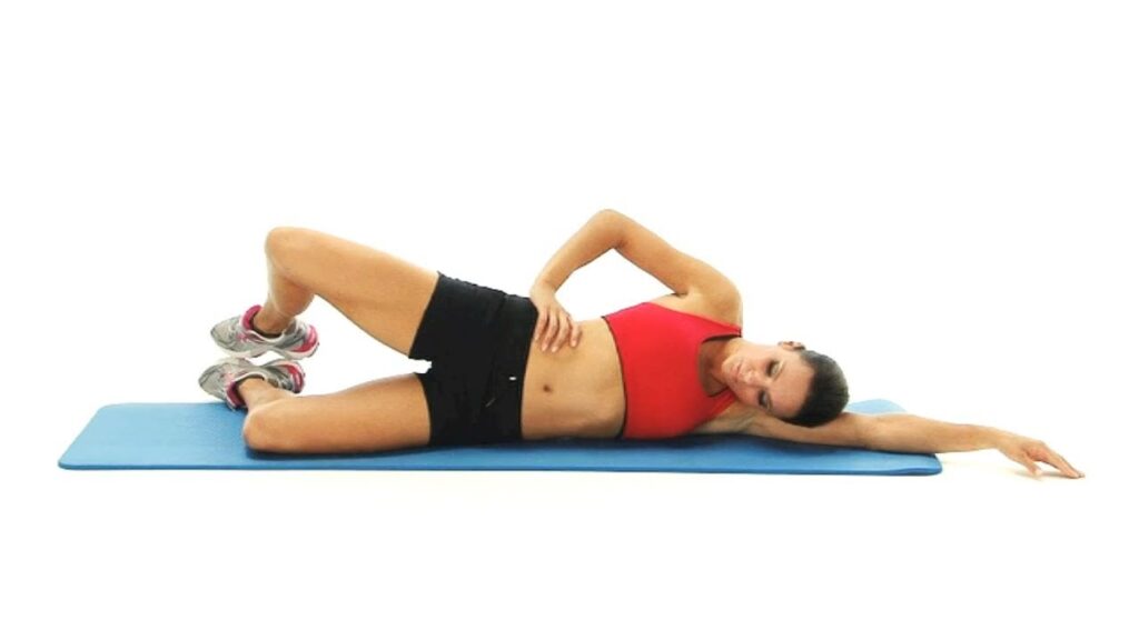
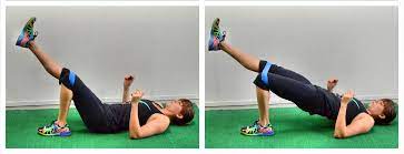
5.Glute bridge with band:
- The starting position for the exercise is the supine position.
- Place a small & tight resistance band around the calves.
- The patient is Lying on the back & lifting the hips.
- Must be Keep tension in the band & tap the hip joint down to the floor before raising the hip back up again.
- This is important to keep the spine straight & do the movement from the hip joint.
- Repeat this exercise 10 times in 1 session.
- Do this exercise in 3 sessions per day.
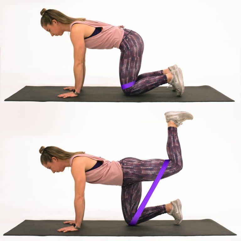
6.Seated hip abduction with resistance band:

- Starting position is the sitting position.
- The patient is Sitting on the floor & placing the resistance band around the calves.
- Bend the knees joint & keep feet on the floor.
- Then Place the hands slightly behind the patient.
- Must be Keep the back straight back & press the legs out to the sides as they externally rotate the hip joint.
- Then Gently & with the control, bring the legs back together.
- Repeat this exercise 10 times in 1 session.
- Do this exercise in 3 sessions per day.

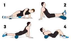
7.Spiky ball roll on glutes
- The starting position for the exercise is the supine position.
- Use the spiky ball to roll out the tight muscles into the buttock.
- Then Roll the ball around to the fleshy part of the buttock for 30 seconds 1 time.
- Do this exercise 10 times in 1 session.
- Perform this exercise in 3 sessions per day.
8.Bridges :

- The starting position of the patient is in the supine position.
- The patient is Lying on to the back with to the knees are bent.
- Then patient feet are touching the floor.
- After that Raise the hips toward the ceiling.
- Must be kept to back into the straight line with the knees joint & shoulders joint.
- Hold this exercise position for 10 seconds.
- Do this exercise 10 times in 1 session.
- Perform this exercise 3 sessions per day.

9.Knee to Chest :
- Starting position is supine.
- Lie onto the back.
- Starting with the left or right knee.
- Use the hands to gently pull by to the bent knee toward the chest.
- Hold for 10 seconds.
- Repeat this knee to chest exercise with to the opposite knee.
- Repeat this exercise 10 times per session & 3 sessions per day.
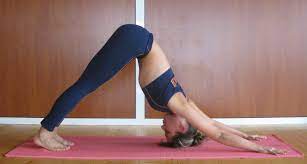
10 Downward-Facing Dog :
- Start of this exercise with on to the hands & knees.
- Then Press into the hands as far as to the lift of the hip joint toward the ceiling.
- Drop to the head down to the bring the ears into the line with the upper arms & chin all to the way into the toward to the chest.
- Bend to the knee joint for the tilt of the pelvis slightly to the forward.
- Then move to the body through to any variations of to the feel for to the appropriate.
- Hold this yoga pose for up to 1 minute.
- do this yoga pose 3 times a day.
11 Pigeon Pose:

- Starting with the all fours limb.
- Move the right knee joint toward to right wrist then place the shin on the floor.
- Then Move to the right ankle toward to left wrist.
- Slide the left leg back, point the toes & keep the hip joint facing forward.
- Then Extend the spine.
- Gently walk the hands forward.
- Hold this position for 5–10 breaths.
- Then Return to the starting position.
- Switch the legs & repeat.
- Do this yoga the 3 times per day.
12. Clam shell exercise
- Lie on your side, with legs stacked & knees bent at a 45-degree angle.
- Rest your head on your lower arm, & use your top arm to steady your frame. …
- Engage your abdominal by pulling your belly button in, as this will help to stabilize your spine & pelvis.
13.Leg bridge exercise:
- Tighten your abdominal & buttock muscles.
- Raise the hips up to create a straight line from your knees to shoulders.
- Squeeze your core & try to pull your belly button back toward your spine.
- Slowly raise & extend one leg while keeping your pelvis raised & level.
- Hold.
- Return to the starting position with the knees bent.
- Perform the lift with the other leg.
14.Standing side bend exercise
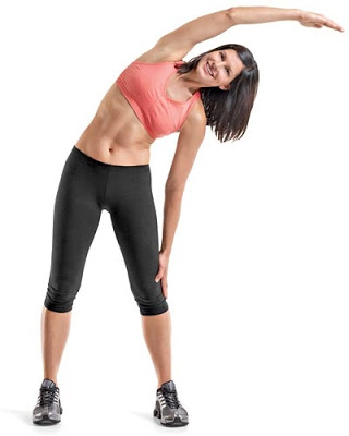
- Using a wall for balance, stand with one side of the body to a wall.
- Cross the leg farthest from the wall in front of the other.
- Place one hand on the wall & the other on the hip. Then lean your upper body away from the wall, & push your hip toward the wall.
- Hold for 20 to 30 seconds, then repeat on the other side
15. Weighted dead lift
- Stand with your feet parallel & hip-width apart. If you feel comfortable, hold light dumbbells.
- Keep your spine long & your gaze forward. Your shoulders should be back & down.
- Squeeze the glutes as you fold from the hips, bending the knees so that your seat reaches back past the heels. Resist the urge to round the spine in order to “give in to the weight.”
- Allow your glutes & belly to control the descent & ascent.
Self-Myofascial Release:
This is a self-massage technique that breaks apart muscle & tissue adhesion that causes sensitivity, pain & stiffness. Use a foam roller to massage your gluteal complex near your outer hip & the head of the femur. Put the foam roller on the floor, & sit on top of it with both feet flat on the floor. Cross your left foot over your right thigh, & put your left hand behind you near the roller. Your weight should shift toward your left hip.

20 Comments