
Biceps femoris: Anatomy, Origin, Insertion, Function, Exercise
Table of Contents
Introduction:
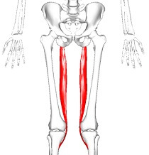
Biceps femoris is the long muscle of the posterior aspect of the thigh. Together with the semitendinosus & semimembranosus muscles, it makes the group of muscles commonly known as the hamstrings.
Biceps femoris muscle runs from the ischial tuberosity, all the way to the proximal portion of the fibula. In doing so the muscle crosses 2 joints; the hip joint & the knee joint. Acting simultaneously on the these joints, biceps femoris has many important functions; flexion & external rotation at the knee joint, extension & external rotation in the hip joint.
As its name suggests, this muscle consists of 2 heads, one lying deep to the other. Each head has a different origin & innervation but they share the same insertion.
Origin of Biceps femoris
Long head: ischial tuberosity
Short head: linea aspera & lateral supracondylar line of the femur
Insertion
Lateral aspect of fibular head
Structure of Biceps femoris
- It has 2 heads of origin:
- The long head arises from the lower & inner impression on the posterior part of the tuberosity of the ischium. This is a common tendon origin with the semitendinosus muscle,& from the lower part of the sacrotuberous ligament.
- The short head, arises from the lateral lip of the linea aspera, between the adductor magnus & vastus lateralis extending up almost as high as the insertion of the gluteus maximus, from the lateral prolongation of the linea aspera to within five cm of the lateral condyle; & from the lateral inter muscular septum.
- The 2 muscle heads joint together distally & unite in an intricate fashion. The fibers of the long head form a fusiform belly, which passes obliquely downward & lateral ward across the sciatic nerve to end in an aponeurosis which is covers the posterior surface of the muscle & receives the fibers of the short head. Inferiorly, the aponeurosis condenses to form a tendon which is predominantly inserts onto the lateral side of the head of the fibula. There is a 2nd small insertional attachment by a small tendon slip into the lateral condyle of the tibia.
- At its insertion the tendon divides into 2 portions, which embrace the fibular collateral ligament of the knee joint. Together, this joining of tendons is generally referred to as the conjoined tendon of the knee.
- The posterior border of the tendon is a thin expansion is given off to the fascia of the leg. The tendon of insertion of this muscle forms the lateral hamstring, the common fibular (peroneal) nerve is descends along its medial border.
Nerve supply
It is a composite muscle as the short head of the biceps femoris develops in the flexor compartment of the thigh and is thus supplied by common fibular branch of the sciatic nerve (L5, S1), while the long head is supplied by the tibial branch of the sciatic nerve (L5, S1).
Variations:
The short head may be absent; additional heads may attaches from the ischial tuberosity, the linea aspera, the medial supracondylar ridge of the femur, or from various other portion. The tendon of insertion may be attached to the Iliotibial band & to retinacular fibers of the lateral joint capsule.
A slip may be pass to the gastrocnemius.
Blood supply
The muscles vascular supply is derived from the anastomoses of several arteries: the perforating branches of the profunda femoris artery, the inferior gluteal artery, & the popliteal artery.
Relations
For its largest part, the biceps femoris muscle runs superficially in the posterolateral thigh and sitting deep only to skin, fat & fascial layers. The exception to this is at its superior side, where it is covered by the gluteus maximus muscle. While the descending from the pelvis into the posterior thigh region, biceps femoris passes on the top of semimembranosus muscle, adductor magnus muscle & the lateral head of gastrocnemius muscle.
Along the way, it is also located superficial to the sciatic nerve and providing protection for it. The sciatic nerve is gives its terminal branch (common fibular nerve) near the insertion of the biceps femoris. The nerve travels to briefly along with the medial border of biceps femoris, adhering to the tendon. This is an important clinical relation when considering injury and performing surgery procedures in this region.
Function of Biceps femoris
- In general, the biceps femoris muscle acts on both the knee & hip joints. Although, due to this attachments, the short head of this muscle acts only on the knee joint while the long head acts on both.
- When acting on the hip joint, biceps femoris produces the motion of hip extension. This action is the strongest when the trunk is bent forward & is to be brought in an upright position. The biceps femoris is also for sometimes described as assisting with external rotation (when the hip joint is in an extended position). When acting on the knee joint, the most prominent action of the biceps femoris muscle is flexion of the leg. This happen when the lower limb is in an anatomical position. In contrast, when the knee is semi-flexed, biceps femoris acts to produce external rotation of the leg at the knee joint.
- Together with other hamstring muscles the biceps femoris stabilizes pelvis, especially during the forward flexion of the trunk occurs. Therefore it has a major role in the gait cycle. Its most important antagonist is the quadriceps femoris muscle which is nearly 3 times stronger than the hamstrings.
Palpation:
- Position the patient in prone lying with the knee in slight flexion
- Begin distally locate the lateral proximal border of the popliteal fossa to locate the insertion of the tendon
- Palpate the hamstrings laterally to locate the biceps femoris
- Move palm toward the ischial tuberosity (proximally) to locate the muscle belly of the biceps femoris
- Length Tension Testing / Stretching
- With one hand, palpate the patient’s ASIS and iliac crest with thumb & index finger
- With the other hand, support the patient’s leg just above the ankle
- Raise the patient’s leg into hip flexion keeping the knee extended, & add internal rotation of the tibia to bias the biceps femoris
- Flexibility of the biceps femoris is exhausted when the innominate starts to rotate posteriorly
- Trigger Point Referral Pattern
- Pain referred from T Rps in the lower half of the biceps femoris (long or short head) focuses on the back of the knee & may extend up the posterolateral area of the thigh as far as the crease of the buttock.
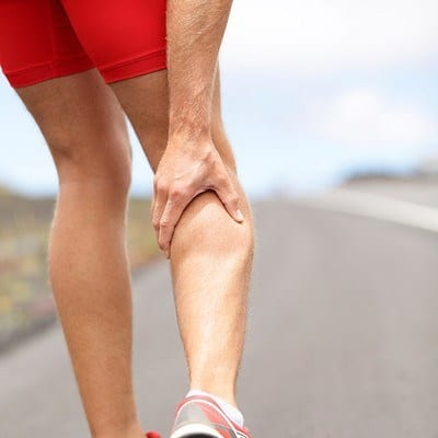
Clinical importance of Biceps femoris:
- The biceps femoris muscle usually has 2 heads, namely short head & long head of biceps femoris. These two heads insert on the head of fibula, where at the site of insertion they divide into two portions by fibular collateral ligaments.
- Any sort of subluxation or dislocation of biceps femoris tendon or abnormal insertion of the tendon, and any or no trauma, meniscal instability can lead to Snapping Biceps Femoris Tendon. It is an unusual condition with symptoms of pain on the lateral side of knee, fibular head will be prominent, painful snap on lateral aspect of knee during active and passive knee flexion at 90 degrees & difficulty in performing ADL’s or sport related activities.
- Furthermore, the exact pathology leading to snapping biceps femoris tendon is still unknown. However, according to researches, occurrence of this condition could be either due to overuse, prolong sporting or congenital.
- While performing sprinting there will be an abrupt raise or alter in speed of running due to which biceps femoris muscle & semitendinosis present at the back of thigh are usually hurt causing over stretching of such muscles. The injury to Long head of biceps femoris tendon & semitendinosis are most common among soccer players leading to sport related injuries.
Stretching exercise of biceps femoris muscle
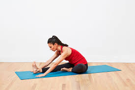
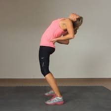
1.Hamstring muscle stretch:
- Sit on the ground with both legs are out straight.
- Extend the arms & reach forward by bending at the waist as far as possible while keeping the knees straight.
- Hold this position for 15-30 seconds.
- Relax back into the starting position.
- Repeat three times.
Be sure to stretch until a moderate pull is felt in the back of the thighs. If you are feel any excessive pain, you might be stop the exercise.
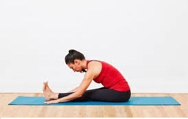
2. Hurdler hamstring muscle stretch:
- Sit on the ground with one leg are out straight.
- Bend the other leg at the knee & position the sole of that foot against the opposite inner thigh.
- Extend the arms & reach forward over the one straight leg by bending at the waist as far as possible.
- Hold this position for 15 seconds.
- Relax and than Repeat with the other leg.
3. Standing hamstring muscle stretch( Both legs are together):
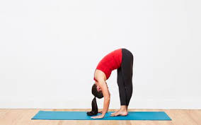
This hamstring stretch is a simple one to do anywhere at the all. It is done in the standing position & stretches both legs at once. Here is how you to do the standing hamstring stretch:
- Stand & cross the right foot in front of the left.
- Slowly lower the forehead to the right knee joint by bending at the waist.
- Keep the both knees straight.
- Hold this position for 15-30 seconds.
- Relax and then Repeat for the other side by crossing the left foot in front of the right.
4. Standing hamstring muscle stretch (one leg at a time)
The one-legged standing hamstring stretch is completely possibly the easiest hamstring stretch to do. It can be done anywhere home, office, or outdoors & it requires no special tools.
- Stand up the straight with one heel resting on a small stool. if it is outside, one can use the curb, but be sure to watch for cars.
- Keep the knee straight.
- Reach both arms up toward the place where the wall & ceiling meet. If outside where there is no wall and ceiling, when simply reach up into the air so that the arms are about even with the ears. Reaching the arms up and as opposed to reaching down toward the foot, will keep the back straight.
- Keep the back straight & One should be bending forward slightly from the hips.
- Reach the forward & feel a stretch in the hamstring behind the thigh.
- Hold this position for 15-30 seconds, & repeat 3 times.
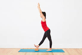
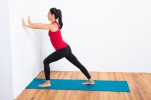
5. Runner’s hamstring stretch
The runner’s stretch is a common flexibility exercise for hamstrings
- Stand one foot from a wall & place the hands on the wall at shoulder height, shoulder width apart.
- Take a step back with 1 leg while pushing into the wall
- Keep the back straight & press the heels into the floor.
- Hold for 15-30 seconds.
- Step forward and the repeat with the other leg.
- Repeat the exercise 3 times on each side.
Strengthening exercise of biceps femoris muscle:
1.Static Hamstring contraction
- Start this exercise in sitting with the knee bent to about 45 degrees.
- Press the heel into the ground tightening the back of thigh.
- Hold for 5 seconds & repeat 10 times as hard as possible pain free.
2.Bridging exercise
- Start this exercise lying on the back in the supine position.
- Gradually, lift the bottom pushing through feet, until the knees, hips & shoulders are in a straight line.
- Tighten the back of the thigh while performing this exercise.
- Hold for 3-5 seconds then slowly lower the bottom of the back down.
- Perform this exercise 3 sets of 10 repetitions provided the exercise is pain free.
3.Single leg bridging
- Lie flat on the back with the knees bent & feet flat on the floor. The feet & knees might be hip distance apart.
- Tighten the abdominal muscles slightly to engage the core and stabilize the lower back.
- Lift up into simple bridge position but as one hold the bridge position, lift 1 foot off the floor & extend the knee as shown.
- Hold for 3-5 seconds, bring the foot back down, & then lower the buttocks to the floor.
4.Hamstring curls with gym ball:
- Lies on the back with the feet & lower calves resting on a gym ball with knees straight & arms crossed over the chest.
- Slowly curl the ball towards the body by bending the knees & then slowly roll the ball away.
- Repeat this exercise for 10 times.
Quadriceps foam rolling:
The foam rolling to quadriceps alone is effective in decreasing the activation of biceps femoris muscle. Foam rolling on patients muscle can lead to alteration in range of motion, performance & muscular co-activation around the particular joint. According to the researcher, when there is simultaneous contraction of the agonist and antagonist muscle, it helps in providing stability to the knee joint. However, if any disturbance occurs to the stability of the joint, the knee particularly goes for injury.
Furthermore, the study portrays about ACL injury, which is more common due to alteration in muscular co-activation, & further research is required to identify the efficacy of foam rolling on activation of muscle around the knee joint.
Soft tissue mobilization:
Performing soft tissue mobilization via IASTM to gluteals, IT band, Quadriceps, adductors & hamstrings, twice per week for 3 weeks helps in improving the knee extension range of motion among patients suffering from decrease in flexibility or decrease movement range due to soft tissue limitations.

8 Comments