Arytenoid muscle
The arytenoid muscles are a group of small muscles that are located in the larynx, or voice box, which is part of the respiratory system. These muscles attach to the arytenoid cartilages, which are paired cartilages that sit on top of the cricoid cartilage in the larynx. The arytenoid muscles help to control the movements of the arytenoid cartilages, which in turn affect the position and tension of the vocal cords.
There are two main types of arytenoid muscles: the posterior cricoarytenoid muscles and the lateral cricoarytenoid muscles. The posterior cricoarytenoid muscles originate from the posterior surface of the cricoid cartilage and insert into the muscular process of the arytenoid cartilages. These muscles are responsible for the abduction, or opening, of the vocal cords during breathing.
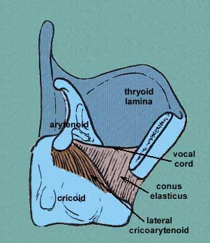
The lateral cricoarytenoid muscles, on the other hand, originate from the upper border of the cricoid cartilage and insert into the muscular process of the arytenoid cartilages. These muscles are responsible for the adduction, or closing, of the vocal cords during phonation or speaking.
Other muscles that attach to the arytenoid cartilages include the transverse and oblique arytenoid muscles, which work together to bring the arytenoid cartilages closer together for vocal cord adduction, and the thyroarytenoid muscle, which runs from the thyroid cartilage to the arytenoid cartilage and helps to control the tension of the vocal cords.
Table of Contents
Origin of Arytenoid muscle
The posterior cricoarytenoid muscles originate from the posterior surface of the cricoid cartilage, which is a ring-shaped cartilage located at the base of the larynx.
The lateral cricoarytenoid muscles originate from the upper border of the cricoid cartilage, near the lateral aspect of the arch of the cricoid cartilage.
The transverse arytenoid muscle originates from the lateral aspect of one arytenoid cartilage and inserts into the lateral aspect of the other arytenoid cartilage.
The oblique arytenoid muscle originates from the muscular process of one arytenoid cartilage and inserts into the apex of the opposite arytenoid cartilage.
The thyroarytenoid muscle has two parts: the vocalis muscle and the muscularis muscle. The vocalis muscle originates from the inner surface of the thyroid cartilage and inserts into the vocal process of the arytenoid cartilage. The muscularis muscle originates from the outer surface of the thyroid cartilage and inserts into the lateral aspect of the arytenoid cartilage.
Insertion of Arytenoid muscle
The posterior cricoarytenoid muscles insert into the muscular process of the arytenoid cartilages, which are small paired cartilages that sit on top of the cricoid cartilage in the larynx.
The lateral cricoarytenoid muscles insert into the muscular process of the arytenoid cartilages as well, but they also have additional fibers that attach to the anterolateral surface of the cricoid cartilage.
The transverse arytenoid muscle inserts into the lateral aspect of the opposite arytenoid cartilage, which allows it to bring the arytenoid cartilages closer together for vocal cord adduction.
The oblique arytenoid muscle inserts into the apex of the opposite arytenoid cartilage, which helps to rotate the arytenoid cartilages medially and bring the vocal cords closer together.
The vocalis muscle of the thyroarytenoid muscle inserts into the vocal process of the arytenoid cartilage, while the muscularis muscle inserts into the lateral aspect of the arytenoid cartilage. Together, these muscles help to control the tension of the vocal cords during phonation or speaking.
Nerve supply
The muscles of the larynx, including the arytenoid muscles, are innervated by branches of the vagus nerve, specifically the recurrent laryngeal nerve and the superior laryngeal nerve.
The posterior cricoarytenoid muscle is innervated by the recurrent laryngeal nerve, which branches off from the vagus nerve and travels along the trachea and esophagus to reach the larynx.
The lateral cricoarytenoid muscle is also innervated by the recurrent laryngeal nerve, but it also receives some innervation from the external branch of the superior laryngeal nerve.
The transverse and oblique arytenoid muscles are both innervated by the recurrent laryngeal nerve.
The vocalis and muscularis muscles of the thyroarytenoid muscle are both innervated by the recurrent laryngeal nerve as well, with the vocalis muscle receiving additional innervation from the internal branch of the superior laryngeal nerve.
Blood supply
The blood supply to the arytenoid muscles comes from several arteries that are part of the laryngeal vasculature. These arteries include:
Superior laryngeal artery: This artery is a branch of the superior thyroid artery, which is a branch of the external carotid artery. The superior laryngeal artery supplies blood to the upper part of the larynx, including the arytenoid cartilages and the muscles that attach to them.
Inferior laryngeal artery: This artery is a branch of the inferior thyroid artery, which is also a branch of the external carotid artery. The inferior laryngeal artery supplies blood to the lower part of the larynx, including the cricoid cartilage and the muscles that attach to it.
Laryngeal branches of the ascending pharyngeal artery: These small arteries arise from the ascending pharyngeal artery, which is a branch of the external carotid artery. The laryngeal branches of the ascending pharyngeal artery supply blood to the laryngeal muscles, including the arytenoid muscles.
The veins of the larynx drain into the internal jugular vein, which returns deoxygenated blood from the head and neck back to the heart.
Function of Arytenoid muscle
The arytenoid muscles are a group of muscles in the larynx that play an important role in regulating the position and tension of the vocal cords during breathing, speaking, and swallowing. The specific functions of the different arytenoid muscles are as follows:
The posterior cricoarytenoid muscles are the only muscles in the larynx that open the vocal cords during inspiration by pulling the muscular processes of the arytenoid cartilages away from each other. This action allows air to flow into the lungs.
The lateral cricoarytenoid muscles are the primary muscles responsible for closing the vocal cords during speech and other vocalizations. They do this by rotating the arytenoid cartilages medially and bringing the vocal processes of the arytenoid cartilages closer together, which causes the vocal cords to come together in the midline.
The transverse and oblique arytenoid muscles work together to adduct the vocal cords and increase their tension, which helps to produce higher-pitched sounds.
The thyroarytenoid muscles, which include the vocalis and muscularis muscles, play a key role in regulating the tension of the vocal cords during speech. The vocalis muscle controls the fine adjustments of the vocal cord tension that produce changes in pitch and intonation, while the muscularis muscle helps to control the overall tension of the vocal cords.
Relation
The arytenoid muscles are located within the larynx, which is a complex structure that connects the pharynx (the upper part of the throat) to the trachea (the windpipe). The arytenoid muscles are situated on top of the cricoid cartilage, which is a ring-shaped cartilage that forms the lower part of the larynx.
The posterior cricoarytenoid muscles attach to the posterior aspect of the cricoid cartilage and the muscular process of the arytenoid cartilages. The lateral cricoarytenoid muscles also attach to the cricoid cartilage, but they have fibers that insert into the anterolateral surface of the cartilage as well as the muscular process of the arytenoid cartilage.
The transverse and oblique arytenoid muscles are both located within the posterior part of the larynx and attach to the lateral and apex of the arytenoid cartilages respectively. These muscles work together to bring the arytenoid cartilages closer together for vocal cord adduction.
The vocalis and muscularis muscles of the thyroarytenoid muscle are located within the vocal cords themselves and insert into the vocal process of the arytenoid cartilage. These muscles play a crucial role in regulating the tension of the vocal cords and controlling the production of sound during speaking and singing.