
Central nervous system (CNS)
Table of Contents
What is a Central Nervous System?
The central nervous system and the peripheral nervous system are two subtypes of the nervous system. The central nervous system is the brain and spinal cord, while the peripheral nervous system includes everything else. The central nervous system’s responsibilities involve receiving, processing, and responding to sensory information.
Responses, sensations, movements, emotions, communication, thought processing, and memory are all controlled by the brain, an organ made of nerve tissue. The meninges, cerebrospinal fluids, and the skull all serve to protect the human brain. The nervous tissue is extremely delicate and can suffer harm by the smallest amount of force. A blood-brain barrier also exists, shielding the brain from any dangerous material that might be floating in the blood.
The spinal cord is a vital aspect of the CNS starting within the vertebral column. The purpose of the spinal cord is to send motor commands from the brain to the peripheral body as well as to transmit sensory information from the sensory organs to the brain. Meninges, cerebrospinal fluids, and bone all protect the spinal cord.
The CNS involves the brain and spinal cord, which are located in the dorsal body cavity. The brain is surrounded by the cranium, and the spinal cord is secured by the vertebrae. The brain and spinal cord connect at the foramen magnum. Cerebrospinal fluid and meninges, which are membranes made of connective tissue, encircle the central nervous system in addition to bone.
Structure
The Brain
The cerebellum, brain stem, diencephalons, and cerebrum comprise the different regions of the brain.
There are four connected lobes, or divisions, within each hemisphere.
- Movement, speech, and a few mental processes like conduct, emotion, memory, and organization are all governed by the frontal lobes.
- The speaking, language, hearing, and memory all depend on the temporal lobes.
- The senses of taste, touch, warmth, and pain, as well as number comprehension, body awareness, and spatial perception, are all significantly influenced by the parietal lobes.
- Clear vision depends on the function of the occipital lobes.
The thalamus and the hypothalamus are located deep within the brain. The thalamus regulates movement and memory in addition to transporting information to and from the lobes. In addition to producing hormones that regulate the pituitary gland’s production of other hormones, the hypothalamus regulates thirst, hunger, and body temperature.
At the base of the brain is where the brainstem is situated. It is crucial for blood pressure, breathing, and the body’s response to danger.
Brain regions
We will now take a closer look at a few particular brain regions:
- Basal ganglia: engaged in the decision-making process over which motor tasks to do, procedural learning, and the regulation of voluntary motor motions. Huntington’s disease and Parkinson’s disease are two illnesses that impact this region.
- Cerebellum: primarily responsible for fine motor control, but also for language and focus. Atherosclerosis, or abnormal motor control, is the main sign of cerebellar injury.
- Broca’s area: Language processing is aided by this little region on the left side of the brain (or occasionally the right in left-handed people). When damaged, a person still has the ability to understand speech but has trouble speaking. An underactive Broca’s area has been linked, on occasion, to stuttering.
- Corpus callosum: an extensive network of nerve cells connecting the left and right hemispheres. It facilitates communication between the two hemispheres and is the biggest white matter structure in the brain. Children with dyslexia have smaller corpus callosums; those who are left-handed, ambidextrous, or musicians usually have larger ones.
- Medulla oblongata: It is involved in involuntary processes including breathing, sneezing, vomiting, and regulating blood pressure. It extends below the skull.
- Hypothalamus: The hypothalamus, which is about the size of an almond and is located slightly above the brain stem, secretes several neurohormones that affect hunger, thirst, and body temperature regulation.
- Thalamus: The thalamus, which is located in the brain’s center, transfers sensory and motor information to the remainder of the cerebral cortex. It has an effect on how sleep, awareness, attentiveness, and consciousness are regulated.
- Amygdala: Located deep within the temporal lobe are two almond-shaped nuclei. They have a role in memory, decision-making, and emotional reactions, especially negative ones.
Spinal cord
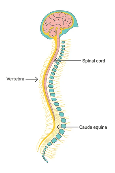
The spinal cord starts at the base of the skull at the foramen magnum and terminates at the first lumbar vertebra. At the foramen magnum, the cord and medulla oblongata are continuous. Similar to the brain, meninges, cerebrospinal fluid, and bone encircle the spinal cord.
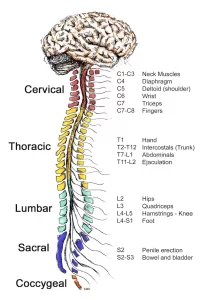
There are 31 segments that make up the spinal cord, and each segment gives rise to two spinal nerves. Numerous spinal neurons at the distal end of the cord branch off beyond the conus medullaris to form a cluster that resembles a horse’s tail. The cauda equina is this. The form of the spinal cord appears round when viewed in cross-section.
The two primary purposes of the spinal cord are:
acting as a channel for impulses entering and leaving the brain during conduction. Through ascending pathways in the spinal cord, sensory impulses are sent to the brain. Motor impulses travel on descending tracts.
Serving as a reflex center. The nervous system’s functional unit is the reflex arc. Reflexes are responses to stimuli that do not need conscious thought and consequently, they happen more quickly than reactions that need thought processes. With the withdrawal reflex, for instance, the affected part is withdrawn by the reflex response before you feel the pain. The spinal cord mediates a great deal of responses without involving the higher brain centers.
White and gray matter
Under a microscope, the neurons and tissue of the peripheral nervous system (PNS) and the central nervous system (CNS) are different from one another. The central nervous system is formed of both white and grey matter. On brain tissue, this is also visible at a macro scale.
Axons and oligodendrocytes make up the white matter, whilst neurons and unmyelinated fibers make up the grey matter. Glial cells, also known as sustaining cells of the central nervous system, are present in both tissues in relatively large numbers (albeit the white matter has more). Various glial cell types perform different roles.
For example, Bergmann glia acts almost as a scaffolding for neuroblasts to climb during neurogenesis, glial cell types perform different roles. For example, Bergmann glia acts almost as a scaffolding for neuroblasts to climb during neurogenesis, while microglia are a specialized type of macrophage that is involved in the brain’s immune system and the removal of various metabolites from the brain.
Astrocytes could be involved in the removal of metabolites as well as the movement of fuel and other beneficial substances from the brain’s capillaries to neurons. After a brain injury, astrocytes proliferate and produce gliosis, a kind of scar tissue that is devoid of living neurons.
Central glial cells
Glial cells, sometimes referred to as neuroglia, are frequently referred to as neurons’ support cells. They outnumber nerve cells in the brain ten to one.
Without glial cells, growing neurons have difficulty forming functional connections and frequently go lost.
There are different types of glial cells in the PNS and CNS, while they are present in both systems. The CNS glial cell types are briefly described as follows:
Astrocytes: These cells bind neurons to their blood supply and have many projections. Additionally, they control the surrounding environment by recycling neurotransmitters and eliminating extra ions.
Oligodendrocytes: in charge of forming the myelin sheath, a thin layer that covers nerve cells and facilitates the rapid and effective transmission of information.
Ependymal cells: These fluid-filled compartments, known as the ventricles of the brain and the spinal cord, produce and discharge cerebrospinal fluid (CSF) and use its whip-like cilia to keep it flowing.
Radial glia: function as support structures for developing nerve cells when the embryo’s nervous system is developing.
Cranial nerves
There are twelve pairs of nerves called cranial nerves, which originate in the brain itself and exit the body through holes in the skull as opposed to passing through the spinal cord. These nerves, which are primarily found in the neck and head, gather and transmit information between the brain and various body parts.
The olfactory and optic nerves, out of these 12 pairs, originate in the forebrain and are regarded as a component of the central nervous system:
Olfactory nerves (cranial nerve I): provide information about smells to the olfactory bulbs located at the base of the brain from the upper nasal cavity.
Optic nerves (cranial nerve II): transport visual data from the retina to the brain’s primary visual nuclei. There are approximately 1.7 million nerve fibers in each optic nerve.
Meninges
Meninges of the CNS
The meninges around the brain and spinal cord have three surfaces. The dura Mater, the outermost layer, is composed of strong, white fibrous connective tissue. The arachnoid, or middle surface, of the meninges, is a thin layer that resembles a cobweb and is attached to the innermost layer by many thread-like strands. There are blood arteries and cerebrospinal fluid in the subarachnoid space, which is located beneath the membrane arachnoid. The pia mater is a name for the meninges’ innermost layer. It is impossible to remove this thin, fragile from the surface of the brain and spinal cord without causing damage to the surface.
Tumors of the nerve tissue around the brain and spinal cord are known as meningiomas.
Doctors sometimes treat meningiomas as though they were malignant in order to relieve symptoms that may arise when a tumor puts pressure on the brain, even though these tumors are typically not likely to spread.
Cerebrum
The cerebrum, which is separated into the two cerebral hemispheres by a deep longitudinal fissure, is the largest and most visible part of the brain. The corpus callosum, an arching band of white fibers that connects the two hemispheres and serves as a communication channel between them, is what connects the two different entities.
There are five lobes in each cerebral hemisphere, four of which are called after the corresponding bones: the frontal, parietal, occipital, and temporal lobes. Within the lateral sulcus is the insula, also known as the Island of Reil, which is the fifth lobe.
Diencephalon
The cerebral hemispheres almost completely encircle the diencephalons, which are situated in the center. It involves the thalamus, hypothalamus, and epithalamus. About 80% of the diencephalons are made up of the thalamus, which is made up of two oval masses of grey matter that act as relay stations for sensory impulses that travel to the cerebral cortex from all other senses but smell. The hypothalamus, a little area located beneath the thalamus, controls a variety of visceral functions and is essential to preserving homeostasis. The diencephalons’ most dorsal region is called the epithalamus. This little gland plays a role in the body’s regular cycles and the development of puberty. Like a biological clock, it is.
Brain Stem
The brain stem is the region connecting the diencephalons and the spinal cord. The midbrain, pons, and medulla oblongata are its three constituent sections. The highest functioning area of the brain stem is the midbrain. The pons are the bulging middle part of the brain stem. This region primarily consists of nerve fibers that form conduction tracts connecting the higher brain centers and spinal cord. The medulla, also known as the medulla oblongata, protrudes inferiorly from the pons. At the foramen magnum, it is continuous with the spinal cord. All ascending (sensory) and descending (motor) nerve fibers that link the brain and spinal cord pass via the medulla.
Cerebellum
The cerebellum, the second largest part of the brain, is located below the occipital lobes of the cerebrum. The cerebellum and other regions of the central nervous system communicate through three paired bundles of myelinated nerve fibers known as cerebellar peduncles.
The cerebellum is located behind the cerebrum (sair-uh-BELL-um). The cerebellum sometimes referred to as the “little brain” due to its resemblance to a smaller form of the cerebrum, controls balance, movement, and coordination.
Other Structures in the Brain
The brain’s many smaller regions perform a variety of crucial functions, including:
The thalamus is responsible for receiving information from the eyes, ears, and fingers regarding the senses, including vision, hearing, and touch.
The hypothalamus regulates the heart rate, thirst, hunger, sleep cycles, and other autonomic functions.
The pituitary gland produces hormones that regulate sexual maturity, growth, metabolism, water and mineral balance, and stress response.
Ventricles and Cerebrospinal Fluid
The brain consists of a network of connected, fluid-filled chambers. The fluid inside these spaces is called cerebrospinal fluid (CSF), and these cavities are the brain’s ventricles.
Neurons
The central nervous system’s fundamental building components are neurons. The body is made up of billions of these nerve cells, which communicate with one another to cause actions and bodily reactions.
The three main components of most neurons are the axon, cell body, and dendrites. There are functional differences among these cells as well. Afferent neurons, efferent neurons, and interneurons are the three different kinds of neurons.
The motor neurons that transmit information from the brain to the peripheral nervous system are known as efferent neurons. Sensory neurons called afferent neurons are responsible for transmitting data from the senses to the brain. Efferent and afferent neurons are connected to the central nervous system by interneurons, which are association neurons.
Protective Structures
The central nervous system (CNS) is shielded by several structures due to its significance. To start, the CNS is completely encased in bone. The cranium shields the brain. The spine, consisting of the vertebrae, encloses the spinal cord.
Meninges are a type of protective tissue that protects the spinal cord and brain. The meninges that cover the brain and spinal cord are composed of three layers:
- Dura mater: This is the outermost layer of the meninges, which is located just beneath the skull and vertebrae, and is named after a Latin phrase that means “hard mother.” Fibrous connective tissue makes up this structure.
- Arachnoid mater: A translucent membrane composed of collagen and elastic fibers that resembles a spider and makes up the second layer of the meninges.
- Pia mater: Originating from the Latin phrase “soft mother,” this shield consists of the innermost layer of the meninges. It is composed of fragile connective tissue that is teeming with microscopic blood vessels that supply the brain with nutrition.
Cerebrospinal fluid, a chemical milieu that provides an additional degree of protection against potential harm to nerve fibers while simultaneously facilitating efficient information transmission, surrounds the entire central nervous system.
Embryology
The adult brain and spinal cord start to form during week 3 of embryological development. The neural plate is formed when the ectoderm begins to thicken. The neutral place then bends inwards, creating the neural groove. Neural bend that migrates laterally flanks the neural groove. The neural groove then grows into the neural tube, which forms the CNS structures.
The neural tube is separated into an anterior and posterior end. The anterior end forms the primary brain vesicles, prosencephalon (forebrain), mesencephalon (midbrain), and rhombencephalon (hindbrain), while the posterior end develops into the spinal cord. The primary brain vesicles continue to differentiate and generate secondary brain vesicles. The forebrain separates to form the telencephalon and diencephalon, and the hindbrain divides to form the metencephalon and the myelencephalon (spinal brain). The midbrain does not divide and remains the mesencephalon. The development of the secondary brain vesicles creates the adult brain structures:
- Telencephalon to cerebrum.
- Diencephalon to hypothalamus, thalamus, retina.
- Mesencephalon to the brain stem (midbrain).
- Myelencephalon to the brain stem (medulla oblongata).
The neural tube’s central region creates ventricles, which are continuous, hollow cavities. During month 6 of gestation, the cerebral cortex changes from a smooth to a wrinkled, complicated appearance; this is due to the continued growth of the cerebral hemispheres. The grooves are called sulci, and the elevated portions of the ridges are called gyri. The convolutions allow for the raised surface area of the brain to fit within the skull. There are regions of white and gray matter all over the brain. Neuronal cell bodies, dendrites, glia, and unmyelinated neurons all develop in the gray matter. Myelinated axons, on the other hand, make up white matter.
The spinal cord, formed from the caudal part of the neural tube, is composed of both gray and white matter. At 6 weeks of gestation, the gray matter starts to aggregate, forming the dorsal alar plate and ventral basal plate. The basal plate is where motor neurons form, whereas the alar plate is where interneurons originate. Dorsal root ganglia, which show information from the periphery to the spinal cord, arise for the neural crest cells.
Development
During the early stages of vertebrate embryonic development, a longitudinal groove on the neural plate progressively deepens. The groove eventually becomes a closed tube known as the neural tube when the ridges on either side of it, known as the neural folds, rise and eventually meet. The neural tube grows by a process known as neurulation. At this stage, developing neural stem cells can be found in the ventricular zone, which is a region of the neural tube walls. Neurogenesis is the process by which neural stem cells, mostly radial glial cells, proliferate and differentiate into neurons, establishing the foundation of the central nervous system.
The neural tube gives rise to the brain and spinal cord. Three brain vesicles, or pockets, originate from the anterior (or “rostral”) region of the neural tube: the prosencephalon at the front, the mesencephalon, and the rhombencephalon, which is located between the mesencephalon and the spinal cord. The prosencephalon then further divides into the telencephalon and diencephalon in the human embryo at six weeks, while the rhombencephalon further divides into the metencephalon and myelencephalon. The posterior, or “caudal,” region of the neural tube gives rise to the spinal cord.
These vesicles differentiate much more as the vertebrate grows. The telencephalon differentiates into the hippocampus, striatum, and neocortex, among other structures, while its cavities become the first and second ventricles. The third ventricle is formed by the diencephalon’s cavity, which contains the thalamus, hypothalamus, thalamus, and epithalamus. The mesencephalon gives rise to various structures such as the tectum, pretectum, and cerebral peduncle, and its hollow develops into the mesencephalic duct, also known as the cerebral aqueduct. The fourth ventricle grows through the cavities of the metencephalon, which provides rise to the pons and cerebellum, and the myelencephalon, which produces the medulla oblongata.
Function
If someone were to ask what the nervous system does, they would have to realize that the central nervous system (CNS) performs a variety of tasks. The human body uses the central nervous system for a variety of purposes, including:
- Receiving sensory input.
- Processing sensory input.
- Reacting to sensory input.
- Cognition.
- Emotional regulation.
- Movement.
- Communication.
- Memory.
Coordination and Movement
The central nervous system is primarily responsible for coordination and integration. The central nervous system (CNS) processes information from multiple sources and combines it to produce a coherent response to external stimuli. For example, the CNS needs visual and integumentary cues (such as surface texture, inclination, obstacle presence, and so on) in order to walk.
The CNS modifies the contraction of skeletal muscle in response to these stimuli. Infants do not need conscious thought or focus once they conversation, or launch an immune response, involving a comparable process of processing complicated information and producing a coordinated response.
Thought and Processing
The brain and the central nervous system (CNS) are thought to be the physical locations of most higher-order mental functions. Mental processes and memory retention are based on connections between neurons. The development of speech, language, and communication is significantly influenced by the brain. Associating abstract sounds and symbols with tangible objects and feelings is the focus of these activities. Neuronal connections in the central nervous system also have a mediating role in motivation, ambition, reward, and satisfaction.
The brain’s limbic system is also in charge of the most fundamental emotions and urges, including pleasure, fear, rage, hunger, thirst, tiredness, and sexual desire. The spinal cord also mediates involuntary reflexes, which shield the body and stop harm from happening right away.
Whether it has to do with breathing, digestion, excretion, circulation, or reproduction, the central nervous system (CNS) affects almost every internal organ system, either directly or indirectly.
Blood supply and Lymphatics
The blood flowing to and from the central nervous system is constantly monitored by the body because of its delicate structure and significance. The circulatory system makes sure that blood is always oxygenated because low oxygen levels can be harmful.
The common carotid arteries branch off of the aorta, which deliver oxygen-rich blood from the heart for distribution. The right and left internal and external carotid arteries, which feed blood to the brain, split out from the common carotid artery.
The vertebral arteries originate in the neck and split as they pass through the foramen magnum to enter the skull. They supply the anterior part of the spinal cord. The basilar artery is structured by the union of the vertebral arteries.
The basilar artery is responsible for supplying blood to the brainstem and cerebellum. Blood will still flow even if one of the arteries isn’t functioning properly thanks to the circle of Willis. The internal carotid and vertebral arteries prepare the circle of Willis.
After being utilized in the central nervous system, blood returns to the lungs to receive oxygen. This is accomplished by several dural venous sinuses:
- Superior sagittal sinus.
- The confluence of sinuses.
- Transverse sinuses.
- Sigmoid sinuses.
- Jugular veins.
- Carotid arteries.
- Superior vena cava.
- Lungs.
Clinical Significance
Wernicke aphasia: The most prevalent cause of Wernicke aphasia is an ischemic or hemorrhagic stroke. The Wernicke region cannot receive oxygenated blood when a stroke occurs in the left middle cerebral artery. A person with Wernicke aphasia is able to make speech and talk clearly. But there’s no meaning to their discourse. They have trouble comprehending spoken words.
Broca aphasia: Brain injuries, brain tumors, or strokes can result in Broca aphasia, sometimes referred to as expressive aphasia. The brain’s supply of oxygen is interrupted when a stroke happens in the Broca region. Damage from the hypoxia is irreparable. When someone develops Broca aphasia, they have trouble speaking. They are not able to produce the words to transmit the message, even though they can understand and know what they want to say.
Traumatic brain injuries: A disturbance to normal brain activity can happen during a sports injury, a car accident, a piercing instrument, or even a blunt object. This is understood as a traumatic brain injury (TBI). The degree of the injury can affect the symptoms of a traumatic brain injury. A contusion, on the other hand, results in long-term brain damage, whereas a concussion only produces momentary vertigo or loss of consciousness. brain stem contusions that cause a coma. TBI can result in cerebral edema and subdural or subarachnoid hemorrhage. Brain blood veins burst as a result of trauma to the brain. The brain tissue is compressed as a result of the blood starting to pool and raise intracranial pressure. The autonomic nervous system is lost when the brain pushes through the skull and onto the spinal cord.
Cerebrovascular Accidents: Strokes, or cerebrovascular accidents, happen when the blood supply to the brain is inadequate for oxygen. Hypoxia is brought on by a lack of oxygen, and brain tissue begins to deteriorate. A blood clot that has moved from one part of the body to the cerebral artery in the brain is often the cause of strokes. Find out what stroke symptoms you get based on where the clot falls. For instance, some patients may have slurred speech, while others may suffer from left-sided paralysis. Because the symptoms of transient ischemic episodes are more transient, they are regarded as mini-strokes. Time is of the essence in any CVA. Physicians have two options if necessary: they can surgically remove the clot or provide tissue plasminogen activator, which breaks it down. How long the oxygen supply to the brain has been cut off directly correlates with the severity of symptoms.
Alzheimer’s disease: Alzheimer’s disease (AD) is a prevalent form of dementia characterized by the deterioration and death of brain cells and neural connections. Cognitive impairment and memory loss are the symptoms of this illness. Alzheimer’s disease progresses over time, with deteriorating symptoms. Aggregations of tau-based neurofibrillary tangles and beta-amyloid plaques have been discovered in the neurons of AD patients. Because of the misfolding of proteins within them, these plaques and tangles induce the death of brain cells. Neural activity in the hippocampus, basal forebrain, and parietal cortex is reduced in AD patients.
Parkinson’s disease: Parkinson’s disease is a disorder of the nervous system that causes the substantia nigra’s dopamine-releasing neurons to deteriorate. Tremors, shaky movements, and loss of balance are brought on by the decline in dopamine levels. Parkinson’s disease develops gradually and typically begins as a hand tremor. As their symptoms worsen, many patients develop bradykinesia, stiffness, a mask-like visage, and a pill-rolling motion in their hands. After considering the patient’s symptoms, medical history, and results of a neurological and physical examination, Parkinson’s disease is diagnosed. The disease has no known cure, however, it is possible to manage how severe the symptoms are. Levodopa can cross the blood-brain barrier and transform into dopamine for usage in the central nervous system. One surgical technique to reduce tremors is deep brain stimulation, which stops aberrant brain activity. Deep brain stimulation, however, has no effect on the disease’s progression.
Huntington’s disease: Huntington’s disease is a genetic brain illness that is degenerative and is caused by a mutation in the huntingtin gene, or HTT. The HTT gene’s CAG region typically repeats up to 35 times. Nonetheless, the CAG section may be repeated up to 120 times in a person suffering from Huntington’s disease. The huntingtin protein builds up in the brain cells as a result of this lengthy CAG stretch, ultimately resulting in cell death. Huntington’s illness first manifests as chorea, jerking involuntary motions, and flapping of the hands. Cognitive impairment happens as the condition worsens. fatally occurs within 15 years of the diagnosis.
Spinal cord traumas: Spinal cord injury symptoms vary depending on the location of the injury. Sensation may be impacted if harm to the sensory tracts takes place. On the other hand, paralysis results from damage to the ventral horns or roots. When nerve impulses do not reach the targeted muscles, the result is flaccid paralysis. Muscle contraction is impossible in the absence of a stimulus. Involuntary contraction of the motor neurons due to irregular stimulation results in spastic paralysis. When the spinal cord is severed between T1 and L1, paraplegia, or paralysis of the lower limbs, results. Damage to the cervical region results in quadriplegia, or paralysis of all limbs.
Poliomyelitis: An inflammation of the spinal cord brought on by the polio virus is known as poliomyelitis. The polio virus can be transmitted from person to person or through contaminated water and food. It causes paralysis by destroying the neurons in the spinal cord’s ventral horn. With the administration of the vaccination, poliovirus infection can be avoided.
Amyotrophic Lateral Sclerosis: The disease known medically as amyotrophic lateral sclerosis (ALS) or Lou Gehrig’s disease is characterized by the death of motor neurons that govern both voluntary and involuntary actions such as breathing, speaking, and swallowing. Regretfully, there is no recognized cause for ALS, and there is also no known treatment. Researchers think that the high levels of extracellular glutamate in ALS patients are linked to cell death. Riluzole is used to lessen the excruciating symptoms of ALS and to slow down the disease’s course since it can interfere with glutamate synthesis.
Multiple sclerosis: Myelin proteins in the central nervous system are attacked by the body in multiple sclerosis, an autoimmune illness that impairs brain-to-body communication. Young individuals are very susceptible to MS, which manifests as discomfort, weakness, loss of vision, and coordination problems. Every patient has different symptoms, each with varying intensity. The body’s immune system is suppressed by medication, which also helps lessen the negative symptoms of this illness.
Summary
As the part of the nervous system that includes the brain and spinal cord, the central nervous system is present in all vertebrates as well as many other higher-order animals. Neurons, or nerve cells, comprise every nerve tissue. Afferent, or sensory, neurons; efferent, or motor, neurons; and interneurons, which link afferent and efferent neurons, are the three different types of neurons.
Through the spinal cord, messages are sent and received by all body parts to and from the brain. All of the nerves that connect the spinal cord to the remaining parts of the body are part of the peripheral nervous system, which is different from the central nervous system.
FAQs
Which seven regions make up the central nervous system?
The seven basic components of the central nervous system, which is comprised of the brain and spinal cord, are commonly understood to be the following: the midbrain, the diencephalon, the pons, the medulla, the cerebral hemispheres, and the cerebellum.
Why is it called CNS?
In addition to breathing, heart rate, hormones, body temperature, and movement, it also regulates thought, emotion, and movement. Because it integrates data from the entire body and controls activity throughout the entire organism, the central nervous system (CNS) is referred to as “central.”
What are the 4 main functions of the central nervous system?
Four fundamental roles of the nervous system
Taking in general sensory data (pressure, temperature, vibration, discomfort, and touch).
Acquiring and documenting distinct sensations (sounds, tastes, smells, visions).
integration and processing of sensory information from many body parts.
formation of responses.
Which two organs make up the CNS?
The central nervous system has two components brain and the spinal cord. Cerebrospinal fluid is used to bathe them and they are enveloped in a thin coating known as meninges (CSF).
Which three nervous system types are there?
ANS peripheral nerve system.
the sympathetic nervous system.
neuronal parasympathetic system.
What is the full form of the CNS?
The central nervous system is known by its full name, CNS. The brain and spinal cord are parts of the CNS. Every bodily function is managed by the brain. The brain is the source of thought, intelligence, and memory.
What are the CNS’s composition and purpose?
The brain, spinal cord, and neurons (or nerve cells) make up the three primary parts of the central nervous system. All three of the CNS’s components work together to process information and regulate the body’s reactions, and each one of them has a significant impact on how the body operates.
What is the structure of the brain?
While all of the brain’s components function together, they each have unique roles to play. Three fundamental brain regions can be identified: the forebrain, midbrain, and hindbrain. The brain stem, the top portion of the spinal cord, and the wrinkly ball of tissue known as the cerebellum are all parts of the hindbrain.
What is the spinal cord?
A long, tube-like band of tissue makes up the spinal cord. Your lower back and brain are connected by it. Nerve impulses are transmitted from your brain to your body via your spinal cord and vice versa. These nerve signals allow your body to move and feel. Your ability to move or function can be impacted by any spinal cord impairment.
What are the layers of the brain?
The innermost, delicate layer is called the pia mater. The arachnoid, a web-like structure that surrounds and cushions the brain, is the layer in the middle. The dura mater is the name for the protective outer layer.
What is the biggest part of the brain?
The brain’s main region, the cerebrum, is made up of the left and right hemispheres. Higher-order processes like touch, vision, hearing, speech, thinking, emotions, learning, and fine motor control are all carried out by it. The cerebrum is positioned above the cerebellum.
What are the three types of neurons?
The three classes of neurons that make up the human nervous system are motor neurons, sensory neurons, and interneurons. These classes are based on the functions of these neurons.
What causes paralysis?
Nervous system disorders result in paralysis. The body’s ability to send motor and movement signals to
different parts of the body is dependent on signals from the brain; injury to the nervous system impairs this ability. Potential causes of paralysis include neurological disorders, autoimmune diseases, spinal cord injuries, brain injuries, congenital conditions, and strokes.
What is the main role of a neuron?
The basic structure blocks of the brain and nervous system are neurons, also known as neurons or nerve cells. Neurons are the cells that receive sensory information from the outside world, give motor commands to our muscles, and convert and relay electrical signals at each stage along the way.
In the CNS, how are the gray and white matter regions organized?
Gray matter makes up the outer cortex of the brain, whereas white matter makes up the inner region. While cell axons are found in the white matter, neurons make up the majority of the gray matter. Glial cells are found in both the white and gray matter of the brain, supporting and shielding the neurons.
Can the central nervous system repair itself?
The delicate structures that comprise the central nervous system are prone to harm, injury, and illness. Due to the intricate nature of these structures, damage is typically irreversible. Scientists are investigating therapies and pharmaceuticals as well as other forms of treatment that might aid in CNS restoration and damage repair.
Reference
- Thau, L. (2022, October 10). Anatomy, Central Nervous System. StatPearls – NCBI Bookshelf. https://www.ncbi.nlm.nih.gov/books/NBK542179/
- Newman, T. (2022, December 12). All about the central nervous system. https://www.medicalnewstoday.com/articles/307076.
- Thau, Lauren. “Anatomy, Central Nervous System.” StatPearls – NCBI Bookshelf, 10 Oct. 2022, www.ncbi.nlm.nih.gov/books/NBK542179.
- Buckley, Gabe. “Central Nervous System.” Biology Dictionary, 15 Jan. 2021, biologydictionary.net/central-nervous-system.
- MSEd, K. C. (2023, May 4). Structure and Function of the Central Nervous System. Verywell Mind. https://www.verywellmind.com/what-is-the-central-nervous-system-2794981.

10 Comments