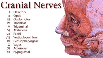
CRANIAL NERVES LIST, ANATOMY, FUNCTION
The cranial nerves are the nerves that come out of the brain. They are often called the “invisible” nerves because they pass through the skull and are not readily visible. They are responsible for signaling the body to do things like talking, chewing, and keeping your eyes focused.
Table of Contents
CRANIAL NERVES LIST
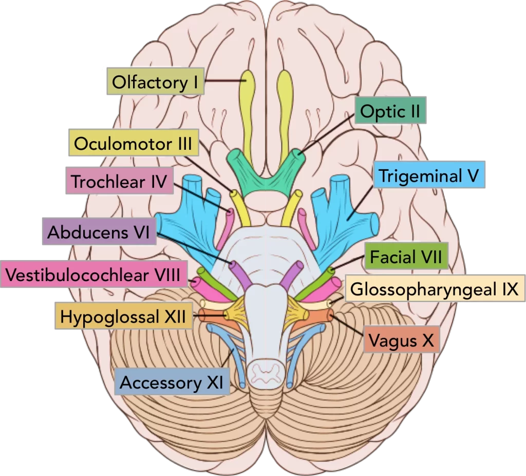
There are 12 pairs of cranial nerves and they are numbered according to their position of where they originate in the inferior surface of the brain.
Cranial Nerve’s function and tests
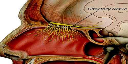
CN I – Olfactory Nerve
function: Smell (olfaction)
test:- Test sense of smell by closing the other nostril and using non-irritating odors like coffee, lemon oil, etc
– Inability to detect smells (Anosmia) = temporal lobe lesions
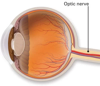
CN II – Optic Nerve
function: Vision
test:- Test visual acuity using a Snellen chart, test central and peripheral vision
– Blindness, impaired vision: far (myopia) and near (presbyopia)
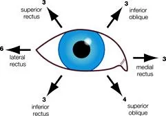
CN III – Oculomotor Nerve
function: Innervates the inferior oblique muscle and medial, inferior, and superior rectus muscles of the eye (move the eye); levator palpebrae superioris muscle (elevate eyelid)
Innervates the sphincter pupillae muscle (constricts the pupil), and the ciliary muscle (accomodate the eye for near vision)
test:- Test pupil equality, size and shape
– Test pupil constriction by shining a light in the eye
– Absence of pupil constriction
– Unequal pupils (anisocoria)
– Horner’s syndrome
CN IV – Trochlear Nerve
function: Innervate the superior oblique eye muscle (moves the eye inferiorly and laterally)
CN VI – Abducens Nerve
function: Innervate the lateral rectus muscle of eye (abducts the eye)
test:- Test extraocular movements
– Observe eye position, presence of strabismus (loss of ocular alignment) or ptosis of eyelid
– Test pursuit eye movement without head movement
– Strabismus and impaired eye movement
– CN III: Ptosis, pupil dilation
– CN IV: Eye cannot look down when adducted
– CN VI: Eye pulled inward, eye cannot look out
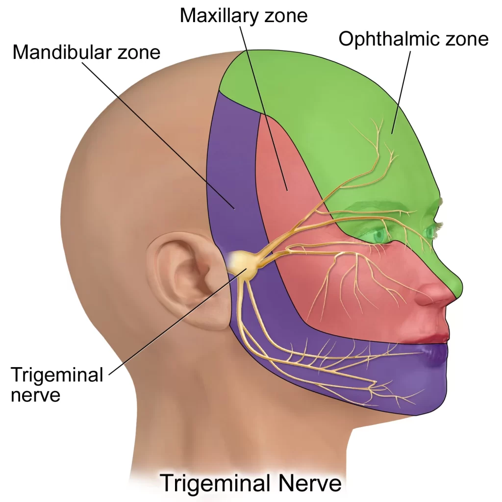
CN V – Trigeminal Nerve
function: Conducts touch, temperature, and pain sensation from the face, nose, mouth, nasal and oral mucosa, anterior two-thirds of the tongue, and anterior scalp; part of auricle of the ear
Innervate the muscles of mastication, mylohyoid, digastric (anterior belly), tensor veli palatini, and tensor tympani
test: Pain and light touch sensation of face (forehead, cheeks, jaw)
– Open and close jaw against resistance
– Test corneal and jaw jerk reflex
– Loss of facial sensation and numbness
– Loss of ipsilateral corneal reflex
– Weakness and wasting of mastication muscles
– Jaw deviation when opened to ipsilateral side
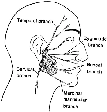
CN VII – Facial Nerve
function:Taste from anterior two-thirds of tongue
Innervate muscles of facial expression, digastric (posterior belly), and stapedius muscle.
Increase secretion from the lacrimal (tear glands) and nasal mucosal glands; submandibular and sublingual.
salivary glands
test:- Test motor function of the facial muscles and look for asymmetry: raise eyebrows, frown, smile, close eyes tightly, puff cheeks, etc.
– Ipsilateral paralysis of facial muscles: unable to close eye, mouth corner droops, difficulty with speech articulation
– peripheral nerve injury (PNI) Bell’s Palsy (CN VII); or facial paralysis due to stroke
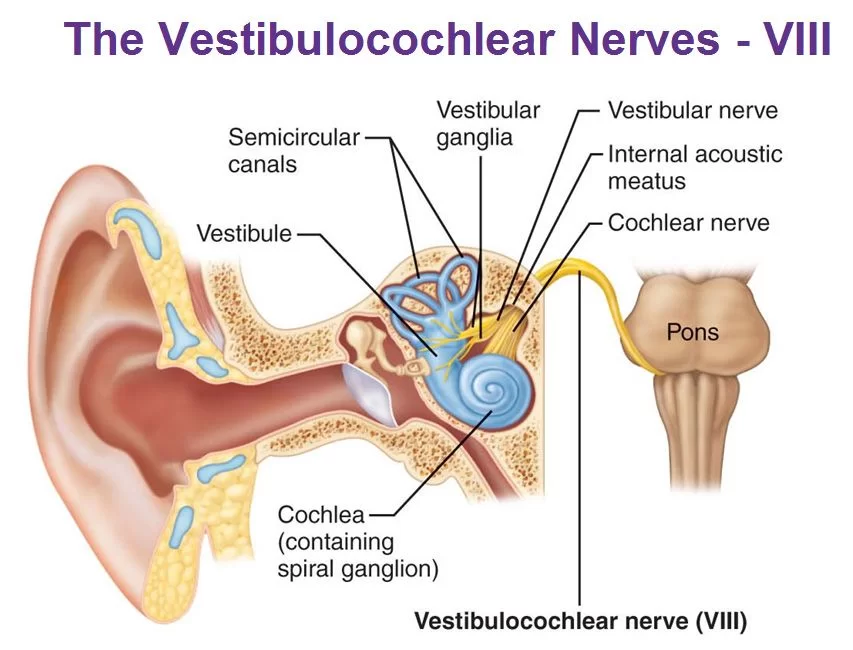
CN VIII – Vestibulocochlear Nerve
function: Hearing (cochlear branch); linear and angular acceleration, or head position in space/equilibrium
test: Test balance
– Gaze instability with head rotations
– Test auditory acuity with a tuning fork placed in the middle on top of the head and check if the sound is equal or louder in one ear (Weber’s test)
– Vibrating tuning fork placed on mastoid bone, then near the ear canal and note hearing acuity (Rinne’s test)
– Vertigo and disequilibrium
– Nystagmus
– Deafness, tinnitus, and hearing loss
– Unilateral conductive loss
– Sensorineural loss: sound heard in good ear
– Conductive loss: sound heard through bone is longer or equal to air
– Sensorineural loss: sound heard longer through air
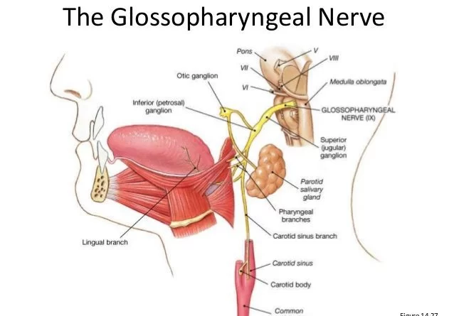
CN IX – Glossopharyngeal Nerve
function: Touch and taste from the posterior 1/3 of the tongue; visceral sensory from the carotid bodies
Innervate the pharyngeal muscle
Increase secretion from the parotid salivary gland
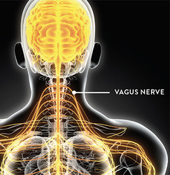
CN X – vagus
function: Visceral sensation (excluding pain) from heart, lungs, abdominal organs, bronchi, trachea, larynx, pharynx, gastrointestinal
tract to the level of the descending colon.
-General sensation from the external acoustic meatus, eardrum, and pharynx
-Innervates pharyngeal and laryngeal muscles and muscles at the base of the tongue
-Innervates smooth muscle and glands of the heart, lungs larynx trachea, and most abdominal organs
test of glossopharyngeal and vagus:
test:- Listen to voice quality
– Test for difficulty swallowing
– Let the patient say “ah” and observe the soft palate elevating and that the uvula remains in the midline
– Examine the gag reflex
– Dysphonia
– Dysphagia
– With paralysis, the palate does not elevate (lesion CN V), with unilateral paralysis there is an asymmetrical elevation
– Absent gag reflex (lesion CN IX, possibly X)
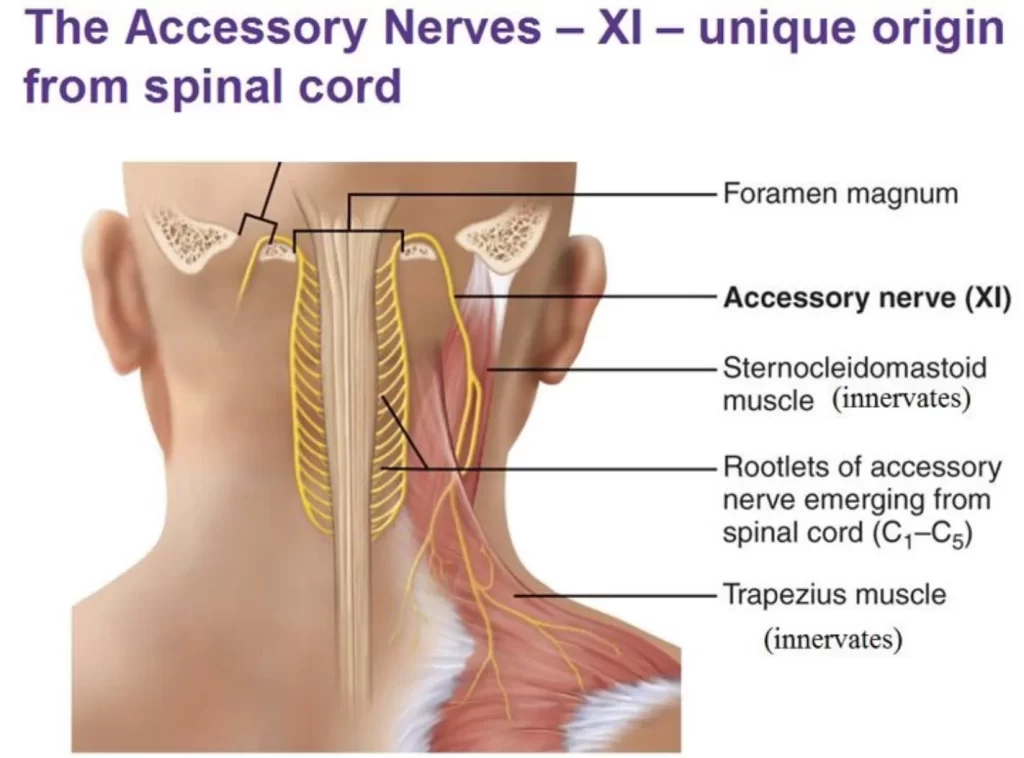
CN XI – Accessory Nerve
function: Innervates the trapezius and sternocleidomastoid muscle
test: Examine muscle bulk
– Test Trapezius and Sternocleidomastoid muscles against resistance
– Atrophy, fasciculations, weakness PNI: shoulder droops and unable to shrug ipsilateral shoulder
– Unable to turn the head to the contralateral side
CN XII – Hypoglossal Nerve
function: Innervates intrinsic and extrinsic tongue muscles
test: Examine protruded tongue: rapid side-to-side movements
– Examine the tongue’s resting position
– Listen to the patient’s word articulations
– Movement impairment: deviation to weak side
– Atrophy or tongue fasciculations
– Dysartrhia (CN X or XII lesions).
FAQs
The trigeminal nerve
The largest cranial nerve and fifth in number (CN V) is the trigeminal nerve. It has three main branches, and its principal purpose is to supply sensory innervation to the face. The ocular (V1), maxillary (V2), and mandibular (V3) nerves are the three distinct branches.
The trochlear nerve
The smallest cranial nerve is the trochlear nerve. The midbrain’s floor is where it starts.
Vagus nerve
The nerve with the most branches is the vagus nerve. It came from the brainstem’s medulla oblongata. Pneumogastric nerve is another name for the vagus nerve. The tenth cranial nerve is this one.
Trigeminal Nerve is the Thickest Cranial Nerve.