
Rib cage
Table of Contents
Introduction
The hard system of the thoracic pit is known as the ribs. All vertebrates are curved, narrow, and flat strips of bone. The rib cage is formed by attaching it dorsally to the vertebrae and ventrally to the breast bone (sternum).
In the human body, the rib confine is a bin-like construction that is shaped from the ribs and their related connections to the sternum and vertebral segment. The rib confine structure houses two essential organs, the lungs, and the heart, and furnishes them with hard assurance from outside injury and injury.
The rib confine is expansible and semi-unbending in nature. The large property lets the cage expand while doing things like breathing. The cage’s expansion is aided by the three false ribs and two floating ribs, which also make breathing easier by allowing the diaphragm to move.
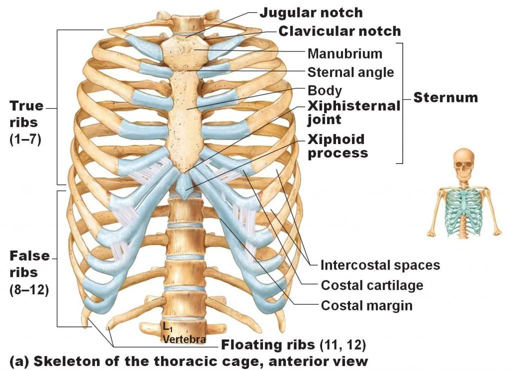
Parts of the rib cage: The thorax, or chest, is made up of the rib cage, a skeletal component. The rib cage includes the sternum, ribs, and vertebral column.
Structure of Ribs
The anatomical characteristics of the ribs typically include:
- Head with two articular facets
- Tubercle
- Neck
- Shaft
- Costal groove
The greater part of the ribs is normal ribs ie they have a large number of highlights. These characteristics are not present in atypical ribs:
- First rib (wide and short, has two coastal scores, and one particular aspect)
- The second rib (slim, long, and has a tuberosity on its predominant surface for the connection of the serratus front muscle)
- Tenth rib (only one articular facet)
- Eleventh and twelfth ribs, each with a single articular facet and no neck
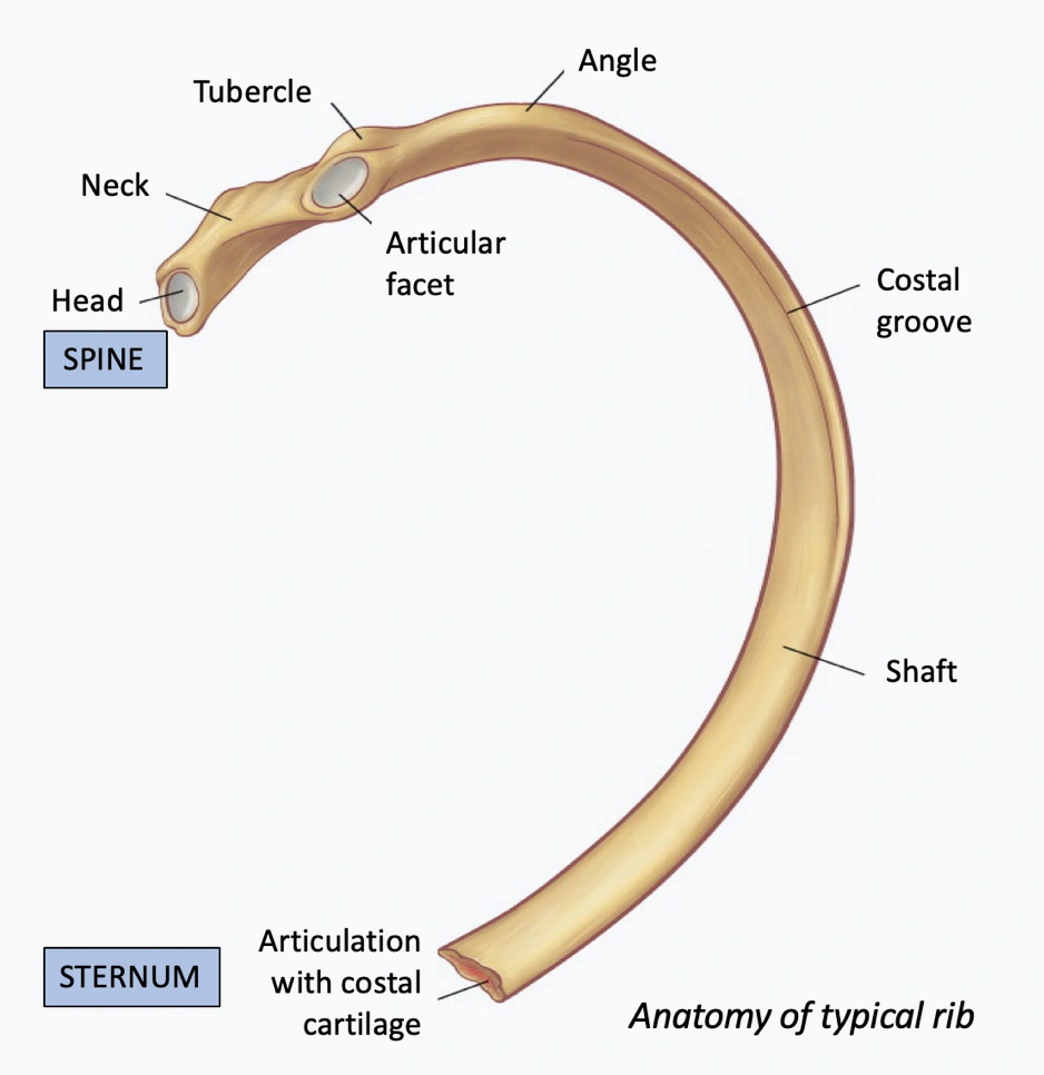
The head, neck, tubercle, and body are the major landmarks, and ribs three to nine are the “typical” ribs.
Head
The crest of the head, a bone wedge that separates the two articular facets of each rib, is the wedge-shaped head of each rib. The smaller of the two facets articulate with the inferior costal facet on the body of the superior vertebra, while the larger of the two facets is for articulation with the corresponding vertebra’s superior costal facet.
Neck
The neck of the rib is by and large mediocre regarding hard tourist spots and is just a level piece of bone that interfaces the top of the rib with the body.
Tubercle
The tubercle is a hard conspicuousness situated at the intersection between the neck and body which projects posteriorly. It has two parts: a smooth articular part that connects to the associated vertebra’s transverse process and a roughened nonarticular part that connects the costotransverse ligament.
Body
The rib has a thin, flat, and curved body, or shaft. The bend turns out to be generally noticeable at the coastal point, which is the point at which the rib turns anterolaterally. Some of the deep back muscles also attach to the ribs at the coastal angle. The costal groove on the internal surface provides some protection and a route for the neurovascular bundle to follow. A cup for the costal cartilage at the body’s end makes it possible for the rib to articulate with the sternum.
Fact: An additional rib called the cervical rib is some of the time tracked down on the highest point of the principal rib as an inborn overdevelopment in around 0.5 to 1% of the human populace.
Anatomy of Ribs
Types of Ribs
There are three distinct types of ribs based on how they attach to the sternum.
- True Ribs: These ribs articulate straightforwardly with the sternum by the costal ligament. True ribs are the first through seventh ribs. A sternocostal joint interfaces these ribs to the sternum for development.
- False Ribs: Because their costal cartilage is attached to the seventh rib’s costal cartilage, these ribs can articulate with the sternum indirectly. Misleading ribs are the eighth, 10th, and tenth ribs. Vertebrochondral ribs are another name for these ribs.
- Floating Ribs: At no point do these ribs articulate with the sternum. Drifting ribs are the 11th and twelfth ribs.
The typical rib and the atypical rib are the two types of ribs. The general structure of typical ribs is normal, whereas the structure of atypical ribs is slightly different.
Typical Ribs
Because of their similar structure and function, ribs three through nine are regarded as typical ribs. The thoracic vertebra for which it is named is where each rib emerges; The third rib and the seventh rib come out of your third and seventh thoracic vertebrae, respectively.
Each rib is made up of three parts: the head, the neck, and the shaft, or body, of the rib.
Your rib head has two distinct areas known as facets and is shaped like a wedge. These aspects articulate with your spinal vertebrae. The upper feature of each rib interfaces with the vertebrae above it, and the lower feature on the top of a rib is associated with its mathematically comparing vertebrae. Your ribs’ costovertebral joints are formed by these articulations.
The head is joined to the shaft by the neck of each typical rib. It is a marginally limited region of the rib bone and contains another feature that verbalizes with the cross-over course of its related vertebrae. The costotransverse joint is the name given to this articulation. As a result, there are three points of articulation between thoracic spinal vertebrae and each typical rib.
A rib has a flat, curved shaft. The costal groove is a small groove that runs along each rib. The vein, nerve, and artery that run along the rib are shielded by this groove. The ribs somewhat pivot as they course around your body, transforming into a ligament called the coastal ligament. In the front of your thorax, this cartilage joins your sternum.
Atypical Ribs
Due to their slightly different structures, ribs one, two, 10, and 12 are all considered atypical ribs.
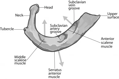
The first rib is a short, thick bone. Its head only has one facet joint, since it arises from the first thoracic vertebrae and there are no thoracic vertebrae above it where it can attach.
The subclavian vein, nerve, and artery are housed in two tiny grooves on the upper surface of the first rib. ( “Below the clavicle,” or collar bone, is where “subclavian” comes from.)
Your first rib is shorter and wider than your second rib. In its head, it has two facet joints that connect it to the first and second thoracic vertebrae. On the second rib, there is a rough spot where the serratus anterior muscle can attach.
Rib number 10 is abnormal because its head just has one feature joint that verbalizes with thoracic vertebrae number 10. The eighth and ninth ribs are above the 10th rib, which runs around your body and connects to a network of cartilage. After that, this cartilage joins your lower sternum. Because they do not connect directly to the sternum, these ribs are also referred to as “false ribs.”
Because they do not attach to the sternum, ribs 11 and 12 are regarded as unusual. They do not attach to you and simply wrap around your thorax. As a result, they are frequently referred to as floating ribs.
Strangely, a person may occasionally have an additional rib above the first rib. A cervical rib is a common name for this. A cervical rib frequently leads to no issues, yet once in a while, it might impede the ordinary capability of the nerves, veins, and conduits close to your collarbone. Thoracic outlet syndrome is a condition that could result from this.
Atypical ribs
The first, second, and tenth to twelfth ribs are considered separately because they are atypical.
First ribs
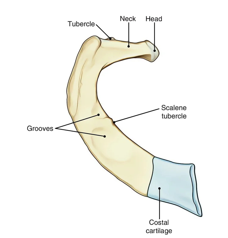
The first rib is the widest and shortest, but all of the ribs have sharp curves. The head just verbalizes with the body of the T1 vertebra and in this manner only one articulatory surface is available. The tubercle, like typical ribs, has a facet that connects it to the transverse process of vertebrae for articulation. The superior surface is distinctive due to the two grooves that permit subclavian vessels to pass through them. The scalene tubercle, to which the anterior scalene muscle is attached, separates these grooves.
Second ribs
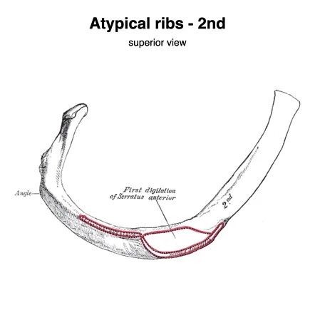
Compared to the first rib, the second is significantly longer and thinner. To enable articulation with the T1 (superior) and T2 (corresponding) vertebrae, the head has two facets. The most unusual thing about it is that it has a roughened tuberosity on its superior surface, which is where the serratus anterior got its start.
Tenth, eleventh, and twelfth ribs
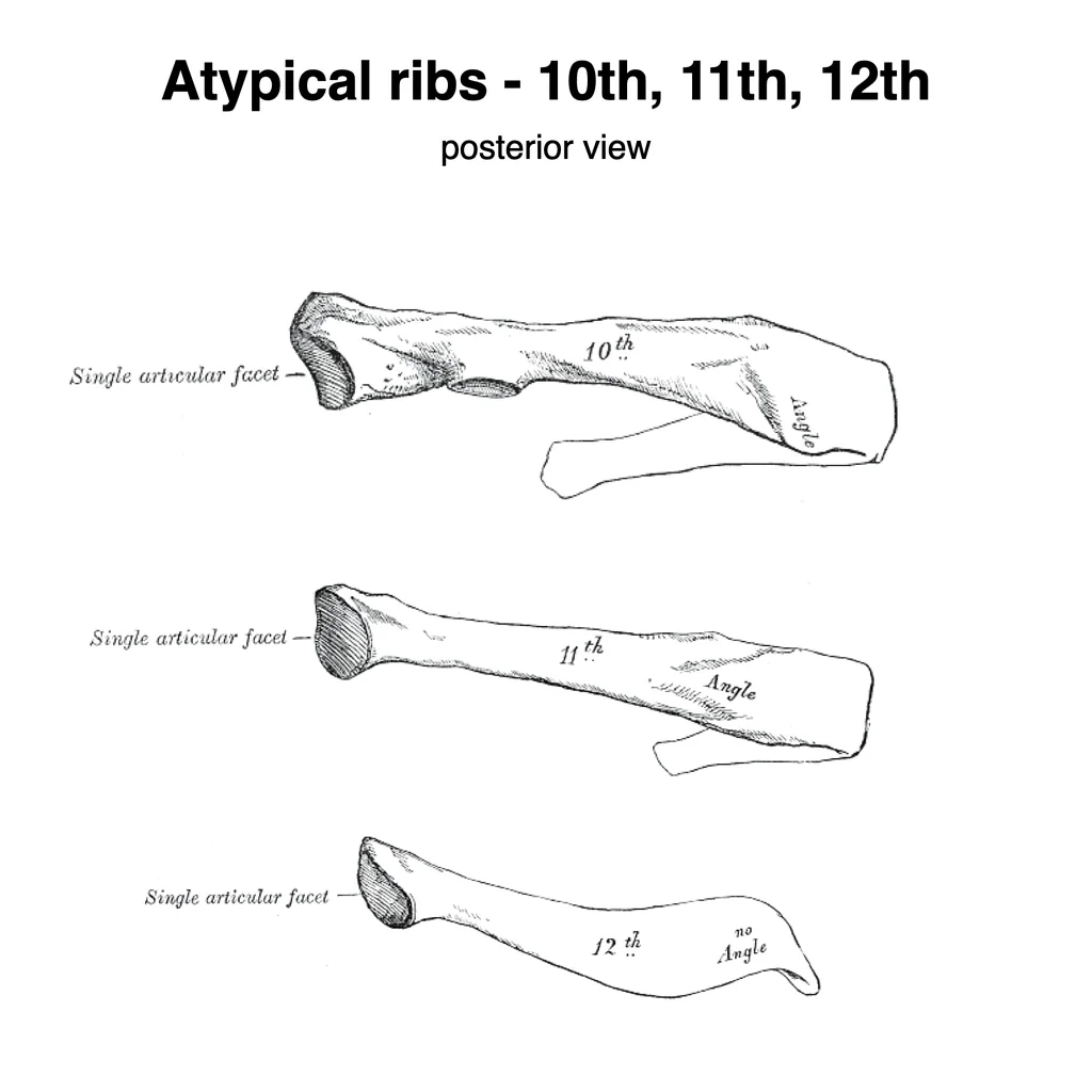
The heads of the tenth to twelfth ribs only have one facet, so they can only move with a single vertebra. Particularly, ribs eleven and twelve lack necks or tubercles and are short.
Functions of the Ribs
There are several functions of your ribs. These functions include:
- safeguard the contents of the mediastinum and thoracic cavity.
- move superiorly, poorly, anteriorly, and posteriorly to work with breathing (their adaptability in their development increments/diminishes the size of the thoracic hole; facilitating respiration by the lungs. The diaphragm, the external intercostals, and the intercartilaginous portion of the internal intercostals control these movements.
- give some muscles a place to come from or attach.
- furthermore, assume a part in erythropoiesis during improvement (upon entering the world, the erythropoiesis destinations transform, it subsides in lengthy bones and endures in level bones, similar to ribs)
The most crucial aspect is safeguarding the contents of the thorax. Your diaphragm, lungs, heart, trachea, esophagus, and numerous muscles, nerves, and vascular structures are all located in your thorax. The ribs give a hard depression that folds over your body, keeping your organs no problem at all inside your body.
At the point when you inhale, your stomach muscle in the lower chest moves descending. Small intercostal muscles near your ribs contract while this happens, moving your ribs up and expanding your thorax.
This development makes a strain differential between the air in your body and the surrounding air outside your body. Your lungs are responsible for gas exchange when ambient air enters them. After that, the diaphragm relaxes, the ribs fall, and the pressure in your thorax rises, forcing air out.
To enable breathing, the ribs in your thorax are essential movers. They move and go about as 12 sets of pail handles, going all over while you relax.
Muscle Attachments
The ribs are associated with several muscles.
- intercostal muscles: situated in the spaces between the coasts.
- diaphragm: emerges from the internal surfaces of the costal ligaments on the 6th rib
- serratus anterior: originates from the first to eighth ribs anterolaterally.
- The superior anterior ribs are the source of the pectoralis major and minor muscles.
- latissimus dorsi: originates from the ninth through twelfth ribs.
- scalenus foremost, back, and medius muscles have connections on the first and second ribs
- rectus abdominis: inserts at the sternum and the 5th to 7th costal cartilage
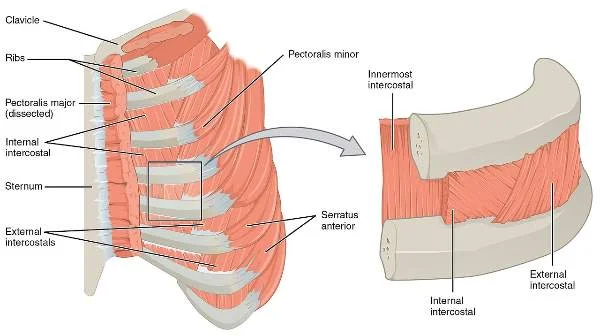
There are eleven pairs of the most superficial external intercostal muscles in the region. They reach out from the parallel line of the coastal depressions to the prevalent edges of the ribs beneath. This muscle’s oblique fibers pass anterior-inferior and wrap around the thoracic wall, reaching from the tubercles to the ribs’ costal cartilages. These muscles help to lift the ribs in motivation.
Internal intercostal muscles
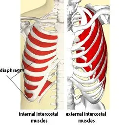
Posterior to the external intercostals are the eleven pairs of internal intercostal muscles. These connect the superior margins of the ribs below with the inferior edge of the costal groove. These muscle fibers pass again in an oblique manner and extend posterior inferiorly. From the parasternal region to the rib angle, these muscles are only present. They depress the ribs during forced expiration.
Innermost intercostal muscles
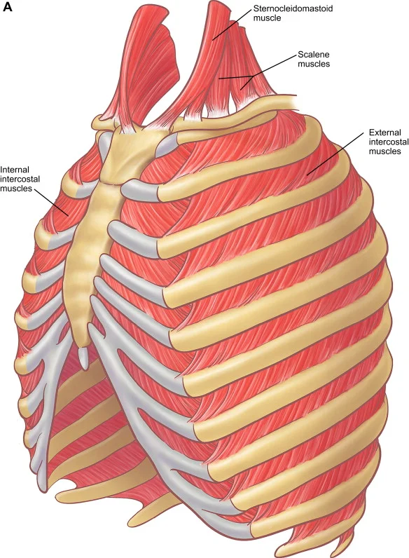
These muscles’ fibers run parallel to those of the internal intercostal muscles. They are mostly found within the lateral thoracic wall and run from the costal groove’s medial edge to the rib’s medial surface below. However, these muscles stand out because the opening between the innermost and internal intercostal muscle layers is where the neurovascular bundles enter. During forced expiration, these muscles aid in rib depression.
Subcostales
Within the same plane as the innermost intercostals are the subcostales muscles. They are remarkable in that they might traverse one or different ribs and become more various inside the substandard locales of the back thoracic wall. They stretch out from one rib’s internal surface to the inward surface of the following or even the rib underneath it. The internal intercostal muscles are supported by this muscle.
Transversus thoracic
Deep within the anterior thoracic wall are these muscles. The lower body of the sternum and the posterior surface of the xiphoid process is where the transverse thoracic muscles originate. The fibers travel superolateral before inserting themselves into the costal cartilages of ribs two to six’s internal surface. The rib depression is made easier by this muscle.
Serratus posterior
The thoracic wall contains this muscle in the posterior region. Superior and inferior fibers separate it. The sub-par filaments start from the spinous cycles of the T11 to L2 vertebrae and append to the lower boundaries of ribs eight to twelve close to the point. The ribs are thought to be depressed by the portion of the muscle. The superior fibers attach to the superior borders of ribs two to four and originate from the spinous processes of the C7 to T3 vertebrae. This piece of the muscle is remembered to raise the ribs.
Levatores costarum
Levatores costarum attaches to the external surface between the tubercle and the angle of the rib below and originates from the transverse processes of vertebrae C7 to T11. Its activity is to aid the rise of the ribs.
Take the quiz below to put what you’ve learned about the muscles of the thoracic wall to the test!
Ligaments
The costotransverse ligament is the primary ligamentous attachment to the ribs. It is a three-part, fairly intricate ligament. The first component, which bridges the space between the rib and the corresponding transverse process, is referred to as “The” costotransverse ligament.
The following gathering of filaments is known as the sidelong costotransverse tendon, which lies posteriorly and joins the cross-over course of the vertebra to the rib, only distal to the tubercle. The last part is the prevalent costotransverse tendon which is a two-layered tendon with the strands orientated at the right points. This passes from the unrivaled edge of the neck of the rib to the cross-over course of the above vertebra.
Development of the Ribs
There are attachments for all ribs to vertebrae. The paired set of paraxial mesoderm known as the somites is what gives rise to the ribs and the vertebral column. Myotome, dermatome, and sclerotome are the three distinct somite types. The sclerotome is the source of the ribs and vertebrae.
Clinical Importance
- In the event of a fall, accident, gunshot wound, or other similar occurrence, the central ribs are more likely to fracture and crack.
- Lifting heavy weights or objects can dislocate the ribs and be extremely painful.
- The inflammation of the costal cartilage, which connects the ribs and sternum, is called costochondritis. It causes pain in the same way that a heart attack does.
- There are also chances of tumor development in the ribs that can be a primary or secondary lesion.
Your ribs may be affected by several conditions. These may include:
Rib fracture
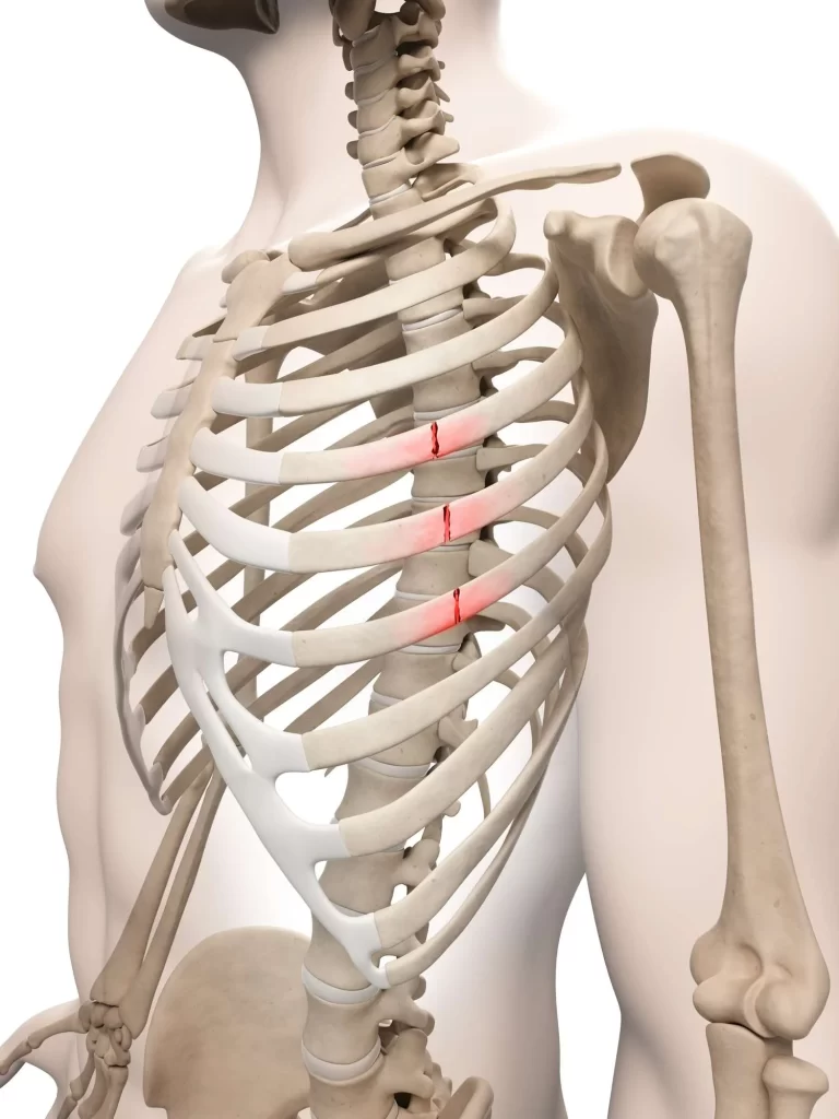
A broken rib can result from severe injury, causing pain, difficulty moving, and difficulty breathing.
Costochondritis: Irritation to the ligament that joins your ribs to your sternum might prompt agony and trouble breathing and keeping up with specific positions.
Rib fractures typically cause chest pain when breathing, coughing, laughing, and most other activities. Most of the time, they are brought on by blows or crushing injuries. Although it is essential to keep in mind that a direct blow can result in a fracture at any point, remember that the weakest part of a rib is just anterior to its angle, making this the most common location of the injury.
The middle rib is the most likely to break, and the broken end can puncture several organs, including the lungs, making it dangerous. Due to their relatively safe position, upper rib fractures are uncommon, but if they do occur, the brachial plexus may be damaged. The diaphragm may also tear as a result of lower rib fractures.
When multiple ribs are fractured, such as in the event of severe trauma, the most common problems typically arise. A flail segment, also known as a flail chest, can occur in this situation if enough ribs are broken. A flail segment occurs when the separated group of ribs moves in the opposite direction of chest wall expansion during inspiration, restricting breathing. If the fragment is huge enough helped ventilation might be required until the ribs have recuperated.
Rib dislocation or subluxation
The most common issues typically arise when multiple ribs are fractured, such as during severe trauma. If enough ribs are broken, this can cause a flail segment, also known as a flail chest. When the separated group of ribs moves in the opposite direction of chest wall expansion during inspiration, limiting breathing, this is known as a flail segment. If the section is colossal enough aided ventilation may be expected until the ribs have recovered
Osteoporosis is bone weakening, often leading to an increased risk of rib fractures as the result of a fall.
Tumor
Although rib tumors are uncommon, the subtle onset of pain may indicate the presence of a benign or malignant tumor.
If you have torment in your mid back or close to your sternum, trouble breathing, or trouble keeping a situation because of muscle fits in your mid back, you might have a rib issue. Visit your doctor right away if you think this is the case. They can evaluate your condition and arrive at a precise diagnosis, allowing you to begin treatment right away.
Since the greater part of your ribs has three marks of connection to your thoracic vertebrae, they are viewed as entirely stable joints and not defenseless to serious injury except if you experience critical injury.
Chest drain insertion
Pneumothorax, pleural effusion, empyema, and postoperatively following thoracic surgery all necessitate this common procedure. It is especially important to align the tube with the superior rib border. This is because the neurovascular bundle of the intercostal nerve, vein, and artery travel along the rib’s costal groove, which is on the inferior border. The risk of causing damage to the surrounding structures is reduced by this positioning.
FAQs
The majority of people are born with 12 ribs on each side, for a total of 24 ribs. Certain individuals are brought into the world with more than 24 ribs. Supernumerary ribs are the name given to these extra ribs. Agenesis of the ribs occurs when a person is born with fewer than 24 ribs.
Out of the twelve ribs, the first is the most superior. It is an anatomical landmark and an unusual rib. It is one of the lines of the predominant thoracic opening. The main structure of the thoracic cage, which covers the thoracic organs, is made up of the ribs. the thoracic organs.
Costochondral joints are the joints in the front of the rib cage where the ribs meet the costal cartilage. They are synchondrosis or primary cartilaginous joints, or hyaline cartilaginous joints. The costal cartilage articulates with a cup-shaped depression on each rib.
chest
There are two sets of twelve ribs that make up the rib cage, which is connected to the sternum, a long, flat bone in the middle of the chest. The ribs are associated with the sternum with a solid, to some degree adaptable material called a ligament.le material called cartilage.
Depending on how they attach to the sternum, the ribs are divided into three groups: true, false, and floating ribs.
The subcostalis, transverse thoracic, and three intercostal muscles (external, internal, and innermost) are the muscles that make up the thorax wall. Each of the intercostal spaces is occupied by eleven pairs of intercostal muscles, which are arranged from superficial to deep.


20 Comments