
Pneumothorax
Table of Contents
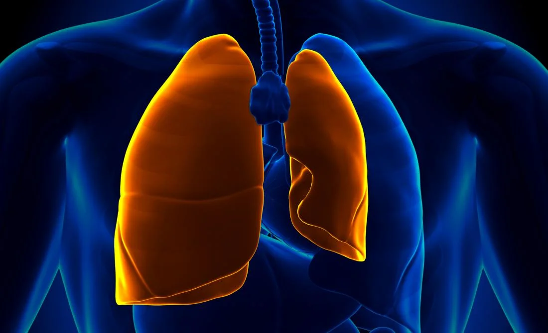
What Is Pneumothorax (Collapsed Lung)?
- Pneumothorax, also known as a collapsed lung, is when air gets between one of your lungs & the wall of your chest. The pressure causes the lung to give away, at least partly.
- When this occurs, you can inhale, but your lung can not expand as much as they should.
- Sometimes, a section of your lung can collapse because of a blockage or less pressure inside the lung, instead of pressure from outside the lung. That is a condition called atelectasis.
Causes and types of pneumothorax
Types of pneumothorax
There are several types of pneumothoraxes. You could have 1 or more at the same time:
- Simple, in which the tissues and organs between your lungs are not moved around.
- Primary spontaneous, when it occurs without any clear cause.
- Secondary spontaneous or disease-related.
- Tension is when air continues to enter the space between your lung and your chest wall, raising the pressure in your chest.
- Traumatic or injury-related.
One way of differentiating them is as follow trusted Source:
- traumatic pneumothorax
- nontraumatic pneumothorax
- primary spontaneous
- secondary spontaneous.
Other subtypes with either traumatic (injurious) or nontraumatic (noninjurious) causes are:
- simple, when it does not harm the position of other structures.
- tension, which harms the position of other structures, like the heart.
- open, when air moves in & out of an open wound in the chest.
Traumatic pneumothorax
- Traumatic pneumothorax occurs later after a certain type of trauma or injury has happened to the chest or lung wall. It can be a small or significant injury. The trauma can damage chest structures & cause air to leak into the pleural space.
Here are certain types of injuries that can cause traumatic pneumothorax:
- trauma (injury) to the chest from the motor vehicle accident
- broken ribs
- a blow to the chest during a contact sport, like a football tackle
- a stab or bullet injury to the chest
- accidental damage during a medical procedure like a central line placement, ventilator use, lung biopsies, CPR, diving, flying, or being at high altitude due to air pressure changes.
- Quick treatment of pneumothorax due to chest trauma is critical as it can conduct in fatal complications like cardiac arrest, respiratory failure, shock, and death.
Nontraumatic pneumothorax
- This type of pneumothorax is known as spontaneous, as it does not result from trauma.
- When primary spontaneous pneumothorax occurs, there is no clear reason why it occurs.
It is more likely rested Source to happen:
- in people who smoke
- during pregnancy
- in people with Marfan syndrome
- in those with a family history of the pneumothorax
- in an otherwise healthy person with a tall & thin body.
Secondary spontaneous pneumothorax can occur if a person has:
- a form of COPD, involving emphysema and chronic bronchitis
- acute or chronic infection, like as tuberculosis or pneumonia,
- lung cancer
- cystic fibrosis
- asthma
- severe acute respiratory distress syndrome (ARDS)
- idiopathic pulmonary fibrosis
- collagen vascular disease
- Inhaling drugs such as cocaine or marijuana can also trigger it.
Tension pneumothorax
Tension pneumothorax is not a classification of pneumothorax yet a term that reflects the severity of pneumothorax. You may experience trusted Sources if you have:
- a blow to the chest
- a penetrating injury
- changes in the pressure when diving, flying, or mountaineering
- a spontaneous pneumothorax progressing to tension-type
- Certain medical procedures.
Pathology
- The pleural cavity is the part between the chest wall and the lungs. If the air enters the pleural cavity, either from the outside (open pneumothorax) or from the lung (closed pneumothorax), the lung collapses and it becomes harmful for the person to breathe.
- Tissue can form a way-valve that allows air to enter the pleural cavity, yet not to escape, overpressure can build up with each breath (tension pneumothorax). This conducts to severe shortness of breath, deviation of the heart, and compression of the vena cava leading to shock.
- There is a loss of intrapleural negative pressure that can result in lung collapse. Due to this, there is a decrease in vital capacity as well as a decrease in PaO2 which is an important consequence of a pneumothorax.
- The decrease in PaO2 results from difficult factors i.e low ventilation-perfusion ratios, anatomic shunts, and alveolar hypoventilation. Most patients that suffer from pneumothorax also have an improvement in alveolar-arterial oxygen tension.
Epidemiology
- Primary spontaneous pneumothorax happens most often in people between ages 18 – 40 and Secondary spontaneous pneumothoraces happen more frequently after age 60 years.
- Prevalence of a pneumothorax in a newborn is a potentially serious problem and it happens in about 1-2% of all births.
- The general person consulting rate for pneumothorax (primary and secondary combined) in the GPRD was 24.0/100 000 each year for men & 9.8/100 000 each year for women.
- Hospital admissions for pneumothorax as a primary diagnosis occurred at a general incidence of 16.7/100 000 per year & 5.8/100 000 per year for men & women, respectively. Mortality prices were 1.26/million per year for men & 0.62/million per year for women.
Causes of pneumothorax
- You can get a pneumothorax in many ways.
Causes involve:
- Lung disease: Tissue that is damaged is more likely to tear, allowing air to leak out. This is especially right with chronic obstructive pulmonary disease (COPD).
- Injury: A broken rib, knife wound, or gunshot injury can puncture your lung. In serious cases, the escaping air can build up pressure on your lung and heart, which might cause life-threatening problems like loss of blood pressure.
- Mechanical ventilation.: This machine that assists you to breathe creates uneven pressure in your chest. As a result, your lung might be collapsed.
- Air blisters: Sacs full of air, known as blebs, may form on the outside of your lung and then burst, creating pressure. This happens most of the time with tall men who are younger than 40 and who smoke.
- Your period: It is rare, but cysts could form inside your chest. Within about 3 days before or after the start of your period, the cysts release blood between the lung and chest.
- Often, someone who has a collapsed lung gets another within one or two years. Smoking can also create the condition more likely. And certain types of pneumothoraces run in families.
Symptoms of pneumothorax
Symptoms can range from mild (slight) to dangerous. If your case is mild (slight), you may not notice a problem. That is why it is important to tell your doctor what’s occurring.
Common symptoms involve:
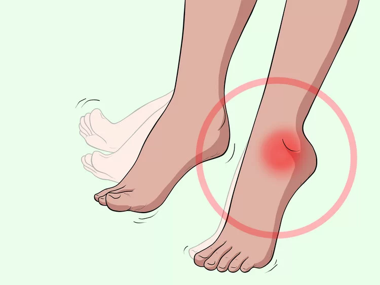
- a sudden, sharp, stabbing pain in the chest
- rapid breathing or shortness of breath (dyspnea)
- turning blue, known as cyanosis
- a rapid trusted Source of heart rate
- low blood pressure
- lung expansion on one side
- an empty sound if you tap on the chest
- an enlarged jugular vein
- anxiety
- fatigue.
Diagnosis of pneumothorax
Your doctor will probably begin with a physical exam. They will listen to your breathing through a stethoscope and tap your chest to find out if it sounds hollow.
You might have tests including:
- X-rays, so your doctor can look at the outline of your lung,
- A CT scan is a series of X-rays that a computer turns into a detailed image of your lung,
- Arterial blood gas or pulse oximetry measures how much oxygen is in your blood,
- EKG, to check how clearly your heart is working.
Risk factors for pneumothorax
The risk factorsTrusted Source was various for a traumatic and spontaneous pneumothorax.
Risk factors for a traumatic pneumothorax include:
- contact sports, like football or hockey,
- employment where there is a risk of falls or other types of injury,
- having a medical procedure that includes the chest or lung area,
- ongoing assisted with respiratory care.
The people at highest risk of a non-traumatic pneumothorax involve those who:
- have a history of smoking,
- have an existing lung condition, such as asthma or chronic obstructive pulmonary disease (COPD),
- have a family history of pneumothorax, which may indicate genetic factors,
- have tall, slim bodies, as this can affect trusted Source pressure at the top of the lung,
- have inflammation in the tiny airways.
Pneumothorax complications
Pneumothorax can conduct to a number of complications, some of which can be life-threatening.
They include:
- respiratory failure or inability in breathing,
- pulmonary edema following treatment for the pneumothorax,
- pneumohemothorax, when blood into the chest cavity,
- pneumopericardium, when air into the cavity around the heart,
- pneumoperitoneum, when are into the space around the abdomen,
- bronchopulmonary fistula, when a passageway opens between the lungs & the space around them,
- heart attack.
Tension pneumothorax can fastly progress to:
- an inability to breathe,
- cardiovascular collapse,
- death.
- It is essential to seek emergency medical help as soon as symptoms arise.
Pneumothorax Treatment
Your treatment will depend on the type of pneumothorax & how serious it is. You may have one or more of these:
- how severe the condition is
- whether the chest cavity is persistent expands
- the cause
- whether it is happened before or has been going on for some time.
- If you have tension pneumothorax or pneumothorax due to any injury, this is a life-threatening emergency. You will need immediate medical care & possibly surgery.
Here are certain treatment strategies:
Observation
- If pneumothorax results from a little injury, it may heal without treatment within a few days. Check with a doctor about previous flying or diving after pneumothorax.
- If you are having trouble breathing, you may require oxygen. Using oxygen can also assist speed trusted Source is the rate at which the lungs reabsorb air from the cavity.
- Your doctor will probably want you to remain in the hospital so they can look at your progress. They treat a collapsed lung by getting clear of the pressure outside the lung so it can inflate again.
- In small cases without any symptoms, the lung can expand again on its own. You may require to breathe oxygen from a container for a short time to assist. Even if your case is mild, it is important to have follow-up visits with your doctor so they can keep track of how you are doing.
Needle aspiration or chest tube insertion or Draining excess air
- If the damage is significant or symptoms are severe, a surgeon may require to remove the air or carry out the surgery.
- Needle aspiration & chest tube insertion are 2 procedures designed to remove excess air from the pleural space in the chest. These can be done at the bedside without needing general anesthesia.
- In needle aspiration, the doctor passes the needle into the cavity & extracts the air using a syringe.
- For a chest tube insertion, the doctor will pass a hollowed tube between your ribs. This allows air to drain & the lung to reinflate. The tube may remain in place for two to five days or longer.
- If your lung has collapsed further, your doctor may use a needle or a tube to release more air from your chest. The tube might be attached to the one-way valve. It could have to remain in place for hours or days.
Physiotherapy Management
Indications for Physiotherapy treatment
- Lung collapse,
- Sputum retention,
- Ventilation/perfusion mismatch (V/Q),
- Increased the work of breathing,
- Blood gas abnormalities,
- Post-operative ITU care.
Goals for Physiotherapy
- To improve ventilation and increases PaO2 levels
- Physical activity (stairs, walking, moderate-intensity aerobic exercise),
- Active cycle of the breathing exercises,
- Sputum removal techniques ex. percussion, cough assist,
- PEP devices,
- Incentive spirometry,
- Non-invasive ventilation (NIV),
- To assist in the sputum removal
- Postural drainage,
- Active cycle of breathing exercises (ACBT),
- Percussion, shaking, & vibrations,
- PEP devices,
- Physical activity (stairs, walking, moderate-intensity aerobic exercise),
- Coughing & huffing (forced expiratory breath),
- Airway suctioning.
- To reduce the work of breathing
- Body positioning,
- Breathing control,
- Relaxation techniques,
- Accessory muscle use.
- Improve exercise tolerance
- Early mobilization and positioning,
- Graded exercise program,
- Breathing exercises.
Physiotherapy outcome evaluation includes
- Respiratory rate,
- O2 saturation,
- Arterial blood gases,
- Additional O2 requirements,
- Auscultation,
- Chest x-ray,
- Mobility status.
Recurrent pneumothorax treatment
- For patients with recurrent pneumothorax later surgical intervention, there are several options. For patients with total or near-total lung collapse, repeat surgical intervention is recommended.
Options involve:
- Repeat mechanical pleurodesis if it is uncertain whether appropriate mechanical pleurodesis was done initially
- Pleurectomy in which the pleura overlying the ribs is usually removed.
- Chemical pleurodesis is when a drug or other agent is used to create an inflammatory response that results in pleurodesis. Talc is the most usually used agent due to its effectiveness. Historically, talc pleurodesis was considered a contraindication to future lung transplantation because of the intense inflammatory response that made surgery very varied.
- Lung transplant
Autologous blood patch
- Your doctor can take blood from your arm & put it down into your chest through a tube. This creates a patch on your lung that stops air leaks.
Surgery or pleurodesis
- Cases involving lung disease, an accident, or repeated collapsed lungs may require surgery. Or you could have a procedure known as pleurodesis. Your doctor uses a needle and tube to put medicine such as doxycycline into your chest. It triggers inflammation, which assists your lung stick to the chest wall & stays inflated.
- The doctor may require you to carry out a more invasive procedure to see what is happening in your lungs, like a thoracotomy or thoracoscopy. During a thoracotomy, your surgeon will make an incision in the pleural space to help them see the problem. During a thoracoscopy, also known as video-helped thoracoscopic surgery (VATS), the doctor inserts a small camera through the chest wall to examine the lung. If you have had repeated episodes of pneumothorax, you may need a little operation to repair any weak areas in the lung where the air is getting through. The doctor may carry out pleurodesis, in which they stick the lung to the inside of the chest wall.
Other surgical options involve:
- sewing blisters closed,
- closing air leaks,
- or removing the collapsed portion of your lung, which is known as lobectomy,
- These interventions can decrease the risk of pneumothorax happening again.
Prognosis
- Up to 50% of patients who suffer from a pneumothorax will have another one or a recurring pneumothorax. However, there is no long-term complications later successful treatment.
Pneumothorax Outlook
- With treatment, many people do not have long-term health effects from pneumothorax. But it can occur again in up to 50% of cases.
- Most cases of primary spontaneous pneumothorax solve with observation or minimal treatment. It is rarely life-threatening. But there is a 30 percent trusted Source chance that this type will recur within 5 years, & the risk of recurrence increases each time it happens.
It may take longer to recover if:
- you have a large pneumothorax,
- you have a secondary spontaneous pneumothorax,
- you have an underlying lung condition,
- pneumothorax results from an injury,
- it is not your first experience with pneumothorax.
- In around 10% of cases, secondary spontaneous pneumothorax is mortal. The risk is higher if you have HIV or coronary obstructive pulmonary disease (COPD). The risk of this type recurring within five years is around 43 percent trusted Sources, and the risk increases each time it occurs.
- Knowing your risk of developing pneumothorax and seeking assistance as soon as symptoms occur can help prevent severe complications.
Pneumothorax Prevention
If you have had a collapsed lung, you require to take extra care of yourself to keep it from happening again. Some tips:
- If you smoke, ask your doctor for assistance quitting,
- If you scuba dive, your doctor might ask you to stop,
- If you have a lung problem, retain up with your medical visits.
Summary
- Pneumothorax is a condition where air gathers between the lungs & the chest cavity. In certain cases, it will go away without treatment. In another, it can be life-threatening. This will turn on the size and cause of the problem.
- There are various types of pneumothorax. Traumatic pneumothorax can occur if someone has an injury to the chest wall or lungs. Nontraumatic pneumothorax can harm people with COPD and other lung diseases, yet it can also affect people without lung disease.
- Treatment aims to remove the air & re-expand the lungs. In certain cases, a surgeon may need to repair the lungs. Pneumothorax can be a life-threatening emergency. Anyone who experiences symptoms, such as a sharp, stabbing pain in the chest, should seek immediate medical assistance.
FAQs
Sharp, stabbing chest pain that worsens when trying to breath in.
Shortness of breath.
Bluish skin caused by a lack of oxygen.
Fatigue.
Rapid breathing and heartbeat.
A dry, hacking cough.
A small pneumothorax may go away on its own over time. You may only need oxygen treatment and rest. The provider may use a needle to allow the air to escape from around the lung so it can expand more fully. You may be allowed to go home if you live near the hospital.
A pneumothorax can be severe, depending on how much air is trapped in the pleural space. A small amount of trapped air can usually resolve by itself, provided there are no other complications. Larger amounts of trapped air can be serious and lead to death if medical treatment is not obtained.
Treatment options may include observation, needle aspiration, chest tube insertion, nonsurgical repair or surgery. You may receive supplemental oxygen therapy to speed air reabsorption and lung expansion.
If your pneumothorax is caused by an underlying lung condition or chest trauma, you are more likely to need treatment. If the pneumothorax is small, the leak usually heals itself and the trapped air is gradually absorbed by your body. This normally takes 1-2 weeks.
Oxygen. High flow oxygen (>28%) should usually be given to individuals with a pneumothorax in order to maintain adequate oxygenation (saturation >92%) to vital organs.
A collapsed lung feels like a sharp, stabbing chest pain that worsens on breathing or with deep inspiration. This is referred to as “pleuritic” because it comes from irritation of nerve endings in the pleura (inner lining of the rib wall).
The average amount of time to stay in the hospital with a pneumothorax is 5 to 7 days.


3 Comments