Pott’s spine
Table of Contents
What is pott’s spine disease?
Tuberculous spondylitis also known as Pott’s disease, refers to vertebral body osteomyelitis & intervertebral discitis from tuberculosis (TB). The spine is the most often part of musculoskeletal tuberculosis, and commonly related symptoms are back pain, kyphotic deformity of the spine, lower limb weakness, & paraplegia.
Pott disease is tuberculosis of the spine that occurs usually due to hematogenous spread from other sites, often the lungs. Percival Pott was the first person to describe the classic description of spinal tuberculosis in 1779; hence, spinal TB was called ‘Pott’s Disease. Tuberculosis of the spine is one of the oldest determined diseases of mankind and is the common extrapulmonary form of TB.
The morbidity & mortality rate because of spinal TB is higher than other infections in developing countries with dense populations.
Lower thoracic and lumbar vertebrae are the most common areas of spinal TB followed by middle thoracic and cervical vertebrae. The second cervical to the seventh cervical region is reportedly involved in 3 to 5% of cases and atlantoaxial articulation is less common. In TB, the involvement of posterior elements because of TB is not so uncommon. The lamina was the most commonly involved site followed by pedicles, articular processes, spinous processes, & transverse processes.
It leads to a kind of tuberculous arthritis of the intervertebral joints. The infection can spread from two adjacent vertebrae through the adjoining intervertebral disc space. If only one vertebra is affected, the disc is normal, but if two vertebras are affected, the disc, which is avascular, cannot receive nutrients and collapses. In a procedure called caseous necrosis, the disc tissue dies, leading to vertebral narrowing & eventually vertebral collapse and spinal damage. A dry soft-tissue mass often forms and superinfection is rare.
The spread of infection from the lumbar vertebrae to the psoas muscle, causing abscesses, is common. Lately, the disease has shown a significant inclination in developed nations, particularly among the immunosuppressed population secondary to global migration and travel. There has been an ominous, increasing trend in the occurrence of multidrug-resistant bacterial strains of tuberculosis in developing nations over the past decades, a tough challenge to the global community.
Accordingly, the disease continues to exist as a major, global public health menace to date.
What are the causes of pott’s spine disease?
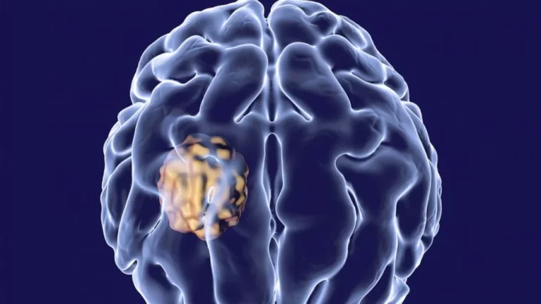
Pott’s disease can take place when air that contains the tuberculosis-causing bacteria Mycobacterium tuberculosis is inhaled into the lungs. From the lungs, an infection can spread into the spine. The spread is hematogenous which is transmitted through the blood.
Someone with active TB infections can spread through the air with aerosol droplets by spitting, sneezing, coughing, speaking, etc. Each sneeze can release up to 40,000 aerosol droplets, & it only takes one infected droplet to spread TB.
Some known predisposing factors for TB include prolonged exposure to infected patients, immunodeficiencies such as HIV, alcohol, drug abuse, overcrowding, malnutrition, poverty, and lower socio-economic situation.
What is the course of pott’s spine disease?
Spinal involvement is generally a result of the hematogenous spread of Mycobacterium tuberculosis into the dense vasculature of the cancellous bone of the vertebral bodies. The primary infection area is either a pulmonary lesion or an infection of the genitourinary system.
Spread occurs either through the arterial or the venous route. An arterial arcade, in the subchondral part of each vertebra, is derived from anterior & posterior spinal arteries; this arcade forms a rich vascular plexus. This vascular plexus facilitates the hematogenous spread of the infection to the paradiscal regions.
Batson’s paravertebral venous plexus in the vertebra is a system without valves that allows the free flow of blood in both directions depending upon the pressure generated by the intra-abdominal & intrathoracic cavities after strenuous activities like coughing. The spread of the infection through the intraosseous venous system might be responsible for central vertebral body lesions. In patients with noncontiguous vertebral tuberculosis, again it is the vertebral venous system that spreads more infection to multiple vertebrae.
Spinal tuberculosis is initially apparent in the anterior inferior part of the vertebral body. Afterward, it spreads into the central part of the body or disc. Paradiscal, central and anterior, damages are the common types of vertebral involvement. In the central lesion, the disc is not involved, & the collapse of the vertebral body produces vertebra plana. Vertebra plana indicate complete compression of the vertebral body.
In younger patients, the disc is primarily included because it is more vascularized. In old age, the disc is not primarily involved because of more age there is avascularity present. In spinal tuberculosis, there is the involvement of more than a vertebra because its segmental arteries bifurcate to supply blood to two adjacent vertebrae.
The spread of the disease beneath the anterior or posterior longitudinal ligaments includes multiple contiguous vertebrae. A lack of proteolytic enzymes in mycobacterial infections is recommended as the cause of the subligamentous spread of infection.
In spinal tuberculosis, characteristically, there is the destruction of the intervertebral disc space and the adjacent vertebral bodies, the collapse of the spinal elements, & anterior wedging leading to the characteristic angulation and gibbus (palpable deformity because of the involvement of multiple vertebrae) formation.
The upper lumbar & lower thoracic spine are the most often involved sites. More than one vertebra is typically affected, & the vertebral body is more often affected than the posterior arch.
In younger patients, the disc is primarily involved because of its more vascularization. In older age, the disc is not primarily involved because of its age-related avascularity. In spinal TB, there is the involvement of more than one vertebra because its segmental arteries bifurcate to supply two adjacent vertebrae.
The spread of the infection beneath the anterior or posterior longitudinal ligaments involves multiple contiguous vertebrae. A lack of proteolytic enzymes in mycobacterial infections has been suggested as the cause of the subligamentous spread of infection.
In spinal TB, characteristically, there is the destruction of the intervertebral disc space and the adjacent vertebral bodies, collapse of the spinal elements, & anterior wedging leading to significant angulation and gibbus formation.
Paraplegia is the most disastrous complication of spinal tuberculosis. It has been divided into two groups: early-onset paraplegia & late-onset paraplegia.
Early onset paraplegia develops in the active stage of spinal TB and it has required active treatment. This type of paraplegia has a better prognosis & is often seen in adults with Pott’s spine. In all these patients, paraplegia is caused by the formation of debris granulation tissue, and pus, because of the destruction of bone and intervertebral disc. Destruction of the anterior vertebral column causes subluxation & subsequent dislocation of the spine.
Concertina collapse is a compression fracture without the involvement of the intervertebral disc that may occur because of extensive tuberculous destruction. Concertina collapse bulges into the parenchyma of the spinal cord.
Intrinsic factors lead to meningomyelitis by direct involvement of the spinal cord, surrounding meninges & roots, or by including the blood vessels supplying the spinal cord. In addition, some rare causes of paraplegia involve infective thrombosis of arteries supplying the spinal cord & nonosseous intramedullary or extramedullary tuberculoma of the spinal cord.
Late-onset paraplegia is a neurological complication that occurs after a variable period in a patient with recovered tuberculosis. Late-onset paraplegia might develop within 2-3 decades after active infection. It is frequently related to marked spinal deformities.
What are the symptoms of pott’s spine disease?
The distinctive clinical features of spinal tuberculosis include local pain, tenderness, stiffness & spasm of the muscles, a cold abscess, gibbus, & a prominent spinal deformity. The cold abscess slowly occurs when tuberculous infection extends to adjacent ligaments & soft tissues. A cold abscess is featured by a lack of pain and other signs of inflammation.
The development of spinal tuberculosis condition is slow and insidious. The total period of the illness varies from a few months to a few years, with the average disease period ranging from four to eleven months. Generally, patients look for advice only when there is severe pain, marked deformity, or neurological symptoms.
Constitutional symptoms are present in approx 20–30% of cases of osteoarticular tuberculosis. The classical constitutional features of tuberculosis indicating the presence of active disease are malaise, loss of weight and appetite, night sweats, evening rise in temperature, generalized body aches, and fatigue.
The symptoms of POTS include but are not limited to lightheadedness (occasionally with fainting), difficulty thinking & concentrating (brain fog), fatigue, blurry vision, intolerance of exercise, headache, palpitations, tremors, and nausea.
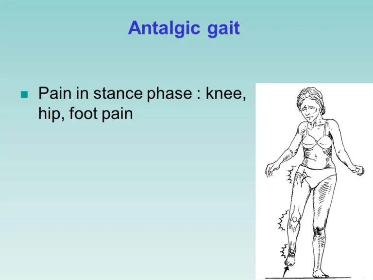
Back pain is the most often presented symptom of spinal tuberculosis. The depth of pain varies from constant mild dull aching to severe disabling. Pain is typically localized to the area of involvement & is most common in the thoracic region. The pain might be aggravated by spinal motion, coughing, & weight bearing, because of advanced disc disruption and spinal instability, nerve root compression, or pathological fracture. Chronic back pain as the only symptom was observed in half of the cases of spinal tuberculosis.
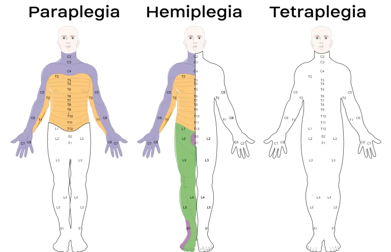
Neurologic deficits are usual with the involvement of thoracic & cervical regions. If it is left untreated, early neurologic involvement might progress to complete paraplegia or tetraplegia. Paraplegia may occur at any time & during any stage of the vertebral disease. The level of spinal cord inclusion determines the extent of neurological manifestations.
In cervical spinal tuberculosis, patients appear with symptoms of cord or root compression. The earliest signs are pain, weakness, & numbness of the upper and lower extremities, eventually progressing to tetraplegia.
If the thoracic or lumbar spine is included, upper extremity function stays normal while lower-extremity symptoms progress over time eventually leading to paraplegia. Patients with cauda equina compression because of lumbar and sacral vertebral damage have weakness, numbness, & pain, but have decreased or absent reflexes among the affected muscle groups. This is in contrast to the hyperreflexia seen with spinal cord compression along with bladder involvement is known as a cauda-equina syndrome.
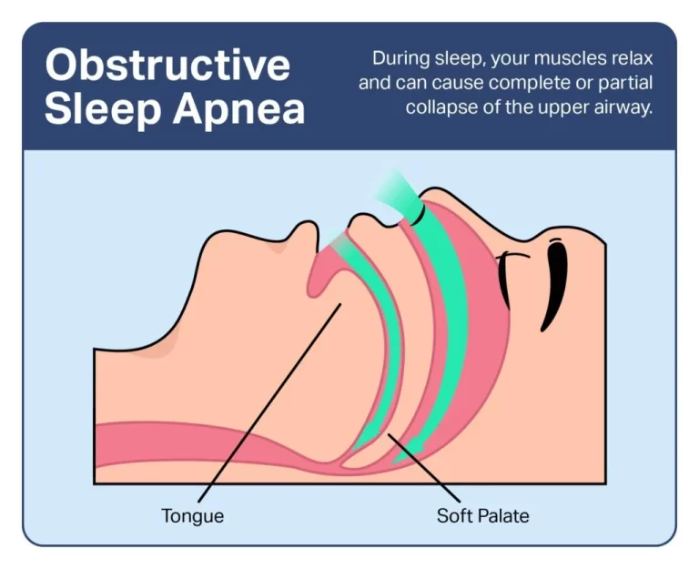
The development of a cold abscess around the vertebral lesion is another characteristic sign of spinal tuberculosis. Abscess formation is common & can grow to a very big size. The area of a cold abscess depends on the region of the vertebral column affected. In the cervical region, the pus accumulates posterior to the prevertebral fascia to form a retropharyngeal abscess. The abscess might track down to the mediastinum to enter into the trachea, esophagus, or pleural cavity. A retropharyngeal abscess can produce considerable pressure effects such as respiratory distress, dysphagia, or hoarseness of voice. In the thoracic spine, the cold abscess generally presents as a fusiform or bulbous paravertebral swelling and might produce posterior mediastinal lumps.
The cold abscesses developed at lumbar vertebrae most commonly present as a swelling in the groin & thigh. An abscess can descend down under the inguinal ligament to appear on the medial aspect of the thigh. Collection of pus can follow the blood vessels to form an abscess in the gluteal region if it follows femoral or gluteal vessels, respectively.
Spinal deformity is a significant feature of spinal tuberculosis. The type of spinal deformity depends on the area of the tuberculous vertebral lesion. Kyphosis is the most common spinal deformity that occurs with lesions involving thoracic vertebrae. The severity of kyphosis depends on how many vertebrae are involved. An increase in kyphotic deformity by 10° or more might be seen in up to 20% of cases, even after the treatment. Atlanto-axial tuberculosis may present as torticollis deformity.
Differential diagnosis of pott’s spine disease?
Common differential diagnosis includes pyogenic spondylitis, osteoporotic, metastasis, multiple myeloma, brucellar spondylitis, & lymphoma. Brucellar spondylitis is commonly found in the middle-aged group. The lumbar spine is frequently included followed by the thoracic and cervical spine. Disc involvement and small paraspinal soft tissue component can be seen; nevertheless, gibbus formation is not found in cases of brucellar spondylitis.
Pyogenic spondylitis can be found at any age; generally lumbar and cervical spines are affected. Destruction of vertebral bodies, intervertebral discs, markedly enhancing lesions, and epidural abscesses can be seen. In pyogenic spondylitis, there is a sparing of posterior elements and usually, no gibbus deformity is seen. In osteoporosis, thoracic vertebrae are frequently affected by the sparing of the pedicles. Apart from the destruction of multiple vertebral bodies, decreased bone density is usually seen in osteoporotic vertebrae.
In metastatic disease, the thoracic area is most commonly involved. The posterior wall of the vertebral body, pedicles, and lamina are involved in metastatic disease; however, intervertebral disc heights are preserved. Intervertebral discs may be affected by lymphoma and multiple myeloma. In elderly patients with vertebral collapse, metastatic disease of the spine should always be contemplated.
How to prevent pott’s spine disease?
Controlling the growth of tuberculosis infection can prevent tuberculous spondylitis and arthritis. Patients who have a positive PPD test but not active tuberculosis might decrease their risk by properly taking medicines to prevent tuberculosis. To successfully treat tuberculosis, patients must take their medications exactly as prescribed.
What is the Diagnostic procedure for pott’s spine disease?
Diagnosis of spinal tuberculosis is made on the basis of the classic clinical presentation along with systemic constitutional manifestation, evidence of past exposure to TB or concomitant visceral TB, & neuroimaging modalities.
Skin tests & hematological tests like complete blood count (CBC), Montoux test, enzyme-linked immunosorbent assay (ELISA), erythrocyte sedimentation rate (ESR), & polymerase chain reaction (PCR) are also done in diagnosing spinal tuberculosis. Bone tissue or abscess samples are acquired to stain for acid-fast bacilli (AFB) and isolate organisms for culture, antibiotic sensitivity, and histopathology; the method extensively used is CT-guided or ultrasonography-guided needle biopsy & aspiration or surgical biopsy.
Plain radiographs
Plain radiographs are usually the initial examination in patients with spinal TB. For radiolucent abrasions to be evident on a plain radiograph, there must be bone mineral loss. In paradiscal type of lesion, the earliest radiological features are the narrowing of the joint space & indistinct paradiscal margin of vertebral bodies.
The disc space narrows because of either atrophy or prolapse into the vertebral body of the disc tissue. With further progression, anterior wedging or collapse occurs, resulting in a variable degree of kyphosis. In the anterior type of lesion, the collection of tuberculous granulation tissue and necrotic material causes the formation of a paravertebral abscess. In the area of the thoracic spine, it is visible on plain radiographs as a fusiform or globular radiodense shadow known as the bird’s nest appearance.
Long-standing abscesses might produce concave erosions around the anterior margins of the vertebral bodies producing a scalloped appearance known as the aneurysmal phenomenon. The anterior type is much more usual in the pediatric dorsal spine. The central type of abrasions presents as destruction, ballooning of vertebral bodies, and concentric collapse. In the appendiceal or neural arch type of lesion, there is the inclusion of the posterior arches such as spinous process, pedicle, lamina, & transverse process as well as lateral masses of the atlas, erosion of the adjacent ribs in the thoracic region, pedicular or laminar destruction, or posterior cortex of the vertebral body with relative thrifty of the intervertebral discs, and a large paraspinal mass.
Computed tomography
Computed tomographic scanning gives more good bony detail for irregular lytic lesions, disc collapse, sclerosis, and disruption of bone circumference than a plain radiograph. CT is more efficacious for explaining the shape & calcification of soft tissue abscesses. Nevertheless, CT is less accurate in explaining the epidural extension of the disease and its result on neural structures.
The pattern of bone destruction as fragmentary, osteolytic, sclerotic, and subperiosteal can be seen well on CT. It is excellent for guiding a percutaneous diagnostic needle in potentially dangerous or relatively inaccessible regions. The existence of calcification within the abscess is practically diagnostic of spinal TB. In the past, CT myelography was one of the modalities used for spinal TB for assessing cord compression, but it has been replaced by MRI nowadays.
Magnetic resonance imaging (MRI)
MRI is done via proceeds non-contrast T1-weighted (T1W), T2-weighted (T2W), and short tau inversion recovery (STIR) sequences in axial, sagittal, & coronal planes after that contrast-enhanced T1W sequences after intravenous administration of gadolinium contrast agent. MRI features of Pott’s spine are abnormal signal intensities showing hypointense on T1W and hyperintense on T2W sequences with the heterogeneous enhancement of the vertebral body.
STIR sequences are useful in distinguishing fluid from fatty components in non-contrast sequences. Characteristic findings contained the destruction of two adjacent vertebral bodies and opposing end plates; destruction of the intervening disc; vertebral body edema; & occurrence of prevertebral, paravertebral, & epidural abscesses MRI plays an important part in the diagnosis of spinal TB with high specificity and sensitivity.
In disparity to most imaging methods, MRI has the advantages of good contrast resolution for bone and soft tissues along with the versatility of direct imaging in multiple planes. With the help of intravenous administration of magnetic resonance contrast agents, MRI was highly accurate in distinguishing granulation tissue from a cold abscess.
MRI can show more extensive involvement than plain films. MRI provided more exact anatomic localization of vertebral & paravertebral abscesses in multiple planes not previously available with more conventional diagnostic methods in patients with suspected TB spondylitis. MRI clearly determined the extent of soft tissue disease and its effect on the theca, cord, and foramen in cases with doubtful CT findings. Both CT and MRI are exceedingly helpful for diagnosis, & tissue aspirate is a good confirmatory method.
Administration of gadolinium with diethylene tri-amine pentaacetic acid (Gd-DTPA) is utilized to assess the extent of soft tissue mass and to differentiate postoperative spondylitis from a normal postoperative course, via showing disc enhancement. Disc magnification occurs infrequently in the normal postoperative course. If it is related to adjacent vertebral bone marrow changes, it should be considered postoperative spondylitis.
Differential diagnosis between pyogenic, tuberculous, fungal, & postoperative spondylitis is difficult; although the pattern of enhancement in TB spondylitis was different from the other cases of spondylitis. MRI imaging is a very useful method for the differentiation of tubercular spondylitis from pyogenic spondylitis.
Kyphosis and cord compressions were the most usual complications. The neurological inclusion is relatively benign if urgent decompression is done at the onset of the disease. Late-onset paraplegia is a neurological complication that occurs after a variable period in a patient with healed tuberculosis of the spine. MRI is extremely useful in diagnosing difficult and rare areas of disease like the craniovertebral junction. It finds the marrow changes, exudative & granulation types, extra and intradural disease, and radiological response to the treatment in the early follow-up period of around 6-8 weeks.
An increase in the prevertebral soft tissue shadow on a radiograph is a useful guide to resort to a CT scan or MRI to diagnose TB of the cervical spine. The anterior convexity and forward displacement of the tracheal shadow of greater than 8 mm from the vertebral bodies in a lateral view of plain X-ray and broadening of the superior mediastinum in an anteroposterior X-ray are helpful indicators of tuberculous involvement at the cervicodorsal region.
Sacroiliac joint TB is rare; its conjunction with vertebral TB is even rarer, with only a few such patients reported. MRI is the most sensitive & specific imaging equipment for diagnosing sacroiliitis at its early stage. Sacroiliac joint tuberculosis can extend advanced stages with extensive joint destruction & periarticular abscesses if diagnosis and treatment are delayed. The addition of a coronal STIR T2-weighted sequence to the routine MRI assessment of patients studied for lumbar disc disease may be useful for recognizing sacroiliac joint pathology at an earlier stage.
Cytological and microbiological confirmation
Etiological confirmation can be made either by demonstration of acid-fast bacilli on a pathological specimen or histological evidence of a tubercle or the mere presence of epithelioid cells on the biopsy material.
Neuroimaging guided-needle biopsy from the affected area is the gold standard technique for the early histopathological diagnosis of spinal tuberculosis. A CT-guided needle biopsy generally yields sufficient material either from the spine itself or from an adjacent abscess. An open biopsy of the spine is generally performed when either closed techniques have proved insufficient or other procedures, such as decompression & possibly arthrodesis, are planned.
In India, fine needle aspiration biopsy done under CT guidance was successful in diagnosing spinal tuberculosis. Surgery might be required to establish the etiological diagnosis. Material obtained from the biopsy should be submitted for cytologic, histologic, & bacteriologic studies. Smear positivity for acid-fast bacilli might be seen in up to half of cases and culture positivity in most cases. Nevertheless, as with respiratory tuberculosis, culture is not the gold standard for diagnosing spinal tuberculosis because mycobacterial bacilli are not readily detected from extrapulmonary sites.
Histologic tests confirm the diagnosis of spinal tuberculosis in approx 60% of patients. The most common cytological findings observed are epithelioid cell granulomas, granular necrotic background, & lymphocytic infiltration. Scattered multinucleated and Langhans’ giant cells might be seen in up to half of cases. False-negative results of the biopsy are common and, therefore, diagnosis of spinal tuberculosis must be made on grounds of clinical manifestations and radiology when bacteriology proves negative.
Polymerase chain reaction and other immunological tests
Conventional microbiological methods like Ziehl–Neelsen staining for acid-fast bacilli and culture of Mycobacterium tuberculosis on Lowenstein Jensen media have low sensitivity and specificity. In addition, culturing Mycobacterium tuberculosis is time-consuming, taking 6–8 weeks for the growth to appear. So, mainly the diagnosis of tuberculosis depends on histological evidence.
The polymerase chain reaction has shown very promising results for the early and rapid diagnosis of the disease. This method is able to detect as few as 10–50 tubercle bacilli in various clinical samples. This test offers better accuracy than a smear and can be performed at a greater speed than cultures.
The QuantiFERON-TB Gold assay finds cell-mediated inflammatory responses in vitro to tuberculosis infection by measuring interferon-gamma harvested in plasma from whole blood incubated with the Mycobacterium tuberculosis-specific antigens. In a study, some consecutive patients with vertebral collapse underwent a battery of investigations including the QuantiFERON assay.
The patients were classified as having tuberculosis on the basis of a positive smear or culture, a biopsy consistent with tuberculosis, or a therapeutic response to antituberculosis chemotherapy. Tuberculosis was diagnosed in some patients, and few had a vertebral collapse that was attributable to other causes. In this examination, sensitivity was estimated as 84% and specificity was 95%.
Other tests
Erythrocyte sedimentation rate (ESR) is usually raised many folds in the majority of patients with spinal tuberculosis. ESR decreases to normal or near normal when the active tuberculous lesion is controlled.
In pyogenic infection, leucocytosis parallels with increased ESR, while in patients with spinal tuberculosis, there is markedly elevated ESR with a normal white blood cell count(WBC).
What is the treatment plan for pott’s spine disease?
Medical treatment for pott’s spine disease:
In patients with spinal tuberculosis, antitubercular treatment should be initiated as early as possible. Antituberculous treatment frequently needs to be instituted empirically, much before an etiological diagnosis is established. In poor countries, etiological diagnoses might not be established at all. In patients with established complications of spinal tuberculosis, surgery may also be needed. Sequelae like kyphosis require surgical intervention.
Almost all antituberculous drugs penetrate well into tuberculous vertebral abrasions. The distribution of antituberculosis drugs such as rifampin, isoniazid, and pyrazinamide was evaluated in pretentious vertebral tissues of spinal tuberculosis. In patients without vertebral sclerotic walls around the tuberculous foci, the isoniazid concentrations in foci were of bactericidal levels.
Levels of rifampin & pyrazinamide in foci corresponded to the minimal inhibitory concentrations of each drug, individually. The sclerotic bone of the affected vertebra played a role in blocking the antituberculosis drug’s penetration. In another study, three drugs resulted in an effective bactericidal concentration level in osseous tissues around the foci of spinal tuberculosis excluding the 4 mm of osseous tissue surrounding the sclerotic wall. The results recommended that osseous tissues within 4 mm surrounding the sclerotic wall should be removed during the surgery.
Antituberculous treatment
Various studies have shown that the majority (82–95%) of patients with spinal tuberculosis respond very well to medical treatment. The treatment feedback is apparent in form of pain relief, a decrease in neurological deficit, and even correction of spinal deformity.
Patients with potentially dangerous craniovertebral junction tuberculosis also retaliate satisfactorily to medical treatment. Patients with medically resistant spinal tuberculosis need a careful reassessment of the differential diagnosis before surgery is planned surgery.
Therapeutic regimen:
World Health Organization (WHO) suggests a category-based treatment for tuberculosis. Spinal tuberculosis falls down under category 1 of the WHO treatment category. The category-1 antituberculosis treatment regimen is classified into two phases: an intensive or initial phase and a continuation phase. In the 2-month intensive phase, antituberculous therapy involves a combination of four first-line drugs: isoniazid, rifampicin, streptomycin, & pyrazinamide. In the continuation phase, two drugs isoniazid and rifampicin are allowed for 4 months.
Due to the serious risk of disability and mortality and because of difficulties in assessing treatment response, WHO suggests 9 months of treatment for tuberculosis of bones or joints.
The American Thoracic Society suggests 6 months of chemotherapy for spinal tuberculosis in adults and 12 months in children.
The British Thoracic Society suggests 6 months of daily treatment with rifampicin and isoniazid, supplemented in the starting 2 months with pyrazinamide and either ethambutol or streptomycin, regardless of age.
Although 6 months of treatment is considered sufficient, many experts still prefer a duration of 12–24 months or until radiological or pathological confirmation of regression of disease occurs. To neglect poor compliance, directly observed treatment and short-course regimens might be administered. There is no definite role for corticosteroids in spinal tuberculosis excluding cases of spinal arachnoiditis or nonosseous spinal tuberculosis.
Supportive measures:
Common supportive measures, together with prolonged recumbency and rest, formed the basis of treatment for a patient with tuberculosis of the spine before the period of antituberculous chemotherapy. Sanatorium care was formerly the avenue of treatment for patients with pulmonary & bone tuberculosis.
Today, the majority of patients with bone tuberculosis are healed with ambulatory care without prolonged recumbency and rest. Although cast or brace immobilization was a classic form of treatment, it was found to be inefficient and has usually been abandoned.
Surgical treatment for pott’s spine disease:
There is debate about the precise role of surgery in the management of spinal tuberculosis. This difference of opinion goes back to 1960 when Hodgson and Stock advocated surgical treatment, & Konstam and colleagues advised conservative treatment. Nevertheless, many experts feel that not either all cases of tuberculosis of the spine should be treated conservatively, or not all cases require surgery.
Approx 40% of the cases of tuberculosis of the spine with paraplegia show recovery with antituberculous treatment, rest, and traction. Tuli proposed a ‘middle-path regimen’ for the treatment of spinal tuberculosis. It recommended conservative treatment with multi-drug chemotherapy and surgery reserved for specific indications.
In some circumstances, nevertheless, surgery appears to be beneficial and might be indicated. Potential benefits of surgery were quicker relief of pain, less kyphosis, immediate relief of compressed neural tissue, a higher percentage of bony fusion, quicker bony fusion, less relapse, earlier return to previous activities, & less bone loss. It might also prevent late neurological problems because of kyphosis of the spine if fusion has not occurred. One expert recommended that indications for surgery were pan-vertebral lesions, severe kyphosis, refractory disease, an evolving neurological deficit, & clinical deterioration or lack of clinical improvement.
Two types of surgical procedures are performed. One is the debridement of the infected material. In this form of surgery, no effort is made to stabilize the spine. The other process is debridement with stabilization of the spine. This is a more extensive process and the reconstructions are performed with bone grafts. Stabilization might also be done using artificial materials like carbon fiber, steel, or titanium.
The combined surgical and medical treatment gave excellent results. The surgical treatment contains extensive posterior decompression/instrumented fusion and three-level posterior vertebral column resection, followed by anterior debridement/fusion with cage reconstruction. For a patient with progressive Pott’s paraplegia and severe kyphotic deformity, for whom medical treatment failed, posterior vertebral column resection, multiple-level posterior decompression, and instrumented fusion, followed by an anterior interbody fusion with cage were utilized to decompress the spinal cord, restore sagittal alignment, & debride the infection.
Currently, the treatment of spinal tubercular infections requires a multidisciplinary team that includes infectious disease experts, neuroradiologists, and spine surgeons. The key to successful management is early identification and timely and judicious surgical intervention, the decision of which needs to be taken in view of clinical-radiological compression of the spinal cord and nerve roots, age of the patient, and responsiveness of antitubercular therapy (ATT).
Anterior decompression or corpectomy, strut grafting/ posterior column shortening, or posterior instrumented stabilization
Indications:
- neurologic deficit
- acute severe paraplegia
- with panvertebral involvement with or without subluxation or dislocation
- spinal instability
- kyphosis correction > 60° in adult
- progressive kyphosis in a child
- ≥3 vertebrae involved in the thoracic spine
- children ≤ 7 years affected in T/TL spine and ≥ 2 at-risk signs are likely to have progression and should undergo correction
- late-onset paraplegia
- advanced disease with caseation preventing access to antibiotics
- failing of nonoperative treatment after 3 to 6 months
- diagnosis uncertain
- panvertebral lesion
Advantages of surgical treatment:
- less progressive kyphosis
- decreased sinus formation
- earlier healing
- in patients with neurologic deficits, early debridement and decompression led to the improved neurologic recovery
Technical aspects:
- Autogenous and allograft stride grafts are acceptable with good results
- continue medical management with isoniazid, rifampin, and pyrazanamide
- chronic implant colonization is less common in tuberculosis and other granulomatous infections compared to more common pyogenic infections
Halo traction, bone grafting, anterior decompression, anterior plating
Indications:
- cervical kyphosis
Pedicle subtraction osteotomy
Indications:
- lumbar kyphosis
Direct decompression / internal kyphectomy
Indications:
- correction of treated thoracic/thoracolumbar kyphosis
- allows the spinal cord to relocate anteriorly
Physiotherapy treatment for pott’s spine disease:
The aim of treatment is to achieve healing of disease, to prevent, detect early and promptly any complication like paraplegia
Rest- bed rest for pain relief and to restrict further collapse and dislocation of diseased vertebrae

spinal orthosis
indications:
- might be used for pain control and prevention of deformity
- for the cervical spine: Minerva jacket, collar
- taylor’s brace
Patients with Pott’s disease frequently undergo spinal fusion or spinal decompression surgeries to correct their structural deformity and prevent further neurological complications. There are no established guidelines that dictate treatments that will yield positive outcomes in such patients. Nevertheless, treatment regimens should address each patient individually, focusing on any impairments, functional limitations, and disabilities with which they present.
PT Management Post-Spinal Decompression Surgery
- Spinal Stabilization Exercises
- Maitland
- Back School
- Exercise and Strengthening
- When compared with other physical therapy treatments and self-management, spinal stabilization exercises were found to produce significantly more positive ratings in global outcomes. Pain & disability, nevertheless, did not show significant improvement when compared to the other two treatment options.
PT Management Post-Spinal Fusion Surgery
- TENS (Transcutaneous Electrical Neuromuscular Stimulation)
- Aquatic Therapy
- Overground Training (Walking Program)
- Aerobic Exercise
- Trunk Strengthening
- Studies examining the utilization of TENS have shown higher frequencies are more effective in decreasing neuropathic pain. Aerobic exercise, PT, & trunk strengthening interventions have all attained significant decreases in pain, psychological distress, and disability.
What is the prognosis of pott’s spine disease?
Vertebral collapse resulting in kyphosis
Spinal cord compression
Sinus formation
Paraplegia (Pott’s paraplegia)
The prognosis is generally good in patients without neurological deficits and deformities. Various studies show that 80% of cases respond to medical treatment alone in the form of pain relief, improving neurological deficit, and correction of spinal deformity.
In a recently published study among patients with neurologic deficits, significant recovery occurred in 92%, with 74% improving from nonambulatory to ambulatory status. In a study from an endemic country, the majority of patients had severe motor and sensory impairment. Imaging revealed multiple vertebral involvements.
All patients were managed using antituberculous treatment; nevertheless, a few patients required operative treatment as well. Marked clinical improvement was seen in 90% of patients within 6 months of treatment.
FAQ (frequently asked questions)
Are POTS spine considered a disability?
A patient’s POTS may be considered a disability if the patient meets the (Social Security Administration) SSA’s definition of disability and meet a Blue Book listing. If your POTS does, then you might qualify for disability benefits.
Are POTS spine considered autoimmune?
A growing body of evidence suggests that POTS might be an autoimmune disorder. Antinuclear antibodies & elevations of ganglionic, adrenergic, & muscarinic acetylcholine receptor antibodies have all been reported.
What is POTS disease life expectancy?
POTS tuberculosis is not life-threatening, and there is no evidence of reduced life expectancy.
Do POTS spine get worse with age?
The good news is that, although POTS tuberculosis is a chronic condition, about 80 % of teenagers grow out of it once they reach the end of their teenage years when the body changes of puberty are finished. Most of the time, POTS tuberculosis symptoms fade away with the age of 20