Frontalis muscle Origin, Insertion, Function, Exercises
Table of Contents
Introduction
- The frontalis muscle plays an important role in our day-to-day social interactions. As the only muscle that raises the eyebrows, its action goes beyond simply maintaining the brows out of one’s visual field; it is also necessary for conveying emotions & nonverbal communication. The antagonist muscles to the frontalis are the procerus muscle, the corrugator supercilii, & the orbicularis oculi muscle.
The frontalis, corrugator, procerus, & orbicularis muscles all have cutaneous attached & have a confluence at the glabella, & the orbital rim, where their respective motion & forces extended to the skin may cause cutaneous rhytids The balance between these muscles regulates the eyebrow position & shape.
Origin of Frontalis muscle
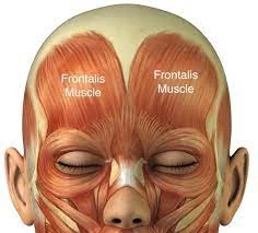
- The origin of the frontalis muscle is the galea aponeurotica, which is a fibrous layer that covers the top of the cranium & extends from the forehead to the back base of the skull.
Insertion
- It attaches as right & left bellies from the skin of the eyebrow, with fibers extending down to interdigitate with orbicularis oculi & procerus.
Structure
- The frontalis muscle is thin, of a quadrilateral form, & intimately adherent to the superficial fascia. It is broader than the occipitalis & its fibers are longer & paler in color. It is situated on the front of the head.
The muscle has no bony attachments. Its medial fibers are continuous with those of the procerus; its intermediate fibers blend with the corrugator & orbicularis oculi muscles, thus attached to the skin of the eyebrows; & its lateral fibers are also combined with the latter muscle over the zygomatic process of the frontal bone.
From these attachments, the fibers are run upward, & join the galea aponeurotica below the coronal suture.
The medial margins of the frontalis muscles are connected together for some distance above the root of the nose; but between the occipitalis, there is a considerable, though variable, interval, inhabited by the galea aponeurotica.
Function of Frontalis muscle
- In humans, the frontalis muscle only distributes facial expressions.
In the eyebrows, its primary action is to lift them (thus opposing the orbital part of the orbicularis), especially when looking up. It also works when a view is too distant or dim. The frontalis muscle is also distributed to wrinkle the forehead.
Embryology
- Like the other muscles involved in facial expression, the frontalis muscle arises from the second pharyngeal arch, which forms between the 3rd eighth weeks of development.
Blood Supply & Lymphatics
- The frontalis muscle collects its blood supply from branches of both the internal & external carotid arteries. The supratrochlear & supraorbital arteries supply the muscle from the inferior margin after they exit the orbit & travel up the forehead. The supraorbital artery can way out from the supraorbital notch/foramen, while the supratrochlear artery pulls out more medially from the orbit. Every branch of the ophthalmic artery, is a branch of the internal carotid artery. Soon after their respective exits, the supratrochlear& supraorbital arteries separate further into both superficial & deep branches. The superficial arteries supply the muscle, galea, & skin, while the deeper layer supplies the periosteum. The arteries form an anastomosis with 1 another to form a highly vascularized network.
Studies looking at the drainage have located that the more lateral part of the forehead most likely drains differently than the medial portion. Regardless, it seems the forehead drains mainly into the preauricular nodes & parotid nodes. - The main venous drainage happens between three veins: the supratrochlear vein being the most medial, then the intermediate supraorbital vein, & finally, the lateral frontal vein. The 3 further connect through a transverse vein that passes superior to the orbit appropriately named the transverse supraorbital vein. The transverse supraorbital vein attaches medially to the angular vein, which eventually drains into the ophthalmic vein & cavernous sinus. It is main to remember this relationship when considering the risk of external facial infections major to potentially harmful intracranial infections/cavernous sinus thrombosis.
Nerve supply
- The muscles of facial expression receive nerve supply from cranial nerve VII (the facial nerve), which separates into 5 main branches: temporal, zygomatic, buccal, marginal mandibular, & cervical. The facial nerve egress the skull at the stylomastoid foramen & its temporal branch crosses over the zygomatic arch, passes through the areolar tissue on the surface of the temporal fascia, & later adds into the frontalis muscle to provide its deep innervation.
- Three branches of the temporal nerve, the anterior, middle, & posterior, are responsible for innervating the orbicularis oculi, frontalis, & corrugator muscles. The temporal (also called the frontal) branch of the facial nerve passes within the superficial layer of the temporoparietal fascia, with the temporal artery just anterior to the nerve. The temporal branch comes to the frontalis muscle into its deeper part. Injury to the temporal branch of the facial nerve is circumvented by either dissecting subcutaneously or just anterior to the superficial part of the deep temporal fascia (the glistening layer in the anterior of the temporalis muscle).
- The supratrochlear & supraorbital nerves pass along their respective arteries, piercing through the frontalis in order to reach the superficial skin. These are branches of the ophthalmic division of the trigeminal nerve & do not directly innervate the muscle, but instead innervate the overlying skin. The medial branches of the supraorbital nerve are superficial & the lateral branches are deep.
Clinical importance
Tissue expansion:
- Tissue expansion is needed when repairing large forehead and nasal defects. Whereas in the neck, expanders are placed in front of the platysma, in the forehead, it is crucial to place expanders under the frontalis muscles, with attachments via incisions constructed within the hairline.
Upper & lower motor neuron facial palsy:
- The nerve supply to the forehead is also responsible when evaluating a patient with a potential stroke. Middle cerebral artery strokes can source contralateral facial paralysis, but these strokes also spare the forehead. This presentation is because the lower motor neurons behind innervating the top 1/2 of the face receive input from both hemispheres, while the lower half does not. If the injury is at the level of the lower motor neuron, as perceived with Bell palsy, then we see wall hemifacial paralysis.
Fillers for grooves at the glabella:
- Fillers are also often placed into the forehead to generate volume & fullness in the horizontal wrinkles at the root of the nose (caused by the procerus muscle) & the verticle “elevenses” at the medial end of each brow, sourced by the corrugator muscles. Knowing the underlying anatomy, and adding the depth & location of the various muscles are main to avoiding devastating complications during injections. For example, too much filler or misplaced filler when targeting the glabella (the area between the eyebrows and above the nose) can conduct in retinal artery occlusion & blindness. inappropriate injection inside an artery, for example, the supratrochlear, could lead to acute necrosis of the tissue. Fillers are never injected with force and in boluses. the further approach we have established to be safe is to pinch the skin upwards with small amounts of filler injected gently.
Utilize botulinum toxin for forehead & frown lines:
- The main anatomy illustrates when dealing with a minimally invasive formula like botulinum injections. Botulinum toxin is a functional tool for treating rhytides & wrinkles & can be injected directly into the frontalis muscle. The decreased muscular contraction conduct to a decrease in rhytid production & a more youthful appearance. This procedure involves various injections into the frontalis. The object is to avoid complete paralysis of the frontalis muscle: 10-20 units of botox distributed across the forehead surrender satisfactory relaxation of the horizontal rhytids.
- When injecting the frontalis, the corrugator & procerus muscles might also be injected. A secure part for frontalis muscle injections is 2 cm above the brow. If run improperly, the toxin can spread to the levator palpebrae superioris & lead to nonpermanent ptosis. Also, it is main to keep the balance between the depressors of the brow & the elevators. A weak frontalis muscle & ruling set of depressors would cause the brow to descend. Also, improper distribution of botulinum into the medial frontalis can pilot to a “Spock” deformity, in which the lateral part of the frontalis is capable of contraction, but the medial part is relaxed.
- The other area of frontalis action especially in women that often goes undertreated is where the frontalis is close to the anterior hairline, which results in important residual high horizontal rhytids.
Improvement of forehead skin:
- The forehead is an area that many people seek to rejuvenate to maintain a youthful appearance. Relaxed skin tension lines, also known as wrinkles, form perpendicular to the basic muscles and& as we age. A trial of nonsurgical choice is also the 1st step to try & alleviate lines, vascular changes, & skin pigment changes. Nonsurgical choices can add creams, peels, abrasives, & lasers.

Strengthening exercise of Frontalis Muscle
- To strengthen the muscle in the forehead slowly hold the skin down with all the fingers to ensure it does not crease, then raise the eyebrows against the resistance. Pause for 20 seconds.
Stretching exercise of Frontalis Muscle
- This stretch is great for relaxing the frontalis, which initiates at the eyebrows & goes to the hairline. This stretch assists to minimize the horizontal lines that can appear on the forehead by relaxing the muscle.
- Maintain the face in a neutral position & place finders on the eyebrows. There are 2 different stretches for these muscles.
1) While pressing softly with the fingers, move the fingers up towards the hairline.
2) From the hairline, repeat moving regards the eyebrows. Move through the stretch & pause for 5 seconds. Rest & repeat. Start out with 5 reps per day & work up to 10 reps per day, by increasing the number of reps each week.
This stretch can also be done by initiating from the middle of the forehead & moving the hands away from each other towards the temples for a horizontal stretch. This is a simple motion that can be done at any time.
FAQ
The frontalis muscles are two large fanlike muscles that extend from the eyebrow region to the top of the forehead.
The frontalis muscles are a pair of vertically oriented muscles in the forehead that lift the eyebrows. When one contracts the forehead muscle it causes elevation of the brows with the simultaneous development of horizontal wrinkles in the forehead.
Frontalis exercises
To relieve tension in your frontalis muscle, sit in a comfortable chair and frown while lowering your eyebrows as much as possible. Hold this position for five seconds then immediately raise your eyebrows as high as possible while smiling
The frontalis muscle originates at the anterior, or front, of the galea aponeurotica, which is near the top of the forehead. From the galea aponeurotica, the frontalis muscle extends down the forehead and attaches, or ends, at the eyebrows and upper nose.
The frontalis muscles are a pair of vertically oriented muscles in the forehead that lift the eyebrows. When one contracts the forehead muscle it causes elevation of the brows with the simultaneous development of horizontal wrinkles in the forehead.
The corrugator supercilii muscle is a small, narrow, pyramidal muscle close to the eye. It arises from the medial end of the superciliary arch and inserts into the deep surface of the skin of the eyebrow. It draws the eyebrow downward and medially, producing vertical wrinkles on the forehead.
A question we’re often asked is can you treat the frontalis muscle on its own? Without the others areas which are the glabella and the obicularis oculi. Generally, we advise against this because of the slightly heavy brow caused.
To tighten forehead skin, first frown as hard as you can, attempting to bring your eyebrows over your eyes and closer together. Then do the opposite and lift your eyebrows as high as possible, opening your eyes widely. Repeat these movements five times
the forehead
Noun. frontalis (plural frontalis) (anatomy) A muscle of the forehead, sometimes considered to be part of the occipitofrontalis muscle.