Superior rectus muscle Anatomy, Origin, Insertion, Function
Table of Contents
Introduction
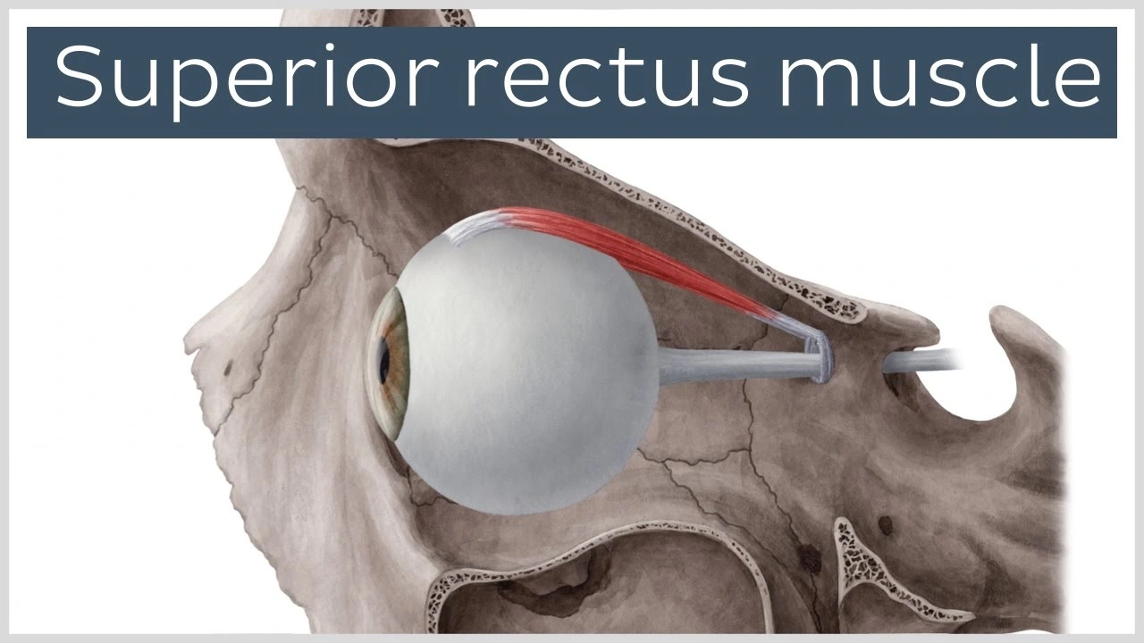
The superior rectus muscle is one of the six extraocular muscles that control eye movement. It is located in the orbit of the eye, and its primary function is to elevate the eye and rotate it medially, or inward. The muscle originates from the common tendinous ring (also known as the annulus of Zinn) and inserts into the superior surface of the eyeball.
- The superior rectus is one of the extraocular muscles. There are a total of four rectus muscles, 2 oblique muscles, & the levator palpebrae superioris. The superior rectus is one of the four rectus muscles, which further include the superior rectus, the medial rectus, & the lateral rectus. The oblique muscles are the superior & inferior obliques. The levator palpebrae superioris muscles are primarily for eyelid elevation.
Origin & insertion of Superior Rectus Muscle
- Superior rectus reach from the common tendinous ring (annulus of Zinn) to the superior side of the eyeball, just posterior to the corneal limbus (corneoscleral junction). The largest part of the superior rectus arises from the superior portion of the common tendinous ring, superolateral to the optic canal. While a smaller part arises from the dura mater (meninges) that surrounds the optic nerve (CN II).
- The muscle then courses anterolaterally, passing over the eyeball’s equator to reach & insert onto the anterior half of the sclera superiorly. The line of insertion is oblique, while medial fibers attach more anterior than lateral.
Structure and Function of Superior Rectus Muscle
- With the head & eyes facing straight ahead, the eyes are presumed to be in primary gaze. From this position, a function from an extraocular muscle produces a secondary or tertiary action. Although the globe can be moved about 50 degrees from its primary position, generally during normal eye movement only 15 degrees of extraocular muscle movement happen before head movement begins.
- The annulus of Zinn is the common origin point for the rectus muscles & spans the superior orbital fissure & orbital apex. It consists of superior & inferior tendons. The superior tendon is worked with the superior rectus muscle and also with the medial rectus & lateral rectus muscles while the inferior tendon works with the inferior rectus muscle & part of the medial rectus & lateral rectus muscles.
- The superior rectus has main action is elevating the eye, which leads the cornea to move superiorly. The superior rectus arising from the annulus of Zinn & courses anteriorly & superiorly over the globe, making an angle of 23 degrees with the visual axis. This 23-degree of angle makes the secondary & tertiary movement of the superior rectus muscle to be adduction & intorsion (incycloduction).
- All of these extraocular muscles have a working insertion point, which is at the nearest point where the muscle 1st contacts the eyeball. This point forms a tangential line from the globe to the muscle origin & is known as the arc of contact. The superior rectus has an arc of exposure of 6.5 mm, as does the inferior rectus.
Relation
- The arising fibers of the superior rectus are located inferolateral to the superior oblique muscle & anteromedially to the levator palpebrae superioris muscle. Since levator palpebrae superioris eventually takes a more medial course, it wrongs the majority of the superior rectus’ superior surface.
- The far distal part of the superior rectus travels superficially to the tendon of the superior oblique muscle. The superior branch of the oculomotor nerve (CN III) courses over the proximal 3rd of the muscle’s inferior surface before it pierces the muscle belly to provide innervation. The lateral & medial sides of the Superior rectus muscle are not related to any specific part but rather are cushioned within the periorbital fat tissue.
- At the site of its insertion, the superior rectus pierced the fascial sheath of the eyeball (Tenon’s capsule), which in turn reflects back on the muscle’s tendon & creates a thin fascial sleeve. This sleeve has an expansion known as the check ligament that joins the superior rectus with the levator palpebrae superioris muscle & ensures that the 2 muscles act simultaneously.
Embryology
- The mesenchyme of the head, including the orbit & its structures, arises in mesoderm & neural crest cells primarily. The extraocular muscles arise from mesoderm, but the satellite & connective tissue of the muscle derive from neural crest cells. Most of the other connective tissue of the orbit is derived from neural crest cells.
Blood Supply & Lymphatics
It receives its blood supply primarily from the superior muscular branches of the ophthalmic artery, with some contribution from two anterior ciliary arteries. The ophthalmic artery is a branch of the internal carotid artery and is the primary blood supply for all of the extraocular muscles, along with the lacrimal artery and infraorbital artery. The superior and inferior muscular branches are the two muscular branches of the ophthalmic artery responsible for supplying the superior rectus muscle.
The venous drainage of the superior rectus muscle is similar to its arterial supply and empties into the superior and inferior orbital veins. There are usually four vortex veins located at the lateral and medial sides of the superior and inferior rectus muscles that drain into the orbital venous system.
It is important to note that the lymphatic drainage of the extraocular muscles, including the superior rectus muscle, is not well-understood. Some studies suggest that lymphatic vessels are present in the connective tissue surrounding the muscles, but the exact drainage pathways are unknown. Further research is necessary to fully understand the lymphatic drainage of the superior rectus muscle.
Nerve supply of Superior Rectus Muscle
In terms of nerve supply, the superior rectus muscle is innervated by the superior division of cranial nerve III (oculomotor nerve). The oculomotor nerve is split into upper and lower divisions, with the upper division supplying the superior rectus and levator palpebrae superioris muscles, and the lower division supplying the medial rectus, inferior rectus, and inferior oblique muscles. The lateral rectus muscle is supplied by cranial nerve VI (abducens nerve), and the superior oblique muscle is supplied by cranial nerve IV (trochlear nerve).
Muscles fiber
- The superior rectus & the inferior rectus are vertical. Each of the rectus muscles arising posteriorly at the annulus of Zinn & courses anteriorly.
- Each of the rectus muscles attaches to the globe at varying distances from the limbus,& the curved line drawn along the insertion points makes a spiral that is known as the spiral of Tillaux. start at the medial side of the globe, the medial rectus connects at 5.5 mm from the limbus, the inferior rectus attaches at 6.5 mm from the limbus, the lateral rectus attaches at 6.9 mm from the limbus, & the superior rectus at 7.7 mm from the limbus. The medial & lateral recti attach along the horizontal meridian, while the inferior & superior recti attach along the vertical meridian.
- The superior rectus is 10.6 mm wide at its attaches to the globe. The tendon is 5.8 mm, even from the arising point. The whole length of the muscle is 41.8 mm.
- Extraocular muscles have a big ratio of nerve fibers to skeletal muscle fibers. The ratio is 1:3-1:5; other skeletal muscles are 1:50-1:125. Extraocular muscles are a particular form of skeletal muscle with a variety of fiber types, including both slow tonic types, which resist fatigue, & saccadic (rapid) type muscle fibers.
Physiologic Variants
Variation
- Variations of the superior rectus muscle are rare. It may rarely have 2 muscle bellies parallel to each other. It may be congenitally absent very rarely.
Clinical importance
- The function of the muscle can be evaluated with the other extraocular muscles during the clinical examination. The movement of the extraocular muscles can be examined by having the patient look in 9 directions beginning with a primary gaze, followed by the 2nd movement (up, down, left, & right) & the last movement (up and right, up & left, down & right, down & left). The clinician can test these positions by having the patient follow the clinician’s finger & trace a wide letter “H” in the air. The superior rectus muscle is isolated by urging the patient to elevate the eye in full abduction.
- Ocular alignment can be tested further using some methods, including cover tests, corneal light reflex, dissimilar image tests, & dissimilar target tests. Since many patients with extraocular muscle abnormalities are young children, the clinician may require to employ various clever means such as using toys and other objects to elicit the cooperation of the child.
- Strabismus, or ocular misalignment, can be sourced by abnormalities in binocular vision or abnormalities of neuromuscular control. Paralysis, Muscle Weakness, or any damage that involves the superior rectus muscle can be included in strabismus.
FAQ
Extraocular muscle
The superior rectus is an extraocular muscle that is inserted at the top of the eye. It moves the eye upward. The inferior rectus muscle is an extraocular muscle that is inserted into the bottom of the eye. It moves the eye downward.
extraocular muscles
The superior rectus muscle is one of the extrinsic muscles of the eye. Being situated outside the eyeball but within the orbit, it belongs to a group known as the extraocular muscles. This category of muscles permits the moves of the eyes within the orbit.
The superior and inferior rectus muscles are two of the six extraocular muscles that control the movement of the eyes.
The superior rectus muscle is responsible for elevating the eye, which means it causes the cornea to move upward. This muscle originates from the common origin point of the rectus muscles called the annulus of Zinn and courses anteriorly and superiorly over the globe, making an angle of 23 degrees with the visual axis. The other movement of the superior rectus muscle is adduction and intorsion (incycloduction).
On the other hand, the inferior rectus muscle is responsible for depressing the eye, which means it causes the cornea to move downward. This muscle also originates from the annulus of Zinn and courses anteriorly and inferiorly over the globe. The other function of the muscle is abduction and extorsion (excycloduction).
The coordinated action of the extraocular muscles is essential for the eyes to move smoothly and accurately, allowing us to see objects in different directions and distances.
The primary function of the superior rectus muscle is to elevate the eye, causing the cornea to move upward. When the superior rectus muscle contracts, it pulls the eye upward and inward, producing a movement known as adduction. Additionally, due to the oblique angle of the muscle relative to the visual axis, the secondary and tertiary actions of the superior rectus muscle are adduction and intorsion (incycloduction), respectively.
The superior rectus muscle works in coordination with the other extraocular muscles to allow the eyes to move smoothly and accurately in different directions and distances, enabling us to perceive and interact with the environment around us.
Damage to the superior rectus muscle can cause various eye movement disorders and visual disturbances, depending on the extent and location of the injury. Some possible consequences of superior rectus damage may include:
Impaired upward eye movement: The primary function of the superior rectus muscle is to elevate the eye. Therefore, if this muscle is damaged, the eye may have difficulty moving upward, resulting in a condition called “superior rectus palsy.” This can cause double vision or strabismus (eye misalignment) when looking upward.
Reduced adduction and intorsion: As the superior rectus muscle also contributes to adduction (inward eye movement) and intorsion (rotation of the eye inward), damage to this muscle may also affect these functions. This can lead to problems with depth perception, balance, and coordination.
Diplopia (double vision): If the superior rectus muscle is damaged, it may not be able to coordinate properly with the other eye muscles, leading to double vision or blurred vision, especially when looking upward.
Head tilting or compensation: In some cases, people with superior rectus palsy or other forms of eye muscle damage may tilt their head to compensate for their visual impairment, which can cause neck pain, discomfort, and postural imbalances.
Treatment for superior rectus damage depends on the underlying cause and severity of the condition. Options may include vision therapy, prism glasses, botulinum toxin injections, surgery, or other interventions to restore proper eye alignment, movement, and function.
The superior rectus & inferior oblique muscles primarily motion the eye upward. The inferior rectus & superior oblique muscles first move the eye downward. The lateral rectus muscle moves the eye through horizontal abduction). The medial rectus muscle motions the eye horizontally medially (adduction).
Conclusion Isolated superior rectus palsy may be sourced by a contralateral midbrain lesion that selectively involves crossing superior rectus nerve fibers.
The superior rectus muscle attaches from the annulus of Zinn. It attaches to the anterosuperior surface of the eye. This attachment has a width of around 11 mm. It is all over 8 mm from the corneal limbus.
The superior rectus is innervated by branches of the ophthalmic artery & the lacrimal artery (1 to 5 pedicles). The medial rectus is innervated by branches of the ophthalmic artery & the inferior muscular artery (5-9 branches).
Oculomotor Nerve (Cranial Nerve III)
Each nucleus consists of subnuclei, which project fibers to the contralateral superior rectus & the ipsilateral inferior oblique, inferior rectus, & medial rectus muscles.

One Comment