Muscles Of Eye
Table of Contents
What is a Muscles Of Eye?
The human eye is a complex organ responsible for vision, consisting of various muscles that facilitate its movement and function. These muscles play a crucial role in controlling the movement of the eye, allowing us to focus on objects, perceive depth and distance, and track moving targets.
The six major muscles that control the movement of the eye are known as the extrinsic eye muscles. These muscles work in coordination to ensure precise and synchronized movements of both eyes, enabling binocular vision and depth perception.
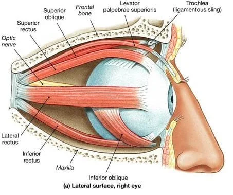
The extrinsic eye muscles (extraocular muscles) include the following:
- Superior rectus muscle
- Inferior rectus muscle
- Lateral rectus muscle
- Medial rectus muscle
- Superior oblique muscle
- Inferior oblique muscle
Each of these muscles has a specific role in controlling the movement of the eye in various directions, such as upward, downward, lateral, and rotational movements. Understanding the function of these muscles is essential in comprehending the complexities of human vision and eye movements.
Introduction
Six extraocular muscles control eye movement. Superior rectus, inferior rectus, lateral rectus, medial rectus, superior oblique, and inferior oblique are the muscles involved. The eye muscles are designed to stabilize and move both eyes. Particular nerve areas in the brain and brainstem connect with each muscle pair (right and left) to coordinate accurate eye movements with minimal cognitive input.
The resting muscle tone of all eye muscles is designed to stabilize eye posture. Specific muscles become more active during motions, while others become less active. Eye movement has its distinct terminology. The movements of the eye include adduction (moving in toward the nose), abduction (moving out toward the ear), elevation (moving up), depression (moving down), intorsion (or incyclorotation; the top of the eye move rotates in towards the nose), and extorsion (moving in towards the ear).
Structure And Function
The ciliary muscle, sphincter pupillae, and dilator pupillae are all intraocular muscles. The ciliary muscle is a ring of smooth muscle that regulates accommodation by changing the curvature of the lens and directing the flow of aqueous humor into Schlemm’s canal. The ciliary muscle is linked to the zonular fibers, which hold the lens in place.
When the ciliary muscle contracts, the strain on the lens decreases, causing it to adopt a more spherical shape to concentrate on close objects. The converse consequence of ciliary muscle relaxation is improved distant focus. Smooth muscle also makes up the sphincter pupillae and dilator pupillae. The pupil’s diameter is constricted by the sphincter pupillae, which encircles the pupil.
Ocular movements include three basic axes: vertical, transverse, and anteroposterior. Adduction (medial movement) or abduction (lateral movement) of the eye arises from rotation around the vertical axis. Elevation (superior motion) or depression (inferior motion) is caused by rotation around the transverse axis. The anteroposterior axis allows the superior pole of the eye to move medially (intorsion) or laterally (extorsion). The anteroposterior axis rotations allow the eye to adjust to the tilting of the head.
The medial rectus muscle is in charge of medial rotation around the vertical axis, while the lateral rectus muscle is in charge of lateral rotation. The superior rectus muscle lifts the eye and helps with adduction and intorsion. The inferior rectus depresses and rotates the eye laterally, contributing to adduction and extorsion. The superior oblique abducts, depresses, and rotates the eye medially, whereas the inferior oblique abducts, elevates, and rotates the eye laterally.
The four rectus muscles are roughly 40mm long and come from the Annulus of Zinn. They are placed a few millimeters from the limbus on the sclera. The inferior oblique arises from the orbital floor and enters the inferotemporal globe through the sclera. The superior oblique begins in the posterior orbit and goes medially before terminating.
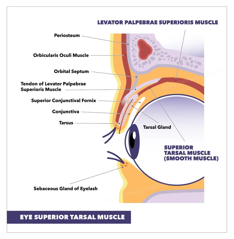
The main skeletal muscle responsible for retracting the upper eyelid is the levator palpebrae superioris. The superior tarsal muscle (Müller’s muscle) is made up of smooth muscle and helps to raise the upper eyelid. The capsulopalpebral fascia and the inferior tarsal muscle are the retractors in the lower eyelid. The principal protractors (closing) of the eyelids are the orbicularis oculi muscles. It is a flat, ringlike band of skeletal muscle that surrounds the front orbit and is divided into three sections: the orbital portion, the palpebral portion, and the lacrimal portion.
Muscles
The extraocular muscles are extrinsic and independent from the eyeball, however, they are located within the orbit. They are responsible for controlling the movements of the eyeball and the superior eyelid.
The levator palpebrae superioris, superior rectus, inferior rectus, medial rectus, lateral rectus, inferior oblique, and superior oblique are the seven extraocular muscles. They are functionally separated into two groups:
- Recti and oblique muscles are in charge of eye movement.
- The movement of the superior eyelid is controlled by the levator palpebrae superioris.
Extraocular eye muscles and their functions
There are four straight (rectus) muscles and two oblique muscles among the extraocular muscles that act together to move the eye from side to side, up and down, and rotate it.
Furthermore, the levator palpebrae superioris (LPS) muscle elevates and maintains the upper eyelid in place.
Three cranial nerves regulate all seven of these eye movement muscles:
- The oculomotor nerve.
- The Trochlear nerve
- The abducens nerve
Recti muscles
The eye has four recti muscles, which all attach to the front portion of the eye (anterior to the equator). These are the muscles:
- Rectus muscle superior
- Rectus medialis muscle
- Rectus lateralis muscle
- Lower rectus muscle
Each of the recti muscles in the eye is derived from the common tendinous ring (also known as the annular tendon or annulus of Zinn), which is a fibrous ring of connective tissue that surrounds the optic nerve and links to the orbit.
The term “rectus” comes from the Latin word for “straight,” indicating that the recti muscles attach directly from the orbit to the sclera of the eye.
Superior rectus muscle
The superior rectus muscle is located near the top of the eye and governs the eye’s upward movement. The oculomotor nerve controls movement of the superior rectus muscle.
Medial rectus muscle
The medial rectus eye muscle pushes the eye inward by attaching to the side closest to the nose. The oculomotor nerve controls the movement of the medial rectus muscle.
Lateral rectus muscle
The lateral rectus eye muscle is located on the side of the eye toward the temple. This muscle is responsible for the eye’s ability to move outward. The abducens nerve allows movement of the lateral rectus muscle.
Inferior rectus muscle
The inferior rectus eye muscle is placed near the bottom of the eye and allows for downward movement of the eye. The oculomotor nerve controls the movement of this muscle.
Oblique muscles
There are two oblique muscles of the eye. These are the muscles:
- Superior oblique muscle
- Inferior oblique muscle
The oblique muscles, unlike the recti muscles, do not join directly to the eye via the common tendinous ring. Oblique muscles, on the other hand, attach angularly to the eye and have different origins.
Superior oblique muscle
The superior oblique eye muscle arises from the sphenoid bone, one of seven bones that comprise the eye socket. The fourth cranial nerve (trochlear nerve) controls it.
The trochlea is a small pulley structure in the eye that connects the superior oblique muscle from the sphenoid bone to the top of the eye, near the nose.
When the eye is in the primary position (typically straight ahead), the superior oblique muscle’s principal function is intorsion. (In other words, it rotates the 12 o’clock position of the cornea’s vertical meridian inward toward the nose.) It also shifts the eye’s line of sight lower and outward.
Inferior oblique muscle
The inferior oblique eye muscle starts near the nose on the front of the orbital floor.
Its primary function is to turn the eye when looking straight ahead (rotate the vertical meridian of the cornea at 12 o’clock toward the ear). It also raises and abducts the eye (shifts the focus upward and outward).
The third cranial nerve (oculomotor nerve) controls the action of the inferior oblique muscle.
Levator Palpebrae Superioris
The only muscle involved in elevating the superior eyelid is the levator palpebrae superioris (LPS). The superior tarsal muscle is a collection of smooth muscle fibers found in a small section of this muscle. The sympathetic nervous system innervates the superior tarsal muscle, as opposed to the LPS.
Attachments: Protrudes from the sphenoid bone’s lesser wing, just above the optic foramen. It connects to the superior tarsal plate of the upper eyelid (a broad connective tissue plate).
Actions: Helps to raise the upper eyelid.
Innervation: The oculomotor nerve (CN III) innervates the levator palpebrae superioris. The sympathetic nervous system innervates the superior tarsal muscle (placed within the LPS).
Clinical Relevance
Cranial Nerve Palsy
Three cranial nerves innervate the extraocular muscles. Damage to one of the cranial nerves will result in muscular paralysis. This will change the affected eye’s resting gaze. As a result, each cranial nerve lesion has its distinct appearance:
Oculomotor nerve (CN III) – A CN III lesion affects the majority of the extraocular muscles. The lateral rectus and superior oblique move the afflicted eye laterally and inferiorly. ‘ Down and out ‘ is the position the eye takes.
The superior oblique muscle will be paralyzed if the Trochlear nerve (CN IV) is damaged. The resting position of the ocular has no discernible effect. The patient, on the other hand, will experience diplopia (double vision) and may develop a head tilt away from the site of the lesion.
Abducens nerve (CN VI) – A CN VI injury paralyzes the lateral rectus muscle. The resting tone of the medial rectus will adduct the afflicted eye.
Horner’s Syndrome
Horner’s syndrome is a set of three symptoms caused by injury to the sympathetic trunk in the neck:
- Denervation of the superior tarsal muscle causes partial ptosis (drooping of the top eyelid).
- Miosis (pupillary constriction) is caused by the dilator pupillae muscle becoming denervated.
- Anhidrosis (lack of sweating) on the ipsilateral side of the face – caused by sweat gland denervation.
- Horner’s syndrome can indicate significant pathologies, such as a lung apex tumor (Pancoast tumor), an aortic aneurysm, or thyroid cancer.
Eye Muscles Exercises
- Blinking exercises act as a rest for your eyes, allowing them to stay fresh and focused for longer periods. Blinking alleviates symptoms of digital eye strain, dry eye, and poor blinking habits, and a ten-second blinking exercise every 20 minutes will be beneficial.
- Slowly move your eyes up and down and repeat three times, then move your eyes slowly from right to left and repeat three times, then relax.
- Another eye movement exercise is to visualize a gigantic figure of 8 8-9 feet away from you, then move your eye in the direction of this infinity loop for around 30 seconds, then swap directions.
- Change your attention by holding your finger a few inches away from you and focusing your gaze on it, then focusing on anything far away and returning your glance to your finger.
- Pushups using a pencil. Holding a pencil/patient’s thumb on the outstretched arm midway between the eyes, instruct the patient to try to keep a single image of the pencil while slowly moving the pencil toward the nose until it is no longer possible to see the pencil in a single image, then instruct the patient to move it slowly away to the closest point where a single image of the pencil is achieved. This exercise is beneficial to those who have symptomatic convergence insufficiency.
Effect Of Eye Muscle Exercises
Who benefits from exercise:
- Digital eye strain occurs when people work on a computer for an extended period, and it can cause dry eyes, eye strain, impaired vision, and headaches.
- Light sensitivity has been improved.
- An operation that necessitates muscular strengthening
- If you have difficulty focusing your eyes to read.
- Inadequate convergence.
- Some youngsters acquire lazy eyes; eye workouts boost the brain’s vision centers.
The effect of oculomotor and gaze stabilization training:
- Ocular-motor exercises improve dynamic visual acuity (DVA) in dynamic sports that have a substantial effect on athletes’ performance, such as basketball; in one study on female basketball players, the intervention lasted four weeks, with six ten-minute sessions each week.
- Gaze and ocular-motor training increase balance and stability in healthy adults, and a study shows that it may be useful in improving balance after stroke.
- These exercises alleviate symptoms of eye fatigue while also strengthening the extraocular muscles.
- Reduce eye strain and enhance blinking.
Summary
The eye muscles are responsible for controlling the movement of the eyeballs and the upper eyelids. There are seven eye muscles, divided into two groups:
- Extraocular muscles: These six muscles control the movement of the eyeballs in all directions. They are called extraocular muscles because they are located outside of the eyeball itself. The extraocular muscles are:
- Superior rectus: Moves the eye upward
- Inferior rectus: Moves the eye downward
- Medial rectus: Moves the eye inward
- Lateral rectus: Moves the eye outward towards the temple
- Superior oblique: Moves the eye upward and outward and rotates it inward
- Inferior oblique: Moves the eye upward and inward and rotates it outward
- Levator palpebrae superioris: This muscle raises the upper eyelid.
The eye muscles are innervated by three cranial nerves: the oculomotor nerve, the trochlear nerve, and the abducens nerve.
Eye muscle problems can cause a variety of symptoms, including double vision, blurred vision, and eye movement difficulties. Some common eye muscle problems include:
- Strabismus (crossed eyes): A condition in which the eyes are not aligned properly.
- Nystagmus: A condition in which the eyes make rapid, involuntary movements.
- Ptosis (droopy eyelid): A condition in which the upper eyelid droops down.
FAQs
What are the symptoms of eye muscle problems?
Symptoms of eye muscle problems can vary depending on the specific condition, but some common symptoms include:
Double vision (seeing two images)
Blurred vision
Eye movement difficulties
Eye strain
Headaches
What are some common eye muscle problems?
Some common eye muscle problems include:
Strabismus (crossed eyes): A condition in which the eyes are not aligned properly.
Nystagmus: A condition in which the eyes make rapid, involuntary movements.
Ptosis (droopy eyelid): A condition in which the upper eyelid droops down.
What causes eye muscle problems?
There are many potential causes of eye muscle problems, including:
Genetics
Birth defects
Trauma
Neurological disorders
Muscle diseases
How are eye muscle problems diagnosed?
Eye muscle problems are typically diagnosed by an eye doctor through a comprehensive eye exam. This exam may include:
Vision testing
Eye movement testing
Examination of the eye muscles
How are eye muscle problems treated?
Treatment for eye muscle problems depends on the specific condition and its severity. Some common treatment options include:
Eyeglasses or contact lenses
Vision therapy
Eye muscle surgery
References
- Ludwig, P. E. (2023, August 7). Anatomy, Head and Neck: Eye Muscles. StatPearls – NCBI Bookshelf. https://www.ncbi.nlm.nih.gov/books/NBK470534/
- The Extraocular Muscles – The Eyelid – Eye Movement – TeachMeAnatomy. (2022, December 22). TeachMeAnatomy. https://teachmeanatomy.info/head/organs/eye/extraocular-muscles/
- Barden, A. (2021, March 2). Eye muscles and their functions. All About Vision. https://www.allaboutvision.com/eye-care/eye-anatomy/eye-muscles/
- Eye Muscles. (n.d.). Vivid Vision. https://www.seevividly.com/info/Physiology_of_Vision/The_Brain/Visual_System/Eye_Muscles
- Eye Muscle Exercise. (n.d.). Physiopedia. https://www.physio-pedia.com/Eye_Muscle_Exercise