
Levator palpebrae superioris muscle Origin, Insertion, Function, Exercise
Table of Contents
Introduction
- The levator palpebrae superioris is a triangular-shaped muscle situated in each upper eyelid. The primary action is elevation & retraction of the upper eyelid. It has an abundant blood supply, which is mainly provided by the ophthalmic & supraorbital arteries (from the internal carotid), anastomosing with branches of the external carotid through angular & superficial temporal arteries via the superior peripheral arcade.
- Lymphatic drainage runs laterally to the preauricular or parotid nodes prior to draining to the deep cervical lymph nodes. It sustains innervation by the superior branch of the oculomotor nerve (cranial nerve III). Problems with the levator palpebrae superioris function or innervation will outcome in upper eyelid ptosis.
Origin of Levator palpebrae superioris muscle
- Levator palpebrae superioris is a triangular muscle that enlarges along the roof of the orbit, from the apex of the orbit to the superior eyelid. It arising s with a short & narrow tendon from the inferior portion of the lesser wing of the sphenoid bone, superior & anterior to the common tendinous ring. The muscle belly gradually widens as it travels anteriorly toward the eyelid.
Insertion
- The muscle fibers infiltrate the upper eyelid, and attach its portion via two aponeurotic fascicles;
- Deep fibers connect to the anterior side of the superior tarsus.
- Superficial fibers radiate via the eyelid & orbicularis oculi to finally attaches to the skin of the superior eyelid.
- The most lateral fibers of the muscle’s aponeurosis attach to the orbital tubercle of the zygomatic bone, whereas the most medial fibers attach to the medial palpebral ligament. Some authors acknowledge another part of the levator palpebrae superioris called the superior tarsal muscle. It is a small slip of the smooth muscle that joins to the underside of the levator palpebrae superioris near its insertion point.
Structure
- The levator palpebrae superioris arise from the inferior surface of the lesser wing of the sphenoid bone, just above the optic foramen. It broadens & decreases in thickness (becomes thinner) & becomes the levator aponeurosis. This part inserts on the skin of the upper eyelid, as well as the superior tarsal plate. It is a skeletal muscle. The superior tarsal muscle, a smooth muscle, is attached to the levator palpebrae superioris & inserts on the superior tarsal plate as well.
Relation
- Levator palpebrae superioris located superior to the superior rectus muscle, which distinguishes it from the eyeball. Both muscles are enclosed by connective tissue sheaths which are combined along their related surfaces. The sheaths are separated only in their anterior portion at the level at which the tendons of these muscles separate. The interval between them is taped by a thick connective tissue plate onto which the superior conjunctival fornix attaches. In inclusion, the levator palpebrae superioris is attached to the check ligament of the superior rectus. All of these connections give to the coordinated simultaneous contraction of these 2 muscles.
- As the levator palpebrae superioris fans out as a wide aponeurosis, the lateral fibers pass between the orbital & palpebral parts of the lacrimal gland to attach to the orbital tubercle. The medial fibers travel adjacent to the reflected tendon of the superior oblique muscle.
Blood supply and lymphatics
- The internal carotid provides most of the levator palpebrae superioris blood supply through the branches of the ophthalmic artery. 4 arterial systems direct blood to the levator palpebrae muscle: the lacrimal, supratrochlear, & supraorbital arteries & muscular branches of the ophthalmic artery. These branches eventually join the superior peripheral arcade, providing blood supply to the superior side of the upper eyelid. The superior peripheral arcade joins medially & laterally with the superior marginal arcade, which provides the blood supply to the margin of the upper eyelid. These 2 arcades form a vast anastomosis with blood from branches of the internal & external carotids. The internal carotid supplies the majority of the blood to the superior peripheral arcade through the branches previously stated. The external carotid provides additional blood to the superior peripheral arcade medially via the angular artery (a branch of the facial artery) & laterally via the superficial temporal artery. This complex anastomosis keeps a rich blood supply to the lateral palpebrae superioris.
- In terms of venous drainage, the muscles of the orbit drain via the superior & inferior ophthalmic veins. The superior ophthalmic vein will eventually drain into the cavernous sinus, & the inferior ophthalmic vein will drain into the pterygoid venous plexus.
- In regards to lymphatic drainage, it was previously thought that the medial upper eyelid drained medially along the angular artery & the lateral upper eyelid lymph drained laterally to the preauricular & parotid nodes. There is a recent change regarding eyelid lymphatic drainage based on lymphoscintigraphy in human subjects. The upper eyelid lymphatics, which would add the levator palpebrae superioris muscles, are believed to entirely drain laterally to the preauricular or parotid lymph nodes, & then to the deep cervical lymph nodes (level II).
Nerve supply
- The levator palpebrae superioris acquire motor innervation from the superior division of the oculomotor nerve. The smooth muscle that arises from its undersurface also known as the superior tarsal muscles is innervated by postganglionic sympathetic axons from the superior cervical ganglion.
Function of Levator palpebrae superioris muscle
- The levator palpebrae superioris muscle elevates the upper eyelids.
- The main function of the levator palpebrae muscle is the retraction and elevation of the superior eyelid & the widening of the palpebral fissure. These functions are limited by many anatomical features of the muscle; Lateral & medial attachments of the muscle’s aponeurosis prevent further elevation of the eyelid once they are stretched maximally. The orbital septum is a fibrous portion of the eyelid that when compressed maximally, prevents further retraction of the eyelid.
- The antagonistic function of the orbicularis oculi counteracts the levator palpebrae superioris since it pulls the eyelid in the opposite direction.
- Levator palpebrae superioris helps the superior rectus & elevates the eyelid when the gaze is directed superiorly. This happens automatically because the fascial sheaths of the levator palpebrae superioris & superior rectus are fused via examination ligament; this is why when the superior rectus contracts, levator palpebrae superioris follow. Levator palpebrae superioris has a fixed tone that is in equilibrium with the opposing tone of orbicularis oculi, thereby preserving the eyes open & limiting the size of the palpebral fissure.
- Being supplied by the sympathetic nervous system, the superior tarsal muscle elevates the eyelid in conditions of a “flight or fight” response. Thus, this muscle causes further widening of the palpebral fissure during moments of excitement, fear, surprise, & others.
Embryology
- The levator palpebrae muscle derives from the mesenchyme of the 2nd pharyngeal arch. It begins formation during the 6th week of gestation & develops from lateral and medial mesodermal extensions of the frontal nasal process. in starting, the levator palpebrae muscles begin development as a portion of the superior mesodermal complex with the superior rectus muscle & the superior oblique muscles. During the 8th week of gestation, the superior mesodermal complex & lower mesodermal complex fuse which is followed by the differentiation of the upper & lower lid structures, including the levator palpebrae muscles.
Clinical importance
Damage to these muscles & their innervation can cause ptosis, which is the drooping of the eyelid. Lesions in CN III can cause ptosis because, without stimulation from the oculomotor nerve, the levator palpebrae cannot oppose the forces of gravity & the eyelid droops.
Ptosis can also result from injury to the adjacent superior tarsal muscle or its sympathetic innervation. Such damage to the sympathetic supply arises in Horner’s syndrome and presents as partial ptosis. It is important to differentiate between these 2 very additional causes of ptosis. This can generally be done clinically without a situation, as each type of ptosis is accompanied by additional specific clinical findings.
The ptosis seen in paralysis of the levator palpebrae superioris is frequently additionally noticeable than that seen due to paralysis of the superior tarsal muscle
Myogenic or neurogenic problems with the levator palpebrae superioris may occur & result in ptosis (drooping of the eyelid).
- Myogenic ptosis may happen due to a failure to convert between the levator palpebrae superioris from the superior rectus muscle. During the surgical correction, a thickened fibrous tissue is also identified instead of 2 differentiated muscles. Also, various myopathies, eyelid trauma (scarring), & overuse/stretching of the eyelid (long-term contact lens use) may cause myogenic ptosis.
- Neurogenic ptosis due to inadequate levator palpebrae superioris innervation may happen from a cranial 3 (oculomotor) nerve palsy, trauma, Guillain–Barré syndrome, & chronic inflammatory demyelinating polyneuropathy.
- Congenital ptosis may happen with dystrophy of the muscular component of the levator muscle, which shows fatty infiltration with poor levator action.
Exercise of Levator palpebrae superioris muscle
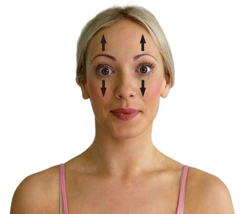
- To strengthen levator palpebrae superioris & to relieve annoying eyelid twitching, you might enact targeted eyelid exercises daily.
- First, close your eyelids as tightly as you can & keep that position for ten whole moments. Then open the eyes as broad as possible & maintain them at that extreme for 10 seconds. Continue to alternate between squeezing the eyes closed & opening them up wide. Conclude a total of 10 sets every day.
Wide eyes
- This exercise is extremely easy to do.
- Open the eyes as broadly as possible for about five seconds.
- Relax the eyes for five seconds.
- resume for 30 seconds.
Brow lifter
- Brow lifter exercise targets the frontalis muscles of the forehead, which attach to the levator muscles of the eyelid.
- Place the hands on the forehead & press against it with light pressure.
- Using the forehead muscles, try to lift the forehead up for 5 seconds.
- Repeat 5 to 10 times.
Rapid lids
- A person can execute this exercise by following the steps below.
- Use 2 fingers to press gently against each temple.
- Open & close your eyes rapidly.
- Resume for about 20 seconds.
Diagonal stretch
- This exercise involves a few simple steps.
- Lightly place 1 finger at the end of the individual eyebrow facing forward.
- Utilizing the facial muscles, look down as far as possible.
- Allow the eyeballs to gaze from one flank to the other like a pendulum.
- resume for about 20 seconds.
Lion’s breath
- This exercise is a type of pranayama breathing that people use in yoga to improve energy and eradicate toxins. It may also help stretch out the muscles closer to the eye & make them more supple.
- Open your eyes as wide as possible while gazing upward.
- At the same time, attach the tongue out & down as distant as it will go.
- Hold for 5 to 10 seconds while exhaling with a “ha” breath.
- Some people may find these exercises worthwhile, while others may notice no difference. do again the exercises several times
- each day exists likely to make the most benefit in those who do see a change.
FAQ
action The main actions of the levator palpebrae muscle are the retraction & elevation of the superior eyelid & widening of the palpebral fissure.
The levator palpebrae superioris is a triangular-shaped muscle situated in each upper eyelid. The primary action is elevation & retraction of the upper eyelid.
LPS is a thin triangular muscle that originates from the inferior surface of the lesser wing of the sphenoid, above & anterior to the optic canal & separated from it by the attachment of the superior rectus.
the oculomotor nerve
The striated levator palpebrae superioris (LPS) muscle is supplied by the oculomotor nerve, & has a common origin with the superior rectus muscle.
Ptosis occurs when the levator palpebrae superioris muscle does not contract correctly. It can also occur when the superior tarsal muscle doesn’t contract correctly. certain kinds of conditions can cause this.
The major action of the levator ani muscle is supporting & raising the pelvic visceral structures. It also helps in proper sexual functioning, defecation, urination, & allowing various structures to pass through it
The two levator ani muscles arise from each side of the pelvic wall, course medially and inferiorly, & join together in the midline.
The levator palpebrae superioris arises on the lesser wing of the sphenoid bone, just above the optic foramen. It broadens & becomes the levator aponeurosis. This part attaches to the skin of the upper eyelid, as well as the superior tarsal plate. It is a skeletal muscle.
2 Comments