
Orbicularis oculi muscle Origin, Insertion, Function, Exercise
Table of Contents
Introduction
- The orbicularis oculi muscle is the face muscle that closes the eyelids.
- It arises from the nasal portion of the frontal bone, from the frontal process of the maxilla in front of the lacrimal groove, & from the anterior surface & short fibrous band, the medial palpebral ligament.
- From this origin, the fibers are direct laterally, forming a broad & thin layer, which occupies the eyelids or palpebrae, surrounds the circumference of the orbit, & spreads over the temple,& downward on the cheek.
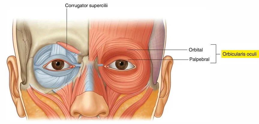
Origin & insertion of Orbicularis oculi muscle
- The Orbicularis oculi muscle is a flat, broad muscle that makes an ellipse around the circumference of the orbit. It is collected of orbital, palpebral & deep palpebral parts, each of which has its own specific set of attachments:
- Orbital part: overlays the orbital rim & originates from the nasal part of the frontal bone, frontal process of maxilla & medial palpebral ligament. The fibers encircle the orbit totally, extending into the soft tissues of adjacent regions. The orbital portion has no bony insertions, but instead, it attaches to various soft tissue structures of the periorbital region. The upper fibers merge with the occipitofrontalis, corrugator supercilii & depressor supercilii muscles, finally inserting into the skin & subcutaneous tissue of the eyebrow. The inferior & medial fibers of the orbital part blend with the levator labii superioris, levator nasolabial & zygomaticus minor muscles. Most peripheral fibers can stretch even further, loosely inserting into the temporal portion of the epicranial aponeurosis.
- Palpebral part: arising from the superficial surface of the medial palpebral ligament. The muscle fibers make the eyelids as they run towards the lateral commissure of the eye. Here, the superior & inferior fibers unite & insert into the lateral palpebral ligament (raphe). The peripheral fibers of the palpebral portion that run along the margins of each eyelid form the ciliary bundle.
- Deep palpebral part: also called the lacrimal part, originates from the lateral surface & lacrimal crest (superior part) of the lacrimal bone. The fibers course laterally, running posterior to the lacrimal sac. Some insert into the superior & inferior tarsi of eyelids, while others continue past the tarsi to attach to the lateral palpebral ligament.
Structure
- There are at least 3 clearly defined parts of the orbicularis muscle. However, it is not clear whether the lacrimal part is a separate section, or whether it is just an extension of the preseptal & pretarsal sections.
Orbital orbicularis
- The orbital portion is thicker & of a reddish color; its fibers form a complete ellipse without interruption at the lateral palpebral commissure; the upper fibers of this portion blend with the frontalis & corrugator.
Palpebral orbicularis
- The palpebral part of the muscle is thin & pale; it arises from the bifurcation of the medial palpebral ligament, forms a series of concentric curves, & is attached to the lateral palpebral raphe at the outer side of the eye. The palpebral portion contains the perceptual & pretarsal muscles. The pretarsal orbicularis is reasoning to be responsible for the spontaneous blink.
Lacrimal orbicularis
- The lacrimal part is a small, thin muscle, about 6 mm in breadth & 12 mm in length, located behind the medial palpebral ligament & lacrimal sac. It arises from the posterior crest & adjacent part of the orbital surface of the lacrimal bone, & passes behind the lacrimal sac, and divides into two slips, upper & lower, which are inserted into the superior & inferior tarsi medial to the puncta lacrimal; occasionally it is very indistinct. The lacrimal orbicularis ease the tear pump into the lacrimal sac.
Functions of Orbicularis oculi muscle
- The muscle acts to close the eye, & is the only muscle capable of doing so. Loss of action for any reason results in an inability to close the eye, necessitating eye drops at the minimum to surgical closure of the eye in extreme cases.
- The palpebral part acts involuntarily, closing the lids slowly as in sleep or in blinking; the orbital part is subject to conscious control. When the entire muscle is brought into action, the skin of the forehead, temple, & cheek is drawn toward the medial angle of the orbit, & the eyelids are firmly closed, as in photophobia. The skin thus drawn upon is pitched into folds, especially radiating from the lateral angle of the eyelids; these folds become permanent in senescence, & form the so-known as “crow’s feet”. The Levator palpebræ superiors is the direct antagonist of this muscle; it raises the upper eyelid & exposes the front of the bulb of the eye. In addition, the orbital & palpebral portions can act independently of each other, as in the furrowing of the brows by contraction of the orbital to decrease glare while maintaining the eyes open by virtue of the relaxation of the palpebral.
- Each time the eyelids are closed through the function of the orbicularis, the medial palpebral ligament is tightened, the wall of the lacrimal sac is thus drawn lateralward & forward so that a vacuum is made in it & the tears are sucked along the lacrimal canals into it. The lacrimal portion of the orbicularis oculi draws the eyelids & the ends of the lacrimal canals medialward and compresses them against the surface of the globe of the eye, thus placing them in the most favorable situation for collecting the tears; it also compresses the lacrimal sac. This part comprises 2 pieces: Horner’s muscle & the muscle of Riolan, the latter helps hold the eyelids together to maintain the lacrimal passage waterproof.
- Associated pathology, similar to a lesion of the facial nerve seen in Bell’s palsy results in the inability to blink or close the ipsilateral eyelid. Subsequent lack of irrigation increases the risk of corneal inflammation & ulcers.
- A number of auxiliary muscles help in cooperating with the eyelid muscles. For example, the corrugator supercilii muscle drags the eyebrows to the bridge of the nose, making a roof over the middle of the forehead & forehead wrinkles, worn importantly to protect the eyes from excess sunlight. The procerus (pyramidal) muscles, in the bridge of the nose, originate from the lower nasal bone to the lower forehead, on each side of the midline. The procerus muscles drag the skin into horizontal wrinkles. The frontalis muscle, which passes from the upper forehead, halfway between the coronal suture (which traverses the top of the skull) & the top edge of the orbit, attaches to the eyebrow skin. Later it drags the eyebrows upward, it is the antagonist of the orbicularis oculi muscle. It is worn in looking up, & increasing vision if there is insufficient light and when objects are far away.
Embryology
- Orbicularis oculi muscle forms from the mesoderm in the eyelids in the 12th week. It is derived from the mesenchyme of the 2nd pharyngeal arch.
Relations
- Orbicularis oculi are situated in the orbital region of the face. It surrounds the orbit & extends into nearby regions of the head; eyelids, eyebrows, temporal & infraorbital area. The muscle overlies corrugator supercilii & it blends inferiorly with the highly variable malaria muscle, when present. Fibers of levator palpebrae superioris pierce orbicularis oculi on their way to the skin of the upper eyelid.
- The orbital portion of orbicularis oculi is pierced by 2 major neurovascular structures; the supraorbital vein & zygomaticofacial nerve. Orbicularis oculi are situated superficial to the palpebral branches of the infraorbital nerve, which also perforated the muscle.
Blood Supply & Lymphatics:
- The orbicularis oculi muscle collects its blood from the branches of the facial artery & superficial temporal artery (Branches of the external carotid artery), and ophthalmic artery (Branch of the internal carotid artery).
- The common carotid arteries originate from the brachiocephalic trunk on the right side & directly from the aortic arch on the left side.
- In the superior border of the thyroid cartilage in the neck, each common carotid artery split into the internal & external carotid arteries.
- The external carotid artery resumes superiorly & splits into a number of branches, adding the facial artery, superior temporal artery, superior thyroid artery, lingual artery, facial artery, ascending pharyngeal artery, occipital artery, maxillary artery, & posterior auricular artery.
- The facial artery then courses from the mandibular area, up the side of the nose, & to the medial canthus of the eye, where it is known as the angular artery.
- Branches of the facial artery & superficial temporal artery innervated the orbicularis oculi muscle.
- The internal carotid artery passes the skull through the carotid canal in the temporal bone.
- It moves through the cavernous sinus & then splits off a major branch known as the ophthalmic artery.
- The ophthalmic artery passes the orbit through the optic canal.
- Within the orbit, it split into various branches, including the lacrimal artery, supraorbital artery, ethmoidal arteries, supratrochlear artery, central retinal artery, ciliary arteries, & muscular branches.
- Some of these branches, such as the lacrimal artery & supraorbital artery, innervated the orbicularis oculi muscle.
Nerve supply
- This muscle nerve supply is from the seventh cranial nerve – the facial nerve.
- The upper half of the orbicularis oculi muscle nerve supply is from the temporal branch of the seventh cranial nerve (facial nerve). In contrast, the lower half of the muscle nerve supply is a zygomatic branch of the seventh cranial nerve (facial nerve).
- Intracranially, the seventh cranial nerve arises in the pons of the brainstem. It passes through the internal acoustic meatus & then the facial canal of the temporal bone. The 7th nerve then goes the skull through the stylomastoid foramen, which is posterior to the styloid process of the temporal bone. It then passes through the parotid gland where it splits into 5 branches, which innervate the muscles of facial expression. These branches are the temporal branch, and zygomatic branch, and buccal branch, marginal mandibular branch, & cervical branch.
- The temporal branch courses superiorly & medially to innervate the upper half of the orbicularis oculi muscle as well as the frontalis muscle & corrugator supercilii muscle. The zygomatic branch courses medially & innervates the lower half of the orbicularis oculi muscle.
Clinical importance
- The involvement of orbicularis oculi in blinking & eyeball hydration can have important clinical consequences in day-to-day living.
- Paralysis of the facial nerve (Bell’s palsy, stroke, trauma, infection) outcome in an inability to close the eye & failure to blink.
- This causes inefficient lubrication & a condition known as dry eye syndrome (keratoconjunctivitis sicca), which manifests with redness, inflammation, irritation, discharge, & fatigue of the eyes.
- These problems can also sing during prolonged computer usage. When revealing visual stressors (glare, small fonts, etc.).
- Blood flow & voluntary contraction of orbicularis oculi increase, the outcome in squinting, less blinking, & tear evaporation.
- The Benign important blepharospasm is a situation in which involuntarily contraction of the orbicularis oculi muscles is the most common symptom. This highly affects the patient’s day-to-day activities like reading & driving, as the patient has difficulty in holding the eyes open to do activities properly.
- Periodic chemodenervation with botulinum toxin injections in the orbicularis oculi muscle can moderate the involuntary contraction of the muscle & the overall gain of the symptoms.
Strengthening Exercise :
Lid Squeeze Exercise :
- Sit/stand with the feet about shoulder-width apart.
- Close your eyes & place the heels of the hands over your eyes & pressed against your eyelids with your fingers resting against the forehead.
- Inhale & tightly squeeze the eyes closed as hard as you can while feeling the eyelids working to overcome the resistance of the hands.
- Hold for 5 seconds & then relax. Repeat 10 times.
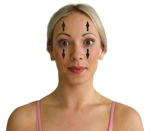
Surprise Exercise:
- When you are surprised, the eyelids & eyebrows raise without the knowing it.
- Sit in a comfortable chair with the feet together & the shoulders directly over the hips.
- Raise your eyebrows & eyelids as far as you can as if you were trying to touch the top of your head with your eyelids & eyebrows.
- Hold this position for five seconds & release the contraction. Repeat 10 times.
Look Around Exercise :
- Sit or stand and look up as far as you can while maintaining your head as still as possible.
- Hold this position for 5 seconds & then look down as far as you can without moving the head.
- Hold this position for 5 seconds & repeat while looking first to the left, then to the right.
- If necessary, you can repeat the entire cycle with your eyes closed as well.

The Partial Wink Exercise :
- You can do the partial wink exercise while standing or seated just about anywhere that you have a few minutes of free time.
- This exercise is done by partially winking one eye at a time & holding the position for a couple of seconds.
- Avoid completely closing your eyes, since this exercise is designed to help you gain more control over the orbicularis oculi muscles.
- Try to do two sets of 50 repetitions with each eye
FAQ
Injury to the orbicularis oculi can outcome from overuse, which may result in headaches, eyestrain, and sinus headaches. The main source for overuse of this muscle is poor eyesight without the actual corrective lenses.
sphincter muscle
The orbicularis oculi muscle is a sphincter of the eyelids. It is a broad & flat muscle spreading into 3 regions. A sphincter muscle closes the edge.
Orbicularis oculi muscle (musculus orbicularis oculi) Orbicularis oculi is a paired facial muscle that surrounds each orbit & the adjacent periorbital region. Together with corrugator supercilii & levator palpebrae superioris, it belongs to the circumorbital and palpebral group of muscles that surround the eye.
Treatment of orbicularis oculi muscle hypertrophy during conventional lower eyelid blepharoplasty is given out. Horizontal resection of a strip of the hypertrophic muscle is carried out, which allows the flattening of the muscular bulge in an anteroposterior site.
Structure & Function
The orbicularis oculi muscle closes the eyelids & assists in pumping the tears from the eye into the nasolacrimal duct system. The orbital section of the orbicularis oculi is more added in the voluntary closure of the eyelid, such as with winking & forced squeezing.
The orbicularis oculi is a muscle in the face that closes the eyelids. It originates from the nasal part of the frontal bone, from the frontal process of the maxilla in front of the lacrimal groove,-& from the anterior surface and borders of a short fibrous band, and the medial palpebral ligament.
The orbicularis oculi originate from the nasal portion of the frontal bone, the frontal process of the maxilla, the medial palpebral ligament, & the lacrimal bone. This muscle inserts onto the lateral palpebral raphe & superior and inferior tarsal plates, which are also known as the eyelids.
The orbicularis muscle is situated just underneath the skin of the eyelid. In general, the muscle attaches to the medial canthal region medially & the lateral canthal region laterally. The orbicularis oculi muscle is divided into2 sections, the orbital & palpebral sections.
Raise the eyebrows up as far as possible& open your eyes widely. Hold this position for 10 seconds, release for 5 seconds, & then repeat ten times. do this exercise daily.
To massage the Orbicularis Oculi, place the thumb over the trigger point just above the eye, as shown in the location video. This will be the most painful spot you will find in that area. Rest your other fingers over your forehead & using the pad of the thumb, apply slow pressure to the trigger point.
One Comment