
Ankle Dorsi Flexion And Planter Flexion
Table of Contents
What is an Ankle Dorsi Flexion And Planter Flexion?
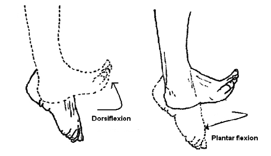
Ankle dorsiflexion and plantarflexion are two essential movements of the ankle joint that play a crucial role in various activities such as walking, running, jumping, and maintaining balance. These movements involve the bending and extension of the foot at the ankle.
Dorsiflexion refers to the movement of pulling the top of the foot toward the shin, which brings the toes closer to the shinbone. This action is primarily controlled by the muscles located in the front of the lower leg, including the tibialis anterior muscle. Ankle dorsiflexion allows the foot to clear the ground during the swing phase of walking, ensuring a smooth and efficient gait pattern.
On the other hand, plantarflexion refers to the movement of pointing the toes away from the shinbone, causing the foot to move downward. This movement is primarily controlled by the muscles located at the back of the lower leg, including the gastrocnemius and soleus muscles. Plantarflexion is crucial for activities such as pushing off the ground during walking or running and maintaining balance while standing on tiptoes.
Both ankle dorsiflexion and plantarflexion are necessary for maintaining a healthy and functional ankle joint. Adequate range of motion in these movements allows for proper foot and ankle alignment, which is essential for efficient movement and injury prevention. Restricted range of motion in either dorsiflexion or plantarflexion can lead to gait abnormalities, reduced performance in sports activities, and increased risk of ankle injuries.
Various factors can affect ankle dorsiflexion and plantarflexion, including muscle strength and flexibility, joint mobility, previous injuries, and certain medical conditions. It is important to maintain a balanced range of motion and strength in both movements through regular stretching and strengthening exercises to optimize ankle function and reduce the risk of injuries.
Ankle Dorsi Flexion
What Is Ankle Dorsi Flexion?
- Ankle dorsiflexion refers to the movement of the ankle joint where the foot is bent up towards the lower leg, in other words, the toes are pulled towards the lower leg. This is the opposite movement of plantar flexion, which directs the foot downward.
Dorsiflexion occurs mainly at the ankle joint, where the tibia (shin bone) and the talus (foot bone) articulate. The primary muscles responsible for ankle dorsiflexion are the tibialis anterior and other muscles in the front of the lower leg. These muscles contract and lift the foot and toes, allowing activities such as walking, running, and climbing stairs.
Adequate ankle dorsiflexion is important for normal walking (walking) and various physical activities. This allows the foot to clear the ground during the swing phase of walking, which ensures proper clearance and prevents toe drag. Insufficient ankle dorsiflexion can lead to gait disturbances, difficulty in activities that require ankle movement, and increased risk of injuries such as ankle sprains.
A variety of range of motion, stretching, and strengthening exercises can be used to improve ankle dorsiflexion. These exercises may be recommended by health professionals such as physical therapists or sports medicine experts. It is important to consult a qualified healthcare provider for proper evaluation and personalized advice on ankle dorsiflexion exercises.
Ankle Dorsi Flexors Muscles
The main muscles responsible for dorsiflexing the ankle, or lifting the foot and toes, are located in the front of the foot. The main muscles involved in ankle dorsiflexion are:
- Tibialis anterior: This muscle is located on the front of the lower leg and is the primary dorsiflexor of the ankle. It runs along the tibia (shinbone) to the inside of the leg and is responsible for pulling the leg up.
- Extensor Hallucis Longus: This muscle is located on the outside of the lower leg. It runs from the fibula (bone in the lower leg) to the big toe. It contributes to the dorsiflexion of the ankle and is also involved in the extension of the big toe.
- Extensor Digitorum Longus: This muscle is located next to the extensor longus on the outside of the calf. It runs from the fibula down to the toes (except the big toe). It contributes to ankle dorsiflexion and is responsible for the extension.
- Peroneus Tertius: This muscle is located on the outside of the lower leg between the extensors. It runs along the fibula to the fifth metatarsal (outside the foot). This helps with ankle dorsiflexion and foot rotation (moves the foot outward).
These muscles work together to lift the foot and toes during ankle dorsiflexion. It is important to note that other muscles, such as the tibialis posterior and flexor hallucis longus, also contribute to ankle dorsiflexion to a lesser extent. However, the muscles mentioned above are the primary ankle dorsiflexion.
Range Of Motion Of Ankle Dorsi Flexion
An ankle dorsiflexion range of motion can vary between individuals, but in general, an ankle dorsiflexion range of about 10-20 degrees beyond neutral is considered normal. In other words, the foot should be able to move up to the calf about 10-20 degrees from the neutral position where the foot is flat on the ground.
Ankle dorsiflexion is usually measured with a goniometer, which is used to assess joint angles. During the measurement, the person usually lies on their back, with the knee extended and the leg relaxed. The goniometer is placed so that one arm is aligned with the lower leg (outside the lower leg) and the other arm with the metatarsal bones (foot bones). The angle formed at the intersection of the hands indicates the area of dorsiflexion of the ankle.
It is important to note that individuals can vary in the extent of ankle dorsiflexion due to factors such as genetics, joint structure, previous injuries, and muscle tension. Limited ankle dorsiflexion can be caused by a number of reasons, including muscle imbalances, joint stiffness, or soft tissue tension. When someone has limitations in ankle dorsiflexion, it can affect their functional activities and movement patterns, potentially increasing their risk of injury or altering their gait.
If you are concerned about the dorsiflexion area of the ankle or experience difficulty or pain with movement, it is recommended to consult a doctor such as a physiotherapist or orthopedist. They can assess your specific situation, provide appropriate interventions and guide you through exercises or therapies to improve ankle dorsiflexion, if necessary.
To check ankle dorsiflexion range of motion:
- Positioning: Ask the person to sit on a firm platform with their legs extended in front of them.
- Stabilization: Keep the foot on the ground or a stable surface, making sure the heel does not come out.
- Alignment: Make sure the leg and foot are in a neutral position, not turned in or out.
- Zero point: Determine the zero point, which is the starting position of the leg without dorsiflexion.
- Measurement: Use a goniometer, a rotary arm, and a device with graduations to measure the dorsiflexion angle.
- Goniometer placement: Place the center of the axis of rotation of the goniometer in the center of the ankle joint with the axis of rotation of the ankle joint.
- Stationary Arm Placement: Align the fixed arm of the goniometer with the leg, pointing towards the hip joint.
- Moving arm placement: Align the moving arm of the goniometer with the leg, pointing toward the toes.
- Range Of Motion: Ask the person to slowly bend their leg up (dorsiflexion) while you hold the goniometer in place. Make sure the leg stays in place during the movement.
- Read the measurement: When the person reaches maximum dorsiflexion, read the degree measurement on the goniometer. This measurement represents the ankle dorsiflexion range of motion.
- Repeat: Take the measurement two to three times to ensure accuracy and consistency.
- Record The Measurements: Save the range of motion measurements for future reference or comparison.
Remember that it is important to be gentle and communicate with the person throughout the process to ensure their comfort and cooperation.
Ankle Dorsi Flexion Test
The ankle dorsiflexion test, also known as the ankle dorsiflexion range of motion test or step-out test, is a simple assessment used to measure the range of motion of the ankle dorsiflexion. This test is usually performed to assess ankle mobility and detect limitations in ankle dorsiflexion.
The ankle dorsiflexion test is usually performed as follows:
Preparations: Find an open space and stand facing a wall or a sturdy object that you can use for support.
Positioning: Place one foot a comfortable distance from the wall/object with toes pointing forward. The other leg can be placed behind the back.
Testing: Keeping the heel of the front foot on the ground, slowly bending forward, and bending the front knee while keeping the foot flat. Gradually move your knee forward towards the wall/object, trying to touch it without lifting your heel.
Measurement: Record the maximum distance (in inches or centimeters) between the wall/object and the big toe. This measurement indicates the extent of ankle dorsiflexion.
Repeat: Do the test with both legs, as the range of motion on the two sides may differ. When performing the ankle dorsiflexion test, it is important to remember a few key points:
- Maintain proper position and alignment during the test.
- Keep the heel of the front foot on the ground at all times.
- Avoid raising the heel or compensating by turning the foot outward (inversion) or inward (inversion).
The ankle dorsiflexion test provides an objective measure of ankle mobility and helps identify limitations that may affect activities such as walking, running, or squatting. If you are concerned about ankle dorsiflexion or mobility issues, it is recommended that you consult a physician, such as a physical therapist or sports medicine specialist, who can provide a comprehensive evaluation and appropriate treatment if necessary.
Exercise For Ankle Dorsi Flexion
There are several exercises that can help improve ankle dorsiflexion. here are some samples:
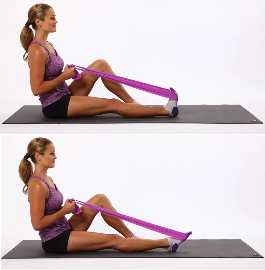
- Towel Stretch: Stretch your legs out in front of you as you sit down on the ground. A towel or a resistance band can be wrapped around the ball of your foot. Gently pull the towel or band toward you, keeping your knee straight. Your leg should begin to stretch in the back. Hold the stretch for 30 seconds and repeat several times on each leg.
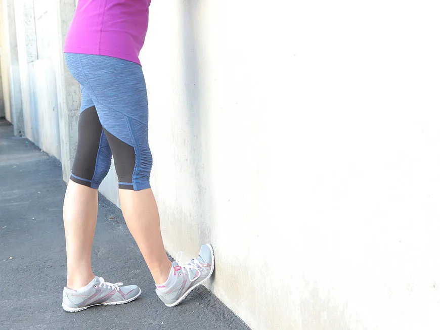
- Wall Calf Stretch: Face a wall and place your hands at shoulder height against it. Step one foot back, keeping it on the ground. Keep the front knee bent and slowly lean forward, feeling the stretch in the calf of the back leg. Hold the stretch for 30 seconds and repeat several times on each leg.
- Ankle Dorsiflexion Stretch: Sit on the edge of a chair or table with your feet hanging off the edge. Point your toes down as far as possible, then slowly raise your toes toward your shins, bending your ankles. Hold the stretch at the top for a few seconds and then release. Repeat this movement several times 10-15 times.
- Heel Raises: Stand with your feet hip-width apart and use a strong base for balance if needed. Slowly rise to your toes and lift your heels as high as possible. Next, gradually bring your heels to the ground. Do several 10-15 repetitions.
- Ankle Alphabet: Sit on a chair with one leg stretched out in front of you. Pretend your big toe is a pencil and “write” the alphabet with your foot in the air. Move slowly and deliberately, using the full range of motion available to you. Repeat with the other leg.
These exercises help stretch and strengthen the muscles involved in dorsiflexing the ankle, which increases the range of motion and improves flexibility. It is important to start slowly and gradually increase the intensity and duration of the exercises according to tolerance. If you have ailments or problems, it is recommended that you consult a health professional, such as a physiotherapist, for guidance and personalized exercise recommendations.
Special Test For Ankle Dorsi Flexion
Health professionals use a number of specialized tests to assess ankle dorsiflexion and identify specific limitations or dysfunctions. Some commonly used special ankle dorsiflexion tests are:
- Silfverskiold test: This test is used to differentiate between gastrocnemius and soleus muscle tension. The person is lying on the ground (picture down), and the knee is bent at a 90-degree angle. The examiner then passively flexes the ankle with the knee dorsiflexed and extended. If ankle dorsiflexion increases significantly when the knee is extended compared to when the knee is flexed, this indicates gastrocnemius muscle strain. If the limitation persists regardless of the knee position, this indicates strain in the plantar muscles.
- Weight-Bearing Lunge Test: In this test, the person stands in a split flea position with the front foot slightly away from the wall. The front knee is placed over the other toe and the person tries to touch the wall with the knee while keeping the heel on the ground. If the knee does not reach the wall without lifting the heel, this indicates limited ankle dorsiflexion.
- Heel-To-Wall Test: The person stands with toes about 5 inches (12 centimeters) from the wall, with both feet pointing straight ahead. Then they bend their knees and try to touch the wall with their knees, keeping their heels on the ground. If the heels cannot stay in contact with the ground, this indicates a limitation of ankle dorsiflexion.
- Ankle Dorsiflexion Range of Motion Test: As mentioned earlier, this test uses a goniometer to measure the range of motion of the ankle dorsiflexion. The person lies supine with the knee extended and the goniometer is pointed at the fibula to measure the angle between the metatarsals. The angle indicates the extent of ankle dorsiflexion.
These specialized tests help healthcare professionals assess ankle dorsiflexion and provide valuable information for diagnosis, treatment planning, and progress monitoring. They are usually carried out by trained professionals such as physiotherapists, sports medicine specialists, or orthopedists. If you are concerned about ankle dorsiflexion, it is best to consult a qualified physician for a thorough evaluation and proper testing.
Ankle Planter Flexion
What Is Ankle Planter Flexion?
- Ankle-plantar flexion refers to the movement of the ankle joint, which involves pointing the foot down, away from the shin. This is the opposite movement of ankle dorsiflexion, which bends the foot up toward the lower leg.
\During plantar flexion of the ankle, the ankle allows the foot to move downward, pointing the toes away from the body. This movement occurs mainly at the ankle joint, where the tibia (shin bone) and talus (foot bone) articulate. The primary muscles responsible for plantar flexion of the ankle are located on the back of the lower leg and include the calf muscles: 1. Gastrocnemius 2. Soleus 3. Plantaris
These muscles contract to direct the leg downward and generate power for activities such as walking, running, jumping, and pushing off the ground. Plantar flexion of the ankle is critical in many functional activities, such as walking on the toes, standing on the balls of the feet, and propelling the body forward, such as when running or jumping. Additionally, it contributes to stability and equilibrium being maintained.
If you have problems or problems with ankle-plantar flexion, it is recommended that you consult a doctor, such as a physical therapist or orthopedist, for a thorough evaluation and appropriate treatment. They can provide personalized advice, exercises, and treatments to correct limitations or dysfunctions associated with ankle-plantar flexion.
Ankle Planter Flexors Muscles
The main muscles responsible for plantar flexing of the ankle, or pointing the foot down, are located on the dorsum of the foot. The main muscles involved in plantar flexion of the ankle are:
- Gastrocnemius: This is the larger muscle of the calf, easily visible as a bulge on the back of the calf. It has two ends, the medial end (inner side) and the lateral end (outer side). The gastrocnemius passes through both the knee and ankle joints and is a powerful plantar flexor. It is particularly active during activities such as running, jumping, and pushing off the ground.
- Soleus: The soleus is a broad, flat muscle located deeper in the calf, below the gastrocnemius muscle. It also crosses the ankle joint and promotes plantar flexion of the ankle. Unlike the gastrocnemius, the soleus is not involved in knee movements. It is very active during activities that require bending the knee, such as walking and squatting.
- Plantaris: This is a thin muscle that runs on the inside of the lower leg, between the gastrocnemius and the sole. It helps in plantarflexion of the ankle, although it is relatively smaller and weaker compared to gastrocnemius and plantarflexion.
These muscles work together to create the force needed to flex the ankle and foot and provide power for activities such as walking, running, jumping, and balancing on the balls of the feet. In addition, they promote the pushing and shoving phase of walking.
It is important to note that other muscles, such as the tibialis posterior and the flexor hallucis longus, can also contribute to ankle-plantar flexion to a lesser extent. However, the muscles mentioned above are the primary plantar flexors of the ankle.
Maintaining the flexibility and strength of these muscles is important for optimal ankle function and performance. If you are concerned or have difficulty with ankle-plantar flexion, it is recommended that you see a doctor, such as a physical therapist or sports medicine specialist, for evaluation and appropriate treatment.
Range Of Motion Of Ankle Planter Flexion
The extent of ankle-plantar flexion, or downward movement of the foot, can vary between individuals. However, the normal range of motion for plantar flexion of the ankle joint is usually considered to be around 40-50 degrees.
During ankle plantar flexion, the foot moves down, away from the shin, causing the toes to point down. This movement occurs mainly at the ankle joint, where the tibia (shin bone) and talus (foot bone) articulate.
A goniometer, which is a tool for assessing joint angles, can be used to measure the range of motion of ankle flexion. A goniometer is usually aligned with the foot and forefoot to measure the angle formed between them during plantar flexion.
It is important to note that individuals can have variations in the plantar flexion of the ankle due to factors such as genetics, joint structure, past injuries, and muscle flexibility. Limited ankle plantar flexion can be caused by a number of reasons, including muscle tightness, joint stiffness, or soft tissue restrictions.
If you are concerned about the plantar flexor area of the ankle or experience difficulty or pain with movement, it is recommended that you consult a doctor such as a physiotherapist or orthopedist. They can assess your specific situation, provide appropriate interventions and guide you through exercises or therapies to improve ankle plantar flexion, if necessary.
You can check the ankle-plantar flexion range of motion as follows.
- Positioning: Ask the person to sit on a firm platform with their legs extended in front of them.
- Stabilization: Keep the foot on the ground or a stable surface, making sure the heel does not come out.
- Alignment: Make sure the leg and foot are in a neutral position, not turned in or out.
- Zero point: Determine the zero point, which is the starting position of the foot without plantar flexion.
- Measurement: Use a goniometer, a device with a rotating arm, and steps to measure the plantar flexion angle.
- Goniometer placement: Place the center of the axis of rotation of the goniometer in the center of the ankle joint with the axis of rotation of the ankle joint.
- Stationary Arm Placement: Align the fixed arm of the goniometer with the leg, pointing towards the hip joint.
- Moving arm placement: Align the moving arm of the goniometer with the leg, pointing toward the toes.
- Range Of Motion: Ask the person to slowly point their foot down (plantar flexion) while you hold the goniometer in place. Make sure the leg stays in place during the movement.
- Read the measurement: When the person reaches maximum plantar flexion, read the degree measurement on the goniometer. This measurement shows the ankle-plantar flexion range of motion.
- Repeat: Take the measurement two to three times to ensure accuracy and consistency.
- Record The Measurements: Save the range of motion measurements for future reference or comparison.
Always be gentle and communicate with the person during the process to ensure their comfort and cooperation.
Ankle Plantar Flexion Test
There is a special test known as the “Ankle Plantar Flexion Test” or “Thompson Test” to assess the plantar flexion of the ankle joint.
- The Thompson test is primarily used to assess the integrity of the Achilles tendon, which is important for plantar flexion of the ankle.
- This is usually done to diagnose a possible rupture of the Achilles tendon. The Thompson test is usually performed as follows:
- The subject usually lies supine (bottom in the picture) on an examination table or bed, with the legs hanging over the edge.
- The examiner observes both feet for noticeable differences in appearance or alignment.
- When the person’s legs are relaxed and hanging, the examiner squeezes the calf muscle of the affected leg or squeezes the calf with his hands.
During this compression, the examiner observes the movement or lack of movement of the foot and ankle. In a normal reaction, squeezing the calf muscle causes the sole of the foot to flex (down). However, if the Achilles tendon ruptures, the affected leg will move little or not at all. This lack of movement is called a positive Thompson test.
It is important to note that the Thompson test is specific to the assessment of the Achilles tendon and not a general assessment of ankle-foot flexion range of motion. If you need a full ankle range of motion, including plantar flexion, a health professional, such as a physical therapist or orthopedist, can perform a more in-depth examination with a goniometer or other specialized tests. If you are concerned about ankle-plantar flexion or suspect an Achilles tendon injury, I recommend seeing a medical professional for an accurate diagnosis and appropriate treatment.
Exercise For Ankle Plantar Flexion
To clarify, ankle plantar flexion refers to a movement in which the foot points down, away from the lower leg. To strengthen and improve ankle flexion, you can add the following exercises to your routine:
- Calf Raises: Stand with your feet hip-width apart and place your hands on a stable surface for support if needed. Stand on your toes and lift your heels as high as possible. Hold the position for a moment and then slowly lower your heels. Do several 10-15 repetitions.
- Seated leg raises: Sit on a chair or bench with your feet on the floor and knees bent at a 90-degree angle. Place a weight, such as a dumbbell, on your thighs near your knees. Press your feet through the balls of your feet to lift your heels as high off the ground as possible. At the peak, pause for a moment before bringing your heels back down. Do several 10-15 repetitions.
- Resistance Band Plantar Flexion: Sit on the ground with your legs out in front of you to do plantar flexion. Wrap the resistance band around the balls of your feet and hold the ends of the strap in your hands. Bend your legs to create tension on the strap, then point your toes as far away from your body as possible. Slowly return to the starting position. Do several sets of 10-15 repetitions.
- Toe walking: Stand tall and lift yourself up on the balls of your feet, walk forward with small steps while maintaining the position of the toe. Focus on maintaining control and balance while tiptoeing for a certain distance or time.
- Stair Calf Raises: Stand on the edge of a step or staircase with the balls of your feet on the step and your heels hanging down. If necessary, cling to a wall or railing for support. Lower your heels below the level of the step, then press your feet through the balls of your feet to lift your heels as high as possible. Do several 10-15 repetitions.
It is important to perform these exercises correctly, starting with a comfortable range of motion and gradually increasing the intensity and resistance as tolerated. If you have ailments or problems, it is recommended that you consult a health professional, such as a physiotherapist, for guidance and personalized exercise recommendations. They can assess your specific needs and provide appropriate exercises to improve ankle-foot flexion safely and effectively.
Special Test For Ankle Planter Flexion
- One common test used to assess ankle plantar flexion is the “heel raise” or “calf raise.” This is usually done as follows.
- Stand near a wall or stable surface that you can use for support if needed.
- Put your feet together, shoulder-width apart, and point your toes forward.
- Slowly rise to your toes, pushing your feet through the balls and lifting your heels off the ground.
- Lift your heels as high as you can to get a full range of motion.
- Hold the raised position for a moment and then slowly lower your heels back to the starting position.
- Repeat the movement several times.
- During this test, you can assess the strength and range of motion of the plantar flexion of the ankle joint.
Summary
Ankle dorsiflexion and plantarflexion are movements of the ankle joint that are essential for walking, running, and maintaining balance. Dorsiflexion involves pulling the top of the foot toward the shin, while plantarflexion involves pointing the toes away from the shinbone. These movements are controlled by muscles in the front and back of the lower leg, respectively.
Dorsiflexion allows the foot to clear the ground during the swing phase of walking and is important for a smooth gait pattern. Plantarflexion is crucial for activities like pushing off the ground during walking or running and maintaining balance while standing on tiptoes.
Having a healthy range of motion in dorsiflexion and plantarflexion is vital for proper foot and ankle alignment, efficient movement, and injury prevention. Restricted range of motion can lead to gait abnormalities, decreased performance, and an increased risk of ankle injuries.
Factors that affect these movements include muscle strength and flexibility, joint mobility, previous injuries, and medical conditions. Regular stretching and strengthening exercises can help maintain a balanced range of motion and strength in both dorsiflexion and plantarflexion, optimizing ankle function and reducing the risk of injuries.
FAQs
Two of the greater not unusual place motives for terrible ankle mobility consist of restricting withinside the joint and/or tightness of the posterior leg muscle mass and Achilles tendon. However, the mechanisms for why those arise can vary.
Proper dorsiflexion lets in for a gold standard foot strike and may make contributions to fewer accidents by placing your foot withinside the great role to take in the surprise of landing. It additionally contributes to decreased touch time, which has been related to greater green running.
For example, harm to the peroneal nerve, or compression of this nerve, may also result in a lack of dorsiflexion. Disorders affecting the muscle mass or nerves of the foot (inclusive of muscular dystrophy or amyotrophic lateral sclerosis) can also result in drop foot.
The soleus, gastrocnemius, and plantaris muscles work together to bend the ankle joint in the plantar direction. Their motion lifts us up off the floor whilst we stand on tip-toe. When balanced towards gravity, equal motion controls our charge of descent.
The gastrocnemius, soleus, and plantaris are the three superficial muscles that make up the major plantar flexors of the foot. Their tendons come together to form the tendo calcaneus or Achilles tendon.
2 Comments