
Ulna Bone
Table of Contents
Introduction
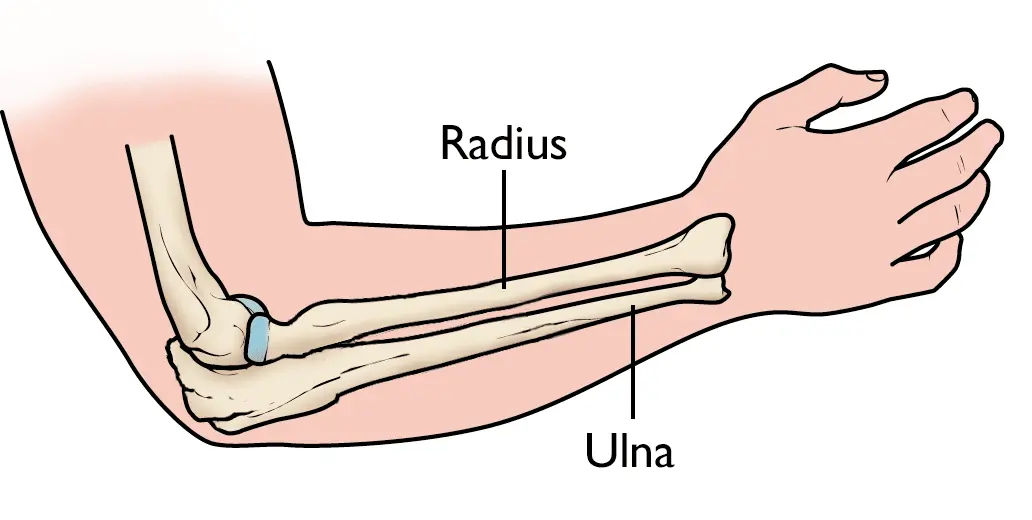
The ulna is one of the 2 long forearm bones that, in junction with the radius, make up the antebrachium. the bone crosses from the elbow to the wrist on the medial side of the forearm when in anatomical position. In comparison to the radius, the ulna is determined to be larger and longer.
It performs as the origin and/or insertion region for more than a dozen muscles and is applied in motions assisted by the elbow and wrist joints. common pathologies are again associated with the bone as several fractures are pathognomonic depending on the place within the ulna, as well as the resulting position of the fractured components
Anatomy position and side determination
The side of the ulna can be determined by holding the bone vertically in such a method that:
The all-around hook-like end is directed upwards.
The sharp crest-like interosseous boundary of the shaft is directed laterally.
The concavity of the hook-like upper end and the coronoid function are fronting forwards.
Features
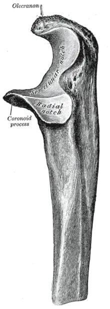
The upper end of the ulna
The upper end of the ulna is supplied and hook-like with the concavity of the curve facing forwards. The concavity of the upper end (trochlear notch) is situated between the big olecranon process above and the small coronoid process below. The upper end has two processes: coronoid and olecranon, and two notches: trochlear and radial notch.
Processes
Coronoid process
Olecranon process
Upper surface
Its rough posterior two-thirds offer insertion to the triceps brachii.
The capsular ligament of the elbow joint is connected anteriorly around its margins.
A synovial bursa is discovered between the tendon of the triceps and the capsular ligament.
Anterior surface
It is soft and forms the upper component of the trochlear notch.
Posterior surface
It makes a subcutaneous triangular location.
A synovial bursa (subcutaneous olecranon bursa) is found between the posterior surface and skin.
Medial surface
Its upper part presents connections to three structures: ulnar head of flexor carpi ulnaris muscle origin. posterior, and tilting rounds of the ulnar collateral ligament. Coronoid process. It has 4 surfaces medial- and lateral or superior-anterior
Superior surface: It is silky and makes up the lower part of the trochlear notch. Anterior surface: It is triangular.
Its lower corner presents an ulnar tuberosity.
Brachialis muscle is inserted into the entire anterior surface including ulnar tuberosity.
The medial margin of the anterior surface is sharp and has a tubercle at its upper end called a sublime tubercle.
The medial margin connects to the next frames beginning with proximal to distal.
Anterior band of ulnar collateral ligament. Oblique band of ulnar collateral ligament. Humero-ulnar head of flexor digitorum superficialis. Ulnar head of pronator teres. Ulnar head of flexor pollicis longus.
Medial surface: It produces (origin) to flexor digitorum profundus.
Lateral surface:
The upper portion of this surface possesses a radial notch for articulation with the head of the radius.
The annular ligament is associated with the anterior and posterior margins of the radial notch.
The lower part of the lateral surface below the radial notch has a depressed area referred to as the supinator fossa, which includes radial tuberosity in the course of supination and pronation.
The supinator fossa is bounded behind the supinator crest. The supinator crest and adjoining part of the supinator fossa give origin to the supinator muscle.
Notches (articular surfaces)
Trochlear notch
It is C-shaped (semilunar) and communicates with the humerus’s trochlea. It has a non-articular strip at the corner of its olecranon and coronoid parts. Its superior, medial, and anterior margins offer a connection to the capsule of the elbow joint.
Radial notch
It articulates forward with the head of the radius to create the superior radio-ulnar joint.
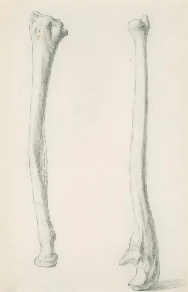
The shaft of the ulna
The extended shaft of the ulna is between the upper and lower ends.
Gradually its thickness reduces from above downwards throughout its height. The lateral border (interosseous boundary) is sharp crest-like.
The Ulna bone’s shaft has three borders (lateral – anterior and posterior) and three surfaces (anterior – medial – and posterior).
Borders
Lateral (interosseous) border
It is the sharpest and is continuous above the supinator crest.
It is ill-defined below.
The interosseous membrane is connected to this border except for its upper part.
Anterior border
It advances from the medial side of the ulnar tuberosity to the base of the styloid process.
It is thick and round. Its upper three-fourth gives origin to flexor digitorum profundus.
Posterior border
It starts with the apex of the triangular subcutaneous location on the back of the olecranon process and drops to the styloid process.
It is subcutaneous throughout and therefore can be palpated along its entire length.
It provides a connection to three muscles by common aponeurosis. The muscles are: FDP, FCU ECU
Flexor digitorum profundus.
Flexor carpi ulnaris. Extensor carpi ulnaris.
Surfaces
Anterior Surface
It is located between the anterior and interosseous borders.
The flexor digitorum profundus comes up from its upper three-fourths.
The pronator quadratus occurs from an oblique ridge on the lower one-fourth of this special surface.
The nutrient foramen is situated a small outside the middle of this surface and is directed upwards.
Medial surface
It is discovered between the anterior and posterior borders.
The flexor digitorum profundus originates from the upper two-thirds of this surface.
Posterior surface
It is located between posterior and interosseous boundaries.
It is divided into the smaller upper part and large lower part by an oblique line, which begins at the junction of the upper and middle third of the posterior border and runs in the direction of the posterior edge of the radial notch.
The area above the oblique line receives insertion of the anconeus muscle.
The area below the oblique line is divided into larger medial and smaller lateral parts by a faint vertical line. The lateral part offers a connection to three muscles from proximal to distal as follows:
Abductor pollicis longus in the upper one-fourth.
Extensor pollicis longus in the middle one-fourth.
Extensor indicis in the next one-fourth.
The distal one-fourth is without any connections.
The lower end of the ulna
The lower end of the ulna is partway expanded and has a head and styloid process. the styloid process is posteromedial to the leader. the ulna surfaces like a line twist with the olecranon process occurring like the upper jaw, the coronoid fossa, the lower jaw, and the trochlear notch in the mouth of the wrenching. the lower end of the ulna is created by the head and styloid process.
Head
It delivers a convex articular surface on its lateral side for declaration with the ulnar notch of radius to make up the inferior radio-ulnar joint.
Its inferior surface is soft and disconnected from the wrist joint by an articular disc of the inferior radio-ulnar joint.
Styloid process
It projects downwards from the posteromedial factor of the head of the ulna. Its tip offers a connection to the medial collateral ligament of the wrist joint. the apex of the triangular articular disc is connected to the depression between the head and base of the styloid process. the tendon of the extensor carpi ulnaris is located in the groove between the back of the head of the ulna and the styloid process.
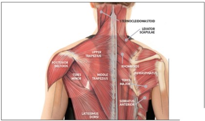
Muscles attachments
The ulna positions as the attachment site for multiple muscles with a myriad of movements. The following are organized in terms of the movement and where on the ulna is the attachment of the muscle’s fibers
Triceps brachii – posterior section of the superior surface of the olecranon anconeus – olecranonBrachialis – the volar surface of the coronoid process. Pronator teres – the coronoid process of the medial surface
Flexor carpi ulnaris – olecranon process
Flexor digitorum superficialis – coronoid process
Flexor digitorum profundus – anteromedial surface
Pronator quadratus – distal anterior shaft
Extensor carpi ulnaris – posterior border
Supinator – proximal ulna
Abductor pollicis longus – posterior surface
Extensor pollicis longus – dorsal shaft
Extensor indicis – posterior distal shaft
Ossification of the ulna bone
The ulna bone ossifies from the 3 main centers: one primary center for the shaft and two secondary centers, one each for the lower end and the upper end.
Primary center
It shows up in the mid-shaft during the 8 weeks of IUL( intra uterine line).
Secondary centers
Upper-end:
formation: 9 years (upper part of the trochlear surface and lid of the olecranon process).
Fusion: 18 years.
The lower end (middle of the head):
Appearance: 6 years.
Fusion: 20 years.
Clinical Importance
When the elbow is fully extended, the tip of the olecranon process and medial and lateral epicondyles of the humerus hinge on the same horizontal line. When the elbow is completely flexed the three bony pinpoints form an equilateral triangle. In the dislocation of the elbow, this connection is disturbed.
Ulna stabilizes the forearm by grasping the lower end of the humerus by its trochlear notch and gives the foundation for the radius to create superior and inferior at supination and pronation radio-ulnar joints. the fracture of the upper third of the shaft of the ulna with dislocation of the radial head at the superior radio-ulnar joint is referred to as Monteggia fracture-dislocations. the fracture of the lower third of the shaft of radius connected with the dislocation of the inferior radio-ulnar joint is referred to as Galeazzi fracture-dislocation. A fracture of the shaft of the ulna as an impact of a direct injury when a night watchman reflexly increases his forearm to ward off the impact of the stick is termed a night-stick fracture
FAQ
The ulna is the long bone in your forearm. It supports you motion of your arm, wrist, and hand. Does your ulna likewise support lots of important muscles, tendons, ligaments, and blood vessels
Muscles Attach to the Ulna
Pronator Teres.
Flexor Carpi Ulnaris.
Flexor Digitorum Profundus.
Flexor Digitorum Superficialis.
Pronator Quadratus.
Triceps Brachii.
Anconeus.
Brachialis.
The ulnar nerve innervates the flexor muscles of the forearm involving the flexor digitorum profundus and flexor carpi ulnaris. It likewise innervates the intrinsic muscles of the hand including the hypothenar, palmaris brevis, lumbricals, and interossei muscles.
musician’s nerve
The Ulnar nerve is also known as the “musician’s nerve” as it handles the good movements of the fingers.
5 Comments