
Talus Bone
Table of Contents
Introduction
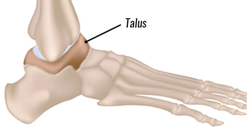
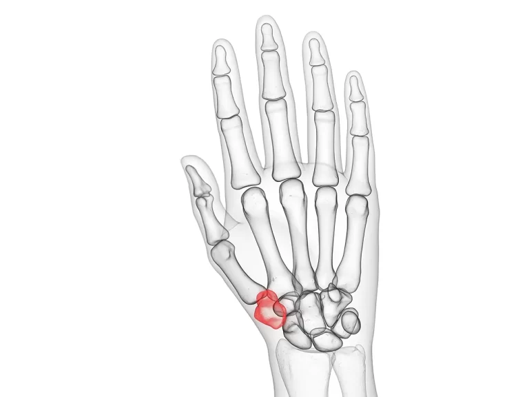
The Talus bone, also known as the ankle bone, is a small bone located between the tibia and fibula in the lower leg. It plays a crucial role in the movement and stability of the ankle joint.
The talus bone has a unique shape that allows it to articulate with both the tibia and fibula, forming the ankle joint. It is responsible for transmitting the weight from the leg to the foot and facilitating movements such as dorsiflexion (lifting the foot upwards) and plantarflexion (pointing the foot downwards).
Anatomy of the Talus Bone
Anatomy of the Talus Bone:
- Shape: The talus bone has a somewhat irregular shape, resembling a cube with rounded edges. It consists of three main parts: the body, neck, and head.
- Body: The body of the talus forms the bulk of the bone and is responsible for bearing most of the weight during activities such as walking or running. It has a convex superior surface that articulates with the tibia, forming the ankle joint. The inferior surface is concave and forms the subtalar joint with the calcaneus bone.
- Neck: The neck of the talus connects the body to the head. It is narrower and acts as a transition between the wider body and the rounded head.
- Head: The head of the talus is located on the medial side of the bone. It is rounded and articulates with the navicular bone, forming the talonavicular joint.
- Articular Surfaces: The talus bone has multiple articular surfaces that allow for various movements and interactions with other bones in the foot and ankle.
Articulation of the Talus Bone
The talus bone has multiple articulations with other bones in the foot and ankle, allowing for various movements and providing stability to the joint.
- Ankle Joint (Talocrural Joint):
The superior articular surface of the talus forms the dome-shaped part of the bone that articulates with the tibia and fibula, forming the ankle joint or talocrural joint. This joint allows for dorsiflexion (upward movement) and plantarflexion (downward movement) of the foot. The talus acts as a hinge between the tibia and fibula, enabling flexion and extension of the foot. - Subtalar Joint:
The inferior articular surface of the talus articulates with the calcaneus bone, forming the subtalar joint. This joint is responsible for the inversion (inward movement) and eversion (outward movement) of the foot. Inversion refers to movement where the sole of the foot turns inward, while eversion refers to movement where the sole of the foot turns outward. The talus bone plays a crucial role in these movements by allowing gliding and rotation between the talus and calcaneus. - Talonavicular Joint:
The medial articular surface of the talus connects with the navicular bone, forming the talonavicular joint. This joint contributes to the overall stability and mobility of the foot. It allows for movements such as adduction (movement towards the midline of the body) and abduction (movement away from the midline of the body) of the foot. The talonavicular joint is important for maintaining proper alignment and function of the medial longitudinal arch of the foot. - Calcaneocuboid Joint:
The lateral side of the talus articulates with the calcaneus bone at a small facet called the calcaneocuboid joint. This joint allows for limited gliding and rotation movements between the talus and calcaneus, contributing to the overall mobility and stability of the foot.
These articulations between the talus bone and other bones in the foot and ankle are facilitated by the presence of articular cartilage. Articular cartilage covers the surfaces of the bones where they come into contact, reducing friction and allowing for smooth movement. The ligaments surrounding these joints provide additional stability and support to the talus bone during weight-bearing and movement.
Functions of the Talus Bone
The talus bone, also known as the ankle bone, is a key bone in the foot and plays several important functions:
- Weight-bearing: The talus bone sits between the tibia and fibula bones of the lower leg and the calcaneus bone of the heel. It acts as a connector, transferring weight from the leg to the foot. The talus bone is responsible for transmitting the body’s weight and forces during activities such as walking, running, and jumping.
- Shock absorption: The talus bone is designed to absorb and distribute forces that occur during weight-bearing activities. Its unique shape and structure help to cushion and distribute the impact forces generated by walking or running, protecting the joints and other structures in the foot and ankle.
- Facilitating movement: The talus bone is involved in various movements of the foot and ankle. It allows for dorsiflexion and plantarflexion at the ankle joint, which are movements that involve flexing and extending the foot. The subtalar joint, formed by the articulation between the talus and calcaneus bones, allows for inversion and eversion movements of the foot. These movements are essential for maintaining balance and stability while walking or engaging in sports activities.
- Maintaining arches of the foot: The talus bone is an important component in maintaining the arches of the foot. It forms a key connection between the hindfoot (calcaneus) and midfoot (navicular), contributing to the stability and flexibility of the foot’s arches. The talonavicular joint, formed by the talus and navicular bones, plays a crucial role in supporting the medial longitudinal arch of the foot, which helps with shock absorption and weight distribution.
- Stability: The talus bone provides stability to the foot and ankle joints. Its articulations with other bones, along with the ligaments that surround these joints, help to maintain proper alignment and prevent excessive movement or instability. The talus bone acts as a hinge between the tibia and fibula, allowing for controlled movement while providing stability to the ankle joint.
In summary, the talus bone is a vital component of the foot and ankle, facilitating movement, absorbing shock, and providing stability. Its articulations with other bones in the foot and ankle allow for various movements and contribute to the overall function and support of the lower limb.
Muscle attachment
Muscle attachment to the talus bone is crucial for the movement and stability of the foot and ankle. Several muscles attach to the talus bone, including:
- Tibialis anterior: This muscle attaches to the medial and superior aspects of the talus bone. It is responsible for dorsiflexion of the foot, which involves lifting the foot and toes towards the shin.
- Extensor hallucis longus: This muscle also attaches to the superior aspect of the talus bone. It is responsible for extending the big toe and assisting in the dorsiflexion of the foot.
- Extensor digitorum longus: This muscle attaches to the lateral and superior aspects of the talus bone. It is responsible for extending the other toes (excluding the big toe) and assisting in the dorsiflexion of the foot.
- Peroneus tertius: This muscle attaches to the lateral aspect of the talus bone. It acts as dorsiflexion and eversion of the foot.
- Flexor hallucis longus: This muscle attaches to the posterior aspect of the talus bone. It is responsible for flexing the big toe and plantarflexion of the foot.
- Flexor digitorum longus: This muscle also attaches to the posterior aspect of the talus bone. It is responsible for flexing the other toes (excluding the big toe) and plantarflexion of the foot.
- Tibialis posterior: This muscle attaches to the medial and inferior aspects of the talus bone. It is responsible for the inversion and plantarflexion of the foot, as well as supporting the arches of the foot.
These muscles work together to produce movements and stabilize the foot and ankle joints. Their attachment to the talus bone allows them to exert force and control movement during activities such as walking, running, and jumping. Proper functioning and coordination of these muscles are essential for maintaining balance, stability, and efficient movement of the foot and ankle.
Blood and Lymph supply
Blood Supply to the Talus Bone:
The talus bone receives its blood supply from several arteries. The main arterial supply to the talus comes from the posterior tibial artery, which branches into the medial and lateral tarsal arteries. These arteries provide blood to the talar body and neck. Additionally, the deltoid artery, a branch of the posterior tibial artery, supplies the medial aspect of the talus.
The blood supply to the talus is important for maintaining the health and viability of the bone. Adequate blood flow ensures that the bone receives oxygen and nutrients necessary for its metabolic activities and healing processes. Impaired blood supply to the talus, such as in cases of avascular necrosis, can lead to bone death and subsequent joint dysfunction.
Lymphatic Drainage from the Talus Bone:
Lymphatic vessels are liable for draining excessive fluid and waste products from tissues. The lymphatic drainage of the talus bone occurs through a network of lymphatic vessels that accompany the blood vessels.
The lymphatic vessels in the foot and ankle region drain into lymph nodes located in the popliteal region behind the knee. From there, lymph is further transported through a series of lymph nodes in the inguinal region and eventually reaches the deep inguinal lymph nodes.
The lymphatic drainage system plays a crucial role in maintaining tissue fluid balance and removing waste products and pathogens from the area. It also aids in immune responses by filtering and processing lymph fluid in the lymph nodes.
Overall, both the blood supply and lymphatic drainage of the talus bone are essential for its proper functioning and overall health. The blood supply ensures adequate oxygen and nutrient delivery, while lymphatic drainage helps maintain tissue fluid balance and removes waste products from the area.
Conditions that affect Talus Bone
There are several associated conditions that can affect the talus bone:
Avascular Necrosis: Avascular necrosis, also known as osteonecrosis, is a condition characterized by the death of bone tissue due to a lack of blood supply. It can occur in any bone, including the talus. When the blood supply to the talus is compromised, the bone tissue begins to die, leading to pain, limited range of motion, and eventually joint dysfunction. Avascular necrosis of the talus is often caused by trauma, such as a fracture or dislocation, or by conditions that disrupt blood flow, such as sickle cell disease or corticosteroid use.

Talus Fractures: Fractures of the talus bone can occur due to traumatic injuries, such as falls or high-impact accidents. These fractures can range from hairline cracks to complete breaks. Talus fractures can cause severe pain, swelling, and difficulty bearing weight on the affected foot. Treatment usually involves immobilization with a cast or boot, and in some cases, surgery may be required to realign and stabilize the bone fragments.
Osteochondritis Dissecans: Osteochondritis dissecans (OCD) is a condition in which a piece of cartilage and underlying bone becomes detached from its normal position within a joint. This can occur in the talus bone, leading to pain, swelling, and joint instability. The exact cause of OCD is unknown, but it is thought to involve repetitive trauma or poor blood supply to the affected area. Treatment options include rest, immobilization, physical therapy, and in severe cases, surgery may be necessary to remove or repair the detached fragment.

Talar Dome Lesions: Talar dome lesions refer to damage or injury to the cartilage covering the top surface of the talus bone. These lesions can be caused by traumatic injuries, such as ankle sprains or fractures, or by repetitive stress on the joint. Talar dome lesions can cause pain, swelling, and a limited range of motion in the ankle joint. Treatment options include rest, immobilization, physical therapy, and in some cases, surgery may be needed to repair or remove the damaged cartilage.
Osteoarthritis: Osteoarthritis is a degenerative joint disease that can affect any joint in the body, including the ankle joint involving the talus bone. Over time, the cartilage covering the talus can wear down, leading to pain, stiffness, and reduced mobility. Osteoarthritis of the talus can be caused by previous injuries, such as fractures or dislocations, or by normal wear and tear associated with aging. Treatment options include pain management, physical therapy, assistive devices, and in severe cases, surgery may be necessary to replace or fuse the affected joint.
It is important to note that these are just a few examples of conditions that can affect the talus bone. Proper diagnosis and treatment by a healthcare professional are essential for managing these conditions and maintaining the health and function of the talus bone.
Rehabilitation of Talus Bone
The rehabilitation of associated conditions of the talus bone depends on the specific condition and its severity. Here is a detailed explanation of the rehabilitation process for each condition:
- Avascular Necrosis: The goal of rehabilitation for avascular necrosis is to relieve pain, improve range of motion, and restore joint function. Initially, treatment may involve immobilization with a cast or boot to protect the affected area and reduce stress on the talus bone. Non-weight-bearing activities, such as swimming or cycling, may be recommended to maintain cardiovascular fitness without putting excessive pressure on the affected foot.
As the condition progresses and pain subsides, physical therapy may be initiated. This typically includes exercises to improve the range of motion, strengthen the surrounding muscles, and enhance stability. Modalities like heat or cold therapy may also be used to manage pain and inflammation.
In severe cases where joint dysfunction occurs, surgical interventions such as core decompression or joint replacement may be necessary. Rehabilitation following surgery will involve a gradual return to weight-bearing activities, guided by a physical therapist or surgeon.
Talus Fractures: The rehabilitation process for talus fractures aims to promote healing, restore normal joint function, and prevent complications such as stiffness or instability. Initially, treatment may involve immobilization with a cast or boot to allow the fracture to heal. During this period, non-weight-bearing exercises may be recommended to maintain muscle strength and flexibility in the rest of the lower limb.
Once the fracture has healed sufficiently, physical therapy can begin. The focus will be on restoring range of motion, strength, and balance. This may involve exercises to improve ankle mobility, strengthen the muscles around the ankle joint, and improve proprioception (awareness of joint position).
As rehabilitation progresses, weight-bearing activities will be gradually introduced. This may include exercises such as walking, jogging, and eventually more dynamic activities like jumping or running. The duration of rehabilitation will vary depending on the severity of the fracture and individual factors.
Osteochondritis Dissecans: The rehabilitation of osteochondritis dissecans aims to reduce pain, improve joint stability, and promote healing of the detached fragment. Initially, treatment may involve immobilization with a cast or boot to protect the joint and allow the fragment to reattach. Non-weight-bearing exercises or activities may be recommended during this period.
Once the fragment has healed or stabilized, physical therapy can begin. The focus will be on restoring range of motion, strength, and joint stability. This may involve exercises to improve ankle mobility, strengthen the surrounding muscles, and enhance balance and proprioception.
In cases where the detached fragment does not heal or causes persistent symptoms, surgical intervention may be necessary. Rehabilitation following surgery will involve a gradual return to weight-bearing activities, guided by a physical therapist or surgeon.
Talar Dome Lesions: The rehabilitation process for talar dome lesions aims to reduce pain, restore joint function, and promote healing of the damaged cartilage. Initially, treatment may involve immobilization with a cast or boot to protect the joint and allow the damaged cartilage to heal. Non-weight-bearing exercises or activities may be recommended during this period.
Once the cartilage has healed or stabilized, physical therapy can begin. The focus will be on restoring range of motion, strength, and joint stability. This may involve exercises to improve ankle mobility, strengthen the surrounding muscles, and enhance balance and proprioception.
In cases where conservative treatment is not effective, surgical intervention may be necessary. Rehabilitation following surgery will involve a gradual return to weight-bearing activities, guided by a physical therapist or surgeon.
Osteoarthritis: The rehabilitation of osteoarthritis in the talus aims to manage pain, improve joint function, and maintain mobility. Treatment options may include pain management techniques such as medications or injections, as well as assistive devices like braces or orthotics to provide support and reduce stress on the joint.
physiotherapy plays an important role in the rehabilitation process. The focus will be on exercises to improve joint range of motion, strengthen the surrounding muscles, and enhance joint stability. This may include flexibility exercises, low-impact aerobic activities, and strength training exercises.
In cases where conservative treatments are not effective, surgical interventions such as joint fusion or joint replacement may be considered. Rehabilitation following surgery will involve a gradual return to weight-bearing activities, guided by a physical therapist or surgeon.
It is important to note that the rehabilitation process for associated conditions of the talus bone is highly individualized. A healthcare professional, such as a physical therapist or orthopedic surgeon, will assess the specific condition, severity, and individual factors to develop a tailored rehabilitation program. Regular follow-up appointments and adherence to the prescribed rehabilitation plan are essential for optimal outcomes.
FAQs
Rehabilitation for each condition depends on the specific condition and its severity. The goals of rehabilitation generally include relieving pain, improving range of motion, restoring joint function, and promoting healing. The specific exercises and interventions used will vary based on the condition.
Common rehabilitation techniques include immobilization with a cast or boot, non-weight-bearing exercises or activities, physical therapy exercises to improve range of motion, strength, and stability, modalities such as heat or cold therapy for pain management, and in some cases, surgical interventions followed by a gradual return to weight-bearing activities.
The duration of rehabilitation will vary depending on the specific condition, severity, and individual factors. It can take time from some weeks to some months. Regular follow-up appointments and adherence to the prescribed rehabilitation plan are important for optimal outcomes.
It is recommended to consult with a healthcare professional, such as a physical therapist or orthopedic surgeon, who specializes in musculoskeletal conditions. They will assess your specific condition and develop a tailored rehabilitation program based on your needs.

3 Comments