

Tibia bone
Table of Contents
Introduction
Your shinbone is called the tibia. It is an essential component of your capacity to stand and move, and it is the second longest bone in your body. Numerous essential muscles, tendons, nerves, and ligaments are supported by your tibia.
Your tibia usually breaks from severe trauma, like a fall or car accident, because it is so strong. If you do sustain a fracture, you will most likely require bone grafting surgery and physical therapy to regain your strength and mobility. Osteoporosis can affect all bones, including your tibia.
The function of Tibia Bone

Your tibia performs several crucial functions, including:
- standing and moving with your weight supported
- Settling you as you move.
- connecting your knees and ankles’ muscles, tendons, and ligaments to the rest of your body.
Location of Tibia Bone
The tibia is the larger of your lower leg’s two bones. The other bone is the fibula or calf bone. From just underneath your knee to your lower leg, the tibia runs. It is medial, which means it is closer to your body than the fibula.
Tibia vs fibula
The tibia and fibula are the two bones that structure your lower leg.
The tibia is longer and, at its top (proximal) end, is part of your knee. At its lower (distal) end, it is part of your ankle. The tibia is farther lateral from the outside of your body than the fibula.
Because it bears weight, the tibia supports your body when you stand or move. The fibula doesn’t hold much weight and generally offers underlying help to your leg.
How does the tibia appear?
Even though it’s one long bone, your tibia is comprised of a few sections. These are some:
Tibia proximal aspect Your knee joint’s bottom is formed by your tibia’s upper (proximal) end. At the proximal end (aspect), there are the following:
- medial Condyle
- lateral Condyle
- Preeminence intercondylar.
Tibia shaft The long part of the tibia that holds your weight and makes up your shin’s structure is called the shaft. It has three sides and is shaped like a prism. Your tibia’s shaft includes the following:
- Anterior border.
- Posterior surface.
- Soleal line.
- Lateral border.
Tibia distal aspect Your ankle joint is topped by your lower (distal) tibia. It connects your calcaneus (ankle bone) and fibula. It contains the:
- Medial malleolus.
- Fibular notch.
When explaining where you are experiencing issues or pain, your healthcare provider will typically make use of all of these components and labels. Your doctor may use some of these terms to describe the location of your injury if you ever suffer a tibial fracture, also known as a broken bone.
Muscles of Tibia Bone
- The Semimembranosus muscle originates from the ischial tuberosity and gets inserted on the medial condyle of the tibia.
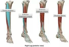
- The Tibialis anterior muscle gets attached to the shaft of the tibia. It originates from the upper 2/3rd area of the lateral surface of the shaft. The primary action of the tibialis anterior is dorsiflexion and inversion of the foot.
- On the upper portion of the medial surface of the shaft of the tibia, sartorius, gracilis, and semitendinosus are inserted.
- The soleus muscle originates from the sole line present on the medial border of the tibia and it primarily performs plantar flexion.
- The popliteus muscle originates from the lateral epicondyle of the femur and gets inserted into the triangular area that is present above the sole line.
Blood supply
The nutrient artery, which is thought to be the largest in the human body, supplies the Tibia bone. The nutrient artery is a branch of the posterior tibial artery, which typically enters the bone through the bone’s posterior surface.
Blood supply The tibia receives blood supply from two different sources: periosteal vessels derived from the anterior tibial artery and a nutrient artery as the primary source Joints Joints The tibia is part of four joints: the tibiofibular joints (knee, ankle, and superior and inferior)
One of the two articulations that connect the tibia to the femur in the knee is referred to as the tibiofemoral component of the knee joint. It is part of the knee joint that supports weight.
Articulations
The articulations between the tibia and fibula are the tibiofibular joints, which allow for a very little movement. The proximal tibiofibular joint is a small plane joint [citation needed]. The undersurface of the lateral tibial condyle and the head of the fibula forms the joint. The joint capsule is supported by the head of the fibula’s anterior and posterior ligaments. The distal ends of the lower limb’s tibia and fibula are joined to the talus at an ankle joint called the synovial hinge joint. such as the talocrural joint. When compared to the smaller fibula’s articulation with the talus, the articulation between the tibia and talus is more significant. Conditions and problems include fractures, osteoporosis, Osgood-Schlatter disease, and Paget’s disease of the bone, which are the most common problems with tibias. [citation needed]
Clinical importance
A “bone fracture” is a broken bone in medicine. Due to their strength, tibias are typically only broken by severe injuries like car accidents, falls, or other traumas. These are some of the symptoms of a fracture:
- Pain.
- Swelling.
- Tenderness.
- Failure to move your leg like you generally can.
- Bruising or discoloration.
- an abnormal bump or deformity on your body.
Go to the hospital right away if you think you have a fracture or have been hurt.
Because bones become weaker as a result of osteoporosis, they are more likely to break suddenly and without warning. Until they break a bone, many people don’t know they have osteoporosis. Regularly, there are no undeniable side effects.
Women, people who were born female, and adults over the age of 50 are more likely to develop osteoporosis. A bone density test, which can detect osteoporosis before it causes a fracture, should be discussed with your doctor. Disease of Osgood-Schlatter: Osgood-Schlatter disease is a condition that causes pain in the knee and upper shin when tendons pull against the top of your shinbone. Typically, adolescents and young children are affected. Another name for it is Jumper’s knee. Some side effects are:
- Swelling.
- Tenderness.
- Pain just below the kneecap.
If your child develops new knee pain, talk to your provider.
Osteitis deformans, also known as Paget’s disease of the bone, is a chronic bone disorder. It causes the affected bones to redevelop continually. Most of the time, people over 50 and of Northern European descent are affected. The signs include:
- Bone or joint pain.
- Larger head size.
- Bowed arms or legs.
- The curvature of the spine.
- Bone fractures.
How are tibias examined?
A bone thickness test is the most well-known procedure for assessing your tibia’s health. It may also be referred to as a DEXA or DXA scan on occasion. The strength of your bones is measured by bone density using low levels of X-rays. It’s a way to look at how much bone loss comes with age.
If you’ve encountered a tibial break your supplier or specialist could require imaging tests, including:
CT scan and x-ray MRI (Magnetic Resonance Imaging).
What are common tibia treatments?
Unless you’ve had a fracture or been told you have osteoporosis, your tibia usually won’t need treatment.
Treatment for a tibial fracture The way your fracture is treated depends on the type and the cause. You will require some form of immobilization, such as a splint or cast, and most likely will need surgery to realign (set) your bone and keep it there so it can heal.
Treatment for osteoporosis Exercise, vitamin, and mineral supplements, and medications are all options for treating osteoporosis.
The majority of the time, you only need to exercise and take supplements to prevent osteoporosis. A treatment plan that is custom fitted to your necessities and your bone well-being will be created with your supplier’s help.
Care
Maintaining your bone and overall health will be easier if you stick to a healthy diet, exercise routine, and regular checkups with your doctor. If you are over 50 years old or have osteoporosis in your family, you should talk about having a bone density scan with your doctor.
To reduce your risk of injury, follow these general safety precautions:
- Always wear your seatbelt.
- For all sports and activities, wear the appropriate safety gear.
- The mess that could block your well-being or that of others ought to be eliminated from your work area and home.
- At home, consistently utilize the right instruments or gear to arrive at things. Never stand on tables, seats, or ledges.
- If you eat right and exercise regularly, you can keep your bones in good shape.
- Use a walker or cane if you have trouble walking or are more likely to fall.
Summary:
When you smash your shin into the edge of your coffee table, you probably only think about your tibia. The tibia is an important part of your ability to stand and move throughout the day, even if you don’t think about it.
At home, consistently utilize the right instruments or gear to arrive at things. Never stand on tables, seats, or ledges.
FAQs
Two long bone in the lower limb is the tibia and the fibula. They connect the knee and ankle and are distinct bones. The larger of the two bones in the lower leg is the tibia, which is the shinbone. The top and bottom of the tibia connect, respectively, to the knee and ankle joints.
Articulations. The knee’s tibial-femoral joint is formed when the tibia and femur articulate proximally. The ankle’s talocrural joint is formed when the talus and tibia articulate distally.
When the tibial nerve is damaged, it causes tibial nerve dysfunction. Numbness, pain, tingling, and weakness of the knee or foot are all possible signs. Fractures or other injuries to the back of the knee or the lower leg frequently cause damage to the tibial nerve.
The fibula is not a bone that can support weight, unlike the tibia. Its primary function is to stabilize the ankle joint by joining with the tibia. Numerous ligament attachment grooves on the distal end of the fibula stabilize and provide leverage during ankle movements.
The tibia, or shinbone, is the long bone in the body that breaks the most frequently. Below and above the ankle, along the length of the bone, a tibial shaft fracture occurs. A significant force typically is required to cause this kind of broken leg.

7 Comments