Lateral Collateral Ligament (LCL)
Table of Contents
Introduction
The LCL, or lateral collateral ligament, is an important ligament located on the outer side of the knee. It provides stability to the knee joint and helps prevent excessive sideways movement of the knee.
The LCL is composed of three distinct layers of tissue. The outer layer is made up of dense connective tissue, which provides strength and stability to the ligament. The middle layer is composed of elastic fibers, which allow the ligament to stretch and contract during movement. The inner layer is made up of synovial tissue, which produces synovial fluid to lubricate the joint.
LCL injuries can occur due to a variety of reasons, including sudden twisting or bending of the knee, direct impact on the knee, or overuse injuries. Symptoms of an LCL injury may include pain, swelling, and difficulty moving the knee. Treatment for LCL injuries may involve rest, ice, compression, elevation, physical therapy, and in some cases, surgery. Recovery time for an LCL injury can vary depending on the severity of the injury and the treatment approach used.
Structure of the Lateral Collateral Ligament
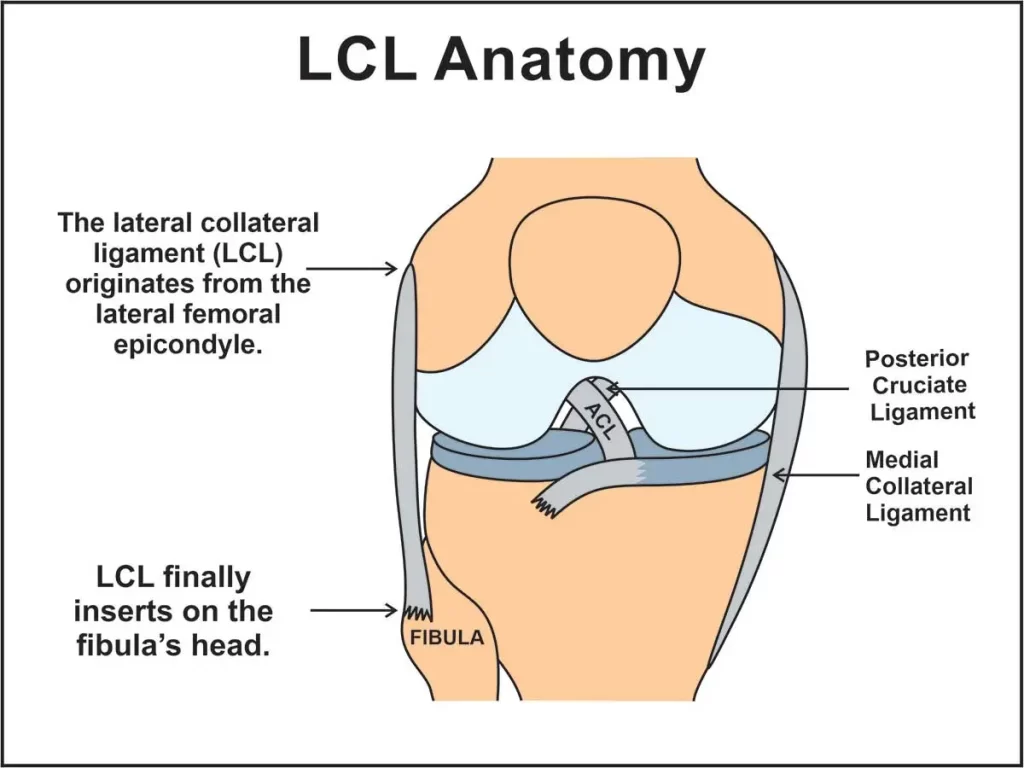
The lateral collateral ligament (LCL) originates from the lateral epicondyle of the femur bone, which is a bony projection on the outer side of the lower end of the thigh bone. Specifically, the LCL originates from a small depression just below the lateral epicondyle called the lateral epicondylar ridge.
From its origin, the LCL runs down and slightly backward to its insertion point on the head of the fibula bone. The fibula bone is a long, thin bone located on the outer side of the lower leg and serves as a site for muscle attachment. The head of the fibula is a rounded bony prominence at the top of the bone that forms part of the knee joint.
The LCL inserts into the fibula bone at two distinct points. The first point of insertion is on the lateral surface of the fibular head, which is a flat area on the top of the bone-facing outward. The second point of insertion is on the posterior aspect of the fibular head, which is a curved surface facing backward.
In the end, the LCL originates from the lateral epicondyle of the femur bone and inserts into two distinct points on the head of the fibula bone. The attachment sites of the LCL are important for providing stability to the knee joint and preventing excessive side-to-side and rotational movements.
Function of the Lateral Collateral Ligament
The lateral collateral ligament (LCL) plays an important role in providing stability to the knee joint. Its main function is to prevent excessive side-to-side movement of the knee joint, also known as varus stress. The LCL works in conjunction with other ligaments and structures in the knee, such as the medial collateral ligament (MCL) and the anterior cruciate ligament (ACL), to maintain proper alignment and stability of the knee joint.
When the knee is subjected to a varus force, such as during a sudden change in direction or a blow to the outer side of the knee, the LCL helps to resist this force and prevent the knee from collapsing inward. This helps to protect the knee joint from injury and maintain its proper alignment.
moreover, to its role in preventing varus stress, the LCL also helps to stabilize the knee joint during rotational movements. When the knee is rotated, such as during pivoting or twisting motions, the LCL helps to control the movement of the tibia bone and prevent excessive rotation that could lead to injury.
Overall, the LCL is an essential structure in maintaining the stability and proper alignment of the knee joint. Without it, the knee would be more susceptible to injury and instability during activities that involve side-to-side or rotational movements.
Blood supply of the Lateral Collateral Ligament
The blood supply of the lateral collateral ligament (LCL) is provided by several small arteries that originate from the genicular arterial network. The genicular arterial network is a complex network of small arteries that supplies blood to the knee joint and its surrounding structures.
The main arteries that supply blood to the LCL are the lateral superior genicular artery and the lateral inferior genicular artery. These arteries branch off from the popliteal artery, which is a major artery located behind the knee joint.
The lateral superior genicular artery runs along the outer side of the knee joint and supplies blood to the upper portion of the LCL. The lateral inferior genicular artery runs along the lower portion of the LCL and supplies blood to the lower portion of the ligament.
In addition to these arteries, there are also several smaller branches that supply blood to the LCL. These include the anterior tibial recurrent artery, which supplies blood to the anterior portion of the LCL, and the fibular collateral artery, which supplies blood to the lateral side of the knee joint.
In the end, the blood supply to the LCL is relatively robust, with multiple arteries providing a steady flow of oxygen and nutrients to the ligament. This helps to ensure that the LCL remains healthy and strong, and is able to perform it is important role in stabilizing the knee joint.
Symptoms of the Lateral Collateral Ligament Injury
Symptoms of an LCL injury can vary depending on the severity of the injury. Common symptoms include:
- Pain: Pain is the most common symptom of an LCL injury. The pain may be mild or severe, and it may be felt on the outside of the knee.
- Swelling: Swelling around the knee is another common symptom of an LCL injury. The swelling may be localized to the outside of the knee.
- Stiffness: Stiffness in the knee may occur after an LCL injury. The knee may feel tight and difficult to move.
- Instability: If the LCL is completely torn, the knee may feel unstable or wobbly. This can make it more difficult to walk or bear weight on the injured leg.
- Bruising: Bruising around the knee may occur after an LCL injury. The bruising may be mild or severe, depending on the severity of the injury.
- Limited range of motion: An LCL injury can cause a limited range of motion in the knee. This can make it difficult to bend or straighten the knee fully.
- Clicking or popping sounds: In some cases, an LCL injury may cause clicking or popping sounds in the knee when it is moved.
Cause of Lateral Collateral Ligament Injury
The lateral collateral ligament (LCL) can be injured by a variety of activities that put stress on the ligament. Some common causes of LCL injuries include:
- Direct impact to the knee: A direct blow to the knee, such as in a car accident or a contact sport, can cause the LCL to stretch or tear.
- Twisting or pivoting movements: Sudden twisting or pivoting movements of the knee, such as those commonly seen in sports like soccer or basketball, can also put stress on the LCL and cause it to tear.
- Hyperextension of the knee: When the knee is forced to bend backward beyond its normal range of motion, it can put excessive stress on the LCL and cause it to tear.
- Sudden changes in direction: Abrupt changes in direction while running or jumping can also cause the LCL to stretch or tear.
- Overuse injuries: Repetitive stress on the LCL, such as from running or jumping, can lead to overuse injuries like tendinitis or inflammation of the ligament.
- Degenerative conditions: As we age, the LCL can become weakened and more prone to injury due to degenerative conditions like arthritis.
It is very important to note that LCL injuries often occur in combination with other knee injuries, such as ACL or meniscus tears. If you suspect that you have injured your LCL, it is important to take medical attention and get an accurate diagnosis and treatment plan.
Treatment of the Lateral Collateral Ligament Injury
Conservative treatment
Conservative treatment for an LCL injury involves non-surgical methods to manage pain, reduce swelling, and promote healing. The following are some common conservative treatments for an LCL injury:
- Rest: Rest is vital for allowing the knee to heal. Avoid activities that cause pain or discomfort, and limit weight-bearing activities as much as possible.
- Ice: The application of ice to the knee can be helpful in reducing swelling and pain. Application of ice should be for 20 minutes at a time, a few times a day.
- Compression: Wearing a compression bandage or brace can help reduce swelling and provide support for the knee.
- Elevation: Elevating the injured leg above the level of the heart can help in reducing swelling.
- Physical therapy: A physical therapist can help you perform exercises that can improve knee strength and range of motion.
- Medications: Over-the-counter pain relievers such as acetaminophen or ibuprofen can be helpful in reducing pain and inflammation.
- Injection therapy: Corticosteroid injections may be used to reduce inflammation and pain in the knee.
It is very important to take in mind that conservative treatment may not be appropriate for all LCL injuries, especially those that are severe or involve other knee injuries. In some cases, surgery may be necessary to repair or reconstruct the LCL. It is important to consult with a medical professional to determine the best course of treatment for your specific injury.
Physiotherapy treatment
Physiotherapy treatment for an LCL injury typically involves a combination of exercises and manual therapy techniques to promote healing, reduce pain and inflammation, and improve knee function. The following are some common physiotherapy treatments for an LCL injury:

Range of motion exercises: These exercises help to improve the flexibility and range of motion of the knee joint. Examples involve knee bends, heel slides, and ankle pumps.
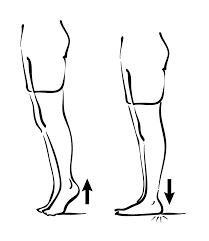
Strengthening exercises: These exercises help to improve the strength of the muscles that support the knee joint, including the quadriceps, hamstrings, and calf muscles. an example involves calf raises, leg presses, and squats.
Balance and proprioception exercises: These exercises help to improve balance and coordination, which can reduce the risk of re-injury. Examples include single-leg stands, wobble board exercises, and step-ups.
Manual therapy techniques: These techniques are used to reduce pain and inflammation in the knee joint. Examples involve joint mobilization, massage, and stretching.
Modalities: Modalities such as ultrasound, electrical stimulation, and ice or heat therapy may be used to reduce pain and inflammation in the knee joint.
Functional training: This involves specific exercises that mimic the movements required for daily activities or sports activities, with the goal of improving overall knee function.
The specific physiotherapy treatment plan will depend on the severity of the injury, as well as individual factors such as age, activity level, and overall health. It is very important to work closely with a physiotherapist to get a personalized treatment plan that uses for your specific needs and goals.
Risk factors
The LCL, or lateral collateral ligament, is a ligament located on the outer side of the knee. It provides stability to the knee joint and helps prevent excessive outward movement of the knee. Like any other ligament, the LCL can be injured due to a variety of factors. Here are some of the risk factors that can increase the likelihood of LCL injury:
- Sports: Athletes who participate in sports that involve sudden changes of direction or contact with other players, such as football, basketball, and soccer, are at a higher risk of LCL injury.
- Overuse: Repetitive stress on the knee joint, such as running long distances or performing jumping exercises, can lead to overuse injuries to the LCL.
- Previous knee injuries: Individuals who have had previous knee injuries, such as ACL tears or meniscus tears, are more likely to experience LCL injuries.
- Poor biomechanics: Abnormal alignments of the knee joint, such as bowlegs or knock-knees, can put extra stress on the LCL and increase the risk of injury.
- Obesity: Excess weight can put additional stress on the knee joint and increase the risk of LCL injury.
- Age: As we age, our ligaments become less elastic and more prone to injury. Older individuals may be more susceptible to LCL injuries.
- Genetics: Some individuals may be born with weaker ligaments, which can increase their risk of LCL injury.
How to reduce the risk of injuries?
Reducing the risk of LCL injuries involves taking steps to protect the knee joint and maintain its stability. Here are some ways to reduce the risk of LCL injuries:
- Maintain a healthy weight: Excess weight can put additional stress on the knee joint, increasing the risk of LCL injury. Maintaining a healthy weight through daily exercise and a balanced diet can help in reducing this risk.
- Wear proper footwear: Wearing shoes that give perfect support and cushioning can help in reducing the effect on the knee joint while physical activity. This can help prevent LCL injuries.
- Practice good biomechanics: Proper alignment and form during physical activity can help reduce stress on the knee joint and prevent LCL injuries. This may involve working with a coach or trainer to learn proper techniques for your sport or activity.
- Warm up and cool down properly: Taking the time to warm up before physical activity and cool down afterward can help prepare the muscles and joints for exercise and prevent injury.
- Strengthen the muscles around the knee: Strengthening the muscles that support the knee joint, such as the quadriceps and hamstrings, can help improve knee stability and reduce the risk of LCL injuries.
- Use proper equipment: Using proper equipment, such as knee pads or braces, can help protect the knee joint during physical activity and reduce the risk of LCL injuries.
- Rest and recover: Taking time to rest and recover after physical activity can help prevent overuse injuries, which can increase the risk of LCL injuries.
By taking these steps, individuals can reduce their risk of LCL injuries and protect the health and stability of their knee joints.
FAQs
The lateral collateral ligament (LCL) is a band of tissue that connects the femur (thigh bone) to the fibula (lower leg bone) on the outer side of the knee joint.
LCL injuries can be caused by direct impact to the outer side of the knee, sudden twisting or pivoting movements, or overuse from repetitive activities.
Symptoms of an LCL injury may include pain, swelling, stiffness, and instability in the knee joint. Some people may also feel a popping or tearing sensation during the time of occurrence of injury.
LCL injuries are typically diagnosed through a physical exam and imaging tests such as X-rays or MRI scans.
Treatment for LCL injuries may include rest, ice, compression, elevation (RICE), physical therapy, and in some cases, surgery. The specific treatment approach will depend on the severity of the injury and individual circumstances.
Yes, LCL injuries can be prevented by maintaining a healthy weight, wearing proper footwear, practicing good biomechanics, warming up and cooling down properly, strengthening the muscles around the knee, using proper equipment, and taking time to rest and recover after physical activity.


One Comment