Tarsals Bone
Table of Contents
Introduction
Tarsal bones are a group of seven small bones located in the ankle joint of the human body. They are responsible for supporting the weight of the body and facilitating movement. The tarsal bones include the talus, calcaneus, navicular, cuboid, and three cuneiform bones (medial, intermediate, and lateral).
The talus bone is the highest of the tarsal bones and connects the leg bones (tibia and fibula) to the foot bones. The calcaneus bone is the largest of the tarsal bones and forms the heel of the foot. The navicular bone is located in the middle of the foot, while the cuboid bone is situated on the outer side of the foot.
The three cuneiform bones are small and wedge-shaped, and are located in the front part of the foot. The medial cuneiform bone is situated next to the first metatarsal bone, the intermediate cuneiform bone is located between the other two cuneiform bones, and the lateral cuneiform bone is located next to the fifth metatarsal bone.
Together, the tarsal bones create the complex structure of the ankle joint, allowing for a wide range of movement and stability. Injuries to the tarsal bones can cause significant pain and mobility issues, and may require medical attention to properly heal.
There are 26 bones in the foot’s skeleton, which can be divided into three categories:
- The tarsals (ankle joint)
- The metatarsus
- The phalanges (bones of the toes)
There are 14 phalanges, 5 tarsal bones, and 7 metatarsal bones.
The forefoot (metatarsals and phalanges), the midfoot (cuboid, navicular, and cuneiforms), and the hindfoot (calcaneus and talus) are the anatomical divisions of the foot.
Wounds to the bones of the foot normally happen in competitors and dynamic people. The majority of injuries occur as a result of sports-related acute trauma.
Due to the absence of endochondral ossification and the presence of their cartilaginous epiphyseal plates, adolescents are particularly susceptible to these injuries. As a result, it is essential to possess fundamental anatomical knowledge of the foot’s bones, including their articulations and attachment sites.
The tarsals are a group of seven articulating bones in each foot that are located between the metatarsus and the lower end of the tibia and fibula of the lower leg. The cuboid, medial, intermediate, lateral cuneiform, and navicular are included for the midfoot, while the talus and calcaneus are included for the hindfoot.
The tarsals make contact with the metatarsal bones, which in turn make contact with the toes’ proximal phalanges. The ankle joint proper is the joint between the tarsus below and the tibia and fibula above.
The calcaneus, which is the weight-bearing bone in the heel of the foot, is the largest bone in the tarsals of humans.
The ankle joint, also known as the talocrural joint, is formed by the superior connection of the talus bone, or ankle bone, to the two lower leg bones, the tibia, and fibula. inferior to the calcaneus, or heel bone, at the subtalar joint. The talus and calcaneus together make up the hindfoot.
The arches of the foot, which absorb shock, are formed by the five irregular midfoot bones—the cuboid, navicular, and three cuneiform bones. Muscles and the plantar fascia link the midfoot to the hind and forefoot.
Structure and Anatomy of Tarsals Bone
The tarsal bone
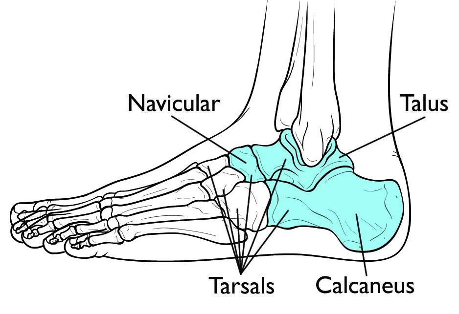
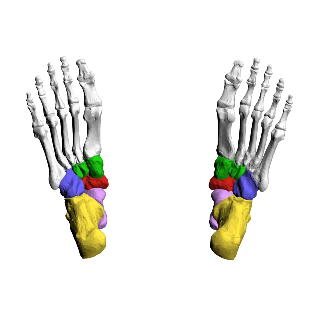
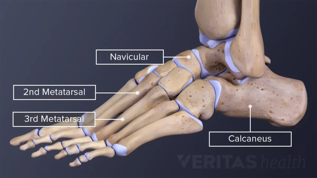
The tarsal consists of 7 bones that make up the posterior aspect of the foot: talus, calcaneus, cuboid, navicular, and three cuneiforms.
The talus and calcaneus of the hindfoot and the cuboid and cuneiform of the midfoot are examples of the tarsal bones. The navicular is the transitional bone between these two gatherings.
As previously stated, some tarsal bones form the midfoot and some form the hindfoot. There are three rows of these foot bones: proximal, distal, and intermediate.
Let us now discuss the 7 bones:
Bones in the Hindfoot (Proximal Tarsal Bones)
The talus and calcaneus bones are the proximal tarsal bones of the hindfoot. They construct a bony framework that encircles the ankle and heel.
- Talus (ankle bone): It is the most superior of the lot and forms the ankle joint articulating with the tibia and fibula.
- Calcaneus (heel bone): It is the heel’s largest tarsal bone, which is underneath the talus.
The tarsal tunnel is an important structure in this area of the foot. It is composed of the talus, calcaneus, and tibia. It connects the foot to the leg’s posterior part and serves as a conduit for numerous tendons, nerves, and blood vessels.
Bones in the Midfoot (Transitional and Distal Tarsal Bones)
In the midfoot, the tarsal bones are again dispersed into two locales: distal and intermediate The navicular is the only bone in the intermediate row of tarsal bones. The distal region is made up of the remaining four, which include the cuboid and the three cuneiforms.
- Navicular: It is a boat-shaped bone that articulates with the calcaneus and the other tarsals.
- Cuboid: It is cuboidal in shape and is in the front (anterior) of the calcaneus, as the name suggests. It is the most level of the distal section, lying on the little toe.
- Cuneiform lateral: It is a wedge-molded bone, lying generally parallel to the three cuneiforms, on the huge toe.
- Cuneiform, Intermediate: Between the lateral and medial cuneiforms is another similar-shaped cuneiform bone.
- Cuneiform: It is a cuneiform that is wedge-shaped and lies in front of the navicular bone (anterior).
An easy way to remember the names of the seven tarsal bones will be through the following sentences, which contain their acronyms.
- Tiger Cub Needs MILC.
Breaking the mnemonic:
T: Talus
C: Calcaneus
N: Navicular
M: Medial cuneiform
I: Intermediate cuneiform
L: Lateral cuneiform
C: Cuboid
Talus bone
Bone (Latin for the lower leg) bone is the most prevalent bone of the bone structure and lays on top of the calcaneus. Three areas of explanation structure the lower leg joint:
- The talus’s superomedial aspect articulates with the tibia’s medial malleolus.
- The tibia’s distal portion articulates with the superior portion of the talus.
- The talus’s lateral aspect articulates with the fibula’s lateral malleolus.
Anteriorly the talus articulates with the navicular bone. The intermediate talus bone and the talus articulate midway.
The talus has a body, head, and neck. Two malleoli are attached to the superior surface, or trochlea, which contributes to the joint’s stability. The trochlea is more extensive anteriorly, prompting a distinction in solidness relying upon joint position. The ligaments are stretched when the joint is in dorsiflexion, resulting in joint stability. Plantarflexion reduces stability because it makes the trochlea narrower.
Calcaneus bone
The biggest and most solid bone in the foot is the calcaneus, which is also known as the impact point bone. The substantialness of the body goes through the tibia, into the bone, and a while later to the calcaneus. On the average portion of the calcaneus that supports the bone, there is a hard bone called the sustentaculum.
For articulation with the talus, the calcaneus’ superior aspect has three articular surfaces. The cuboid articulates with the anterior surface. The calcaneal tubercle is a large weight-bearing region on the posterior aspect of the calcaneus.
A calcaneal fracture can occur during a hard fall on the heel, such as when falling from a ladder. X-ray or CT can be used to confirm a diagnosis based on symptoms (pain, bruising, difficulty walking).
Cuboid bone
The calcaneus end and the posterior cuboid bone articulate. It is the most horizontal bone in the distal line of the tarsal bones. A groove on the lateral and inferior surfaces of the bone, anterior to the cuboid tuberosity, allows a muscle tendon to pass through.
Navicular bone
The navicular bone gets its name because it looks like a small boat. Between the three cuneiforms and the posteriorly positioned head of the talus is the navicular. The navicular bone articulates with the talus proximally, the three cuneiforms distally, and the cuboid laterally on the medial part of the foot.
Cuneiforms bone
Wedge-shaped cuneiform bones can be found. Based on their relative positions, they are referred to as lateral, intermediate, and medial. The transverse curvature of the foot is the result of the narrow inferior surface and broad superior surface of each cuneiform. The respective metatarsals of each cuneiform articulate with the navicular posteriorly.
Articular surfaces
An articulation between the head of the talus and the navicular bone’s proximal or posterior aspect is known as the talonavicular joint.
A navicular articular surface points distally or anteriorly on the head of the talus. It has an overall inferomedial orientation and is oriented inferiorly in the vertical plane and medially in the horizontal plane. In both the horizontal and vertical planes, the surface of the navicular articular joint is ovoid and convex. In contrast, the navicular bone’s corresponding proximal articular surface is concave and oval in the horizontal plane, pointing distally to meet the navicular articular surface.
The anterior (distal) aspect of the calcaneus and the posterior (proximal) aspect of the cuboid are connected by the calcaneocuboid joint.
The cuboid’s articular surface is concave-convex and undulating, with a quadrilateral shape on the calcaneus. It is divided into superior and inferior sections with opposing orientations. The superior portion is arched in similar planes while the inferior portion is curved in even and vertical planes. The cuboid’s articular surface has structural characteristics that are similar but mutually exclusive; it is likewise quadrilateral and undulating, with a curved predominant part and a sunken sub-par part. Because of this matching and mirroring relationship, this joint has a saddle-like shape and a congruent fit between the articular surfaces when compared to the talonavicular joint.
Functions
These bones help the feet withstand the weight of the body by providing mechanical support to the soft tissues in the feet. Together with the other bones in the foot, they form a longitudinal arch that serves as a sturdy platform for supporting weight while standing or moving.
Blood supply
Blood supply to the cross-over tarsal joint is from the parallel tarsal conduit, a part of the dorsal pedal corridor. The latter comes from the artery that runs along the anterior tibia.
Muscles
Tendons connect the muscles in the lower leg to the bones in the foot, where they control the movements of the foot.
The main muscles in the foot that help move are these:
The posterior tibialis: The Tibialis anterior muscle, which supports the foot’s arch: The muscle that permits the foot to move up
Peroneus longus and brevis: Extensors, are the muscles on the outside of the ankle that control movement: Flexors are the muscles that raise the toes so that you can take a step: The muscles that balance out the toes and twist them under
Ligaments
The connective tissues that connect bones together are called ligaments.
Talonavicular ligaments
The talonavicular joint is supported by three main ligaments, despite having a weak joint capsule;
- Inferiorly by the plantar calcaneonavicular ligament,
- laterally by the part of the bifurcate ligament called the calcaneonavicular ligament
- Superiorly by the (dorsal) talonavicular ligament
When taken as a whole, these structures create a substantial supporting socket around the navicular bone that can hold the talus head’s ball. The transverse tarsal joint is also strengthened by doing this.
Plantar calcaneonavicular ligament
The thick but flexible plantar calcaneonavicular ligament has a triangular shape and runs almost transversely across the foot. It extends from the calcaneus’ sustentaculum tali’s anteromedial aspect to the navicular bone’s inferomedial aspect. The calcaneonavicular ligament and medial collateral ligament provide support for the ligament. The dorsal fibrocartilaginous facet of the talus is supported by the plantar calcaneonavicular ligament at the talonavicular joint. Additionally, it stabilizes the foot’s medial longitudinal arch.
Bifurcate ligament
There are two components to the bifurcate ligament: the ligaments of the calcaneonavicular and calcaneocuboid. The calcaneofibular ligament, also known as the talonavicular joint is supported laterally by the calcaneonavicular portion of the bifurcate ligament. It reaches the navicular bone’s lateral aspect from the super interior aspect of the calcaneus. The calcaneocuboid ligament, also known as The dorsomedial aspect of the calcaneocuboid joint is strengthened by the calcaneocuboid part of the bifurcate ligament. It runs from the very inside part of the calcaneus until the dorsomedial point of the cuboid bone. It connects the proximal and intermediate tarsal bone rows that are adjacent.
Talonavicular ligament
Between the plantar calcaneonavicular and calcaneonavicular ligaments is the broad and thin talonavicular ligament. It connects the dorsal surface of the talus to the superior, or dorsal, surface of the neck. The talonavicular joint is supported dorsally by the talonavicular ligament.
Other ligaments
The talonavicular joint is directly protected and strengthened by the aforementioned ligaments. This joint, on the other hand, is supported by several additional ligaments that are further away, indirect, but significant. These include the ankle joint’s medial and lateral collateral ligaments, the ankle’s inferior extensor retinaculum, and the subtalar joint’s talocalcaneal ligaments.
Movements
Subtalar inversion and eversion are the outcomes of the complex motion that the subtalar joint performs in three planes. The subtalar joint, along with the talonavicular and calcaneocuboid joints, converts tibial rotation into forefoot supination and pronation. The joint’s pivot of turn is up 42 degrees from the level plane and 16 degrees medially from the foot’s midline. However, Together, the subtalar facets form an Archimedean spiral or screw in the right foot that is the center of subtalar motion. Because of this, the calcaneus also moves forward along the screw’s axis and in a clockwise direction during subtalar inversion. 20-30 degrees of inversion and 5-10 degrees of eversion constitute the typical subtalar motion. During the gait cycle, functional motion ranges from 10 to 15 degrees, with the heel striking the ground in a slight inversion followed by a quick version.
The transverse tarsal joint, also known as Chopart’s joint, is made up of the talonavicular and calcaneocuboid joints, which are located between the talus and navicular bones and the calcaneus and cuboid bones. It moves in two directions. Eversion and reversal happen around a longitudinal hub that is situated 9 degrees medially from the foot’s longitudinal pivot and 15 degrees vertically from the level plane. The majority of flexion and extension take place around an oblique axis that is 57 degrees anteromedially (forward-inward) and 52 degrees up from the horizontal plane. The talonavicular motion in vitro is 17 degrees pronation-supination and 7 degrees flexion-extension; while the calcaneocuboid movement range is 7 degrees pronation-supination and 2 degrees flexion-extension.
The foot can be either flexible or rigid due to the interactions between the subtalar and transverse talar joints. The fact that the two joints of the transverse joint are parallel when the subtalar joint is in version makes it possible to move this joint. The transverse joint’s axes converge when the subtalar joint is inverted, locking the joint’s movements and rendering the midfoot rigid.
Clinical importance
Tarsal Fractures
A break of any of the tarsal bones in the foot is known as a tarsal bones crack. They are uncommon and can be caused by force from doing the same thing over and over. The calcaneus or navicular bones are typically affected by stress fractures or hairline fractures.
Your doctor may check your vitamin D level if you have a tarsal stress fracture, especially a navicular fracture because vitamin D is necessary for healing.
Fractures of the tarsal bones of the foot are known as tarsal fractures. The anatomic site further subdivides these fractures:
- calcaneal fracture (most common)
- calcaneal tuberosity avulsion fracture
- lover’s fracture
- talar fractures
- talar head fracture
- talar neck fracture
- aviator fracture
- lateral talar (snowboarder) process fracture
- posterior talar process fracture
- navicular fracture
- cuboid fracture
- nutcracker fracture of the cuboid
- cuneiform fracture
- medial cuneiform fracture
- intermediate cuneiform fracture
- lateral cuneiform fracture
Tarsal fracture symptoms
Strong force or foot trauma can result in acute tarsal fractures. The signs include:
- Sudden pain at the time of injury.
- Difficulty bearing weight.
- Loss of function of your foot.
- Tenderness in a particular area, depending on the fractured bone.
- You may also have a deformity.
If your pain is more gradual and worse during weight-bearing, then it is more likely you have a stress fracture.
FAQs
The calcaneus is the biggest and most grounded tarsal bone. It is the tarsal bone that breaks the most, causing about 2% of all fractures. The significance of the calcaneal angles in determining the severity of calcaneus fractures and formulating treatment plans has been emphasized.
The foot’s tarsal bones aid in maintaining the foot’s arches and serve as crucial points of attachment for leg and foot muscles. The bone is the bone that associates the leg with the foot.
The carpal bones of the upper limb are matched by the tarsals. In bipedal locomotion, the tarsal and metatarsal bones of the human foot create a longitudinal arch that is well-suited to carrying and transferring weight. Seven tarsal bones are present in the human ankle.
There are seven explicit entomb tarsal joints (explanations) in the human foot: Subtalar joint. Talocalcaneonavicular joint.