Structure Of The Eye
The eye is a complex organ that consists of several structures that work together to allow us to see. These structures include the cornea, iris, pupil, lens, retina, optic nerve, vitreous humor, sclera, choroid, and ciliary body. Understanding the structure of the eye can help us understand how we perceive light and visual information.
Table of Contents
Structure
The eye of a person is a great organ. Our goal is to understand a complex system with many complicated elements that all work together to produce a final product that practically everyone uses heavily on a daily basis. Despite the fact that we regard our vision the most among all of our senses (it accounts for 80% of all impressions), shockingly few people are aware of how our eyes function.
When we gaze in the mirror, we can distinguish between the portions of the eye that are on the outside and those that are on the interior or farther away. One of the main sense systems in the human body is the eye. Since it is extremely delicate and susceptible to many diseases, protection, and prevention are essential to keep the safe and healthy.
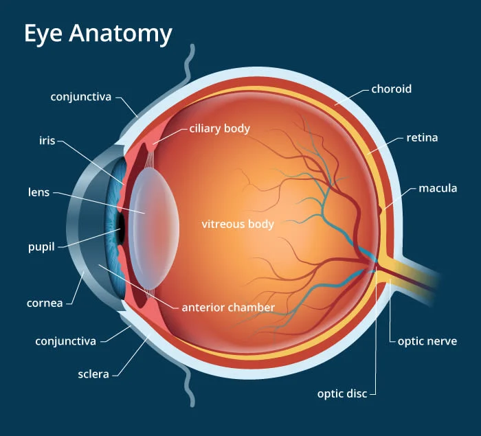
Eye Anatomy
Three layers in the eye.
As listed below, the eyeball has three coatings.
- exterior fibrous layer
- central vascular coat
- inner nervous system
Exterior fibrous layer
The cornea is the anterior, transparent, and one-sixth of the eyeball. The light is refracted into the eye as a result. The conjunctiva, a membrane tissue, extends the cornea even more. The limbus is the region where the conjunctiva and cornea meet. The cornea and sclera combine to produce the fibrous outer layer.
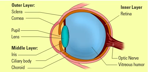
Central vascular coat
The iris, ciliary body, and choroid work together (from anterior to posterior) to generate this covering. Under the sclera is a vascular and colored layer.
Inner nervous system
The retina is what makes up the internal nervous system. An inverted image of the objects seen is sent to the retina. The optic nerve, which connects at the back of the eyeball, carries these images from the retina to the brain.
Visible parts of the eye
Eyelid: Your eyelid shields your eye from debris, sweat, and other contaminants that could harm it. It promotes blinking to maintain the eye well-lubricated and hydrated by opening and closing both voluntarily and involuntarily.
Pupil: The pupil, which is where we view through the eye, enlarges or shrinks depending on the amount of light. The pupil shrinks to let less light in if the environment is exceptionally bright, whereas it expands to let more light in when the environment is darker. This ensures that the proper quantity of light reaches the retina at the rear of the eye and helps us see clearly in a variety of lighting conditions.
Sclera: The white portion of your eye, or sclera, serves as an outer layer of defense. It protects the optic nerve and can provide useful information about the condition of your eyes. For instance, a yellow-tinted sclera may point to liver issues, while a red sclera may signal that your eyes are weary or dry.
Iris: The iris, which is the colored portion of the eye, really regulates the pupil’s size. In other words, it controls how much light enters the eye. The form, pattern, and color of this iris, which is made of muscle and connective tissue surrounding the pupil, are as distinctive as your fingerprints.
Internal parts of the eye
Cornea: The cornea is the transparent surface in front of your eye that allows light to enter. Your iris and pupil are directly covered, adding an additional layer of security. We do laser eye surgery on the cornea because irregularities in your cornea’s curvature are what lead to an eye prescription and your requirement for glasses. Your vision will be better if your cornea has a smoother surface.
Lens: Your lens, which is a component of the eye that enables focus, is situated behind your iris. Light rays passing through the lens are focused so that they strike the retina at the proper angle, changing the focal distance of the eye. As you age, a protein build-up in the eye may cause the lens to become clouded. The term for this is cataract. Fortunately, you can remove your lens and replace it with a synthetic, clear lens to restore your vision.
Aqueous humor: Your eyes continuously manufacture aqueous humor to support healthy ocular pressure and hydrate your cornea. This maintains the health of your eyes, which benefits clear eyesight. It is essential for clear vision and is removed from the eye at the same rate that it is created (when this rate fluctuates, glaucoma develops).
Ciliary muscle: The ciliary muscle, a component of the eye, alters the lens’s shape to enable the eye to focus on various distances. Additionally, it controls the flow of the aqueous humor inside the eye and keeps the lens in the proper place in the middle layer of the eye.
Medial rectus muscle: The medial rectus is the largest of the six extraocular movement muscles in your eye, along with the lateral rectus, superior oblique, superior rectus, inferior rectus, and inferior oblique. It ensures that the eye is properly positioned and pushes the pupil closer to your body’s midline (in the direction of your nose). Strabismus may result from issues with the medial rectus.
Lateral rectus muscle: The muscle known as the lateral rectus is in charge of the eye’s lateral, or sideways, motions, especially those that are away from the midline. Again, esotropia may occur if there are problems with the lateral rectus muscle. Because the muscle that should be moving the eye away from the midline is either too weak or not functioning properly, it twists inward in this type of strabismus.
Retina: The layer of tissue known as the retina is located at the back of the eye. The retina’s main function is to take in light from the lens and transmit signals to the brain so it can convert it into a visual image. Rods and cones are the two categories of photoreceptor cells found in the retina. Cones are in charge of detecting color vision, whereas rods are in charge of detecting motion, dark, and light. Maintaining your retina’s health is essential since issues with the retina can result in loss of eyesight.
Choroid: Between the retina and the sclera in the back of the eye is the choroid, a significant blood artery. It maintains the proper temperature in the eye and feeds the retina’s outer layers. Additionally, it helps the eye work properly by supplying the retina with the proper amount of oxygen and blood flow.
Macula: Your retina’s macula, which has a diameter of about 5 mm, is located in the center of the cell. We will have a clear vision and be able to discern little details if our macula is in good shape. Your central vision is impacted when the macula develops a condition, such as macular degeneration. Your daily life is obviously greatly affected by this, and it may continue to deteriorate until all vision is lost.
Optic nerve: The optic nerve is a component of the eye that carries visual signals from the retina to the brain where they are translated into images. It truly counts as a component of the central nervous system and has over a million nerve fibers. Glaucoma is one of the most prevalent conditions that can harm the visual nerve. When the optic nerve becomes compressed due to increased eye pressure, visual information can no longer be transferred effectively.
Vitreous humor: In your eye, behind your lens but in front of your retina, is a gel-like liquid called the vitreous humor. Floaters are the name for any materials that enter the vitreous humor. While it can be inconvenient to see them in your field of vision, they are usually harmless and can be tiny flecks of blood or clusters of cells. As you become older, your vitreous thins and may even separate from your retina, a condition known as “posterior vitreous detachment”. Even if it worsens floaters, this doesn’t endanger vision.
Function of the Eye:
The eye is an organ that allows us to see and interpret the world around us. Its primary function is to detect and focus light onto the retina, which contains photoreceptor cells called rods and cones. These cells convert the light into electrical signals that are sent to the brain through the optic nerve. The brain then interprets these signals as visual information, which allows us to see and perceive our surroundings.
The eye also has other important functions, including:
- Accommodation: The ability of the eye to change the shape of the lens to focus on objects at different distances.
- Color vision: The ability of the eye to distinguish between different colors of light.
- Depth perception: The ability to perceive the relative distance of objects in 3D space.
- Peripheral vision: The ability to see objects outside of the direct line of sight.
- Protection: The eye is protected by the eyelids, lashes, and tears, which help to keep the eye lubricated and clean.
Overall, the eye is a vital organ that plays a crucial role in our daily lives by allowing us to see, interpret, and navigate our environment.
FAQs
What is the eye’s fundamental structure?
The cornea, iris, pupil, aqueous humor, lens, vitreous humor, retina, and optic nerve are the major structural components of the human eye. Aqueous humor and the clear cornea allow light to enter the eye.
What part of the eye is the most vital?
Retina. The retina is one of the most crucial components of the eye. Ten crucial layers of tissue in the back of your eye called the retina detect light and color and transmit messages to your brain, enabling you to see.
What kinds of eyes are there?
There are six primary eye forms: almond, round, monolid, hooded, downturned, and upturned. Each is gorgeous in its own way. You may have also heard the terms wide-set, asymmetrical, huge, small, close-set, and deep-set used to describe your eyes.
Where exactly is the eye?
On the left and right sides of the face, humans have two eyes. The skull’s orbits are bony spaces where the eyes are located. The whitish sclera, colorful iris, and pupil make up the front portion of the eye that is visible to the naked eye.
Which layer shields the eyes?
The barrier is known as the sclera (SLEER-uh). The cornea, the transparent front surface of the eye, is connected to the hard, fibrous tissue that encircles and protects the eyeball.