
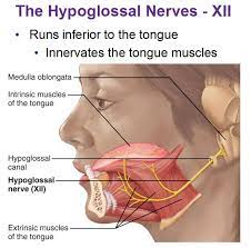
Hypoglossal Nerve-12th Cranial Nerve
The Hypoglossal nerve is the twelth cranial nerve, and innervates all the extrinsic and intrinsic muscles of the tongue, except for the palatoglossus which is innervated by the vagus nerve.
Table of Contents
ANATOMY:
The hypoglossal nerve arises from the hypoglossal nucleus. This is found the full length of the medulla, close to the midline. The roots of the hypoglossal nerve itself arise between the olive and the pyramid. After leaving the hypoglossal canal the hypoglossal nerve receives twigs from C1 and C2 containing general somatic motor fibers, as well as some general sensory fibers from the C2 ganglion. It is these fibers that go on further in the neck to supply the strap muscles.
ANASTOMOSES OF HYPOGLOSSAL:
•Vagus: Several filaments pass between the hypoglossal nerve and the inferior ganglion of the vagus nerve.
•Sympathetic trunk: Via a filament at the exit of the condylar foramen destined either for the superior cervical ganglion or for the carotid filament of the same ganglion.
•Cervical: Opposite the atlas, the nerve is joined by a filament coming from the loop connecting the first and second cervical nerves.
•Trigeminal (lingual nerve): Via a nerve loop formed by the lingual and hypoglossal nerves.
Function of Hypoglossal Nerve
This nerve provides motor control of the extrinsic muscles of the tongue: genioglossus, hyoglossus, styloglossus, and the intrinsic muscles of the tongue. These represent all muscles of the tongue except the palatoglossus muscle.
These muscles are involved in moving and manipulating the tongue. The muscles, attached to the underside of the top and back parts of the tongue, cause the tongue to protrude and deviate towards the opposite side. The hypoglossal nerve also supplies movements including clearing the mouth of saliva and other involuntary activities.
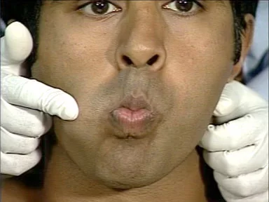
Examination
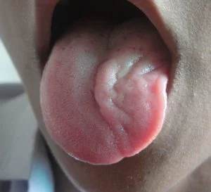
This nerve to be tested, the patient is asked to protrude the tongue in the midline and to move it from side to side. In the presence of a lesion of the hypoglossal nerve, the tongue is seen to deviate toward the side of the lesion, toward the weak half, on the attempted protrusion. This deficit is due to a paralysis of the genioglossus muscle; fasciculations of the tongue might also be observed. At rest, if the nerve is injured a tongue may appear to have the appearance of a “bag of worms” (fasciculations) or wasting (atrophy).
Damage to Hypoglossal Nerve
When the nerve is damaged, the tongue may feel “thick,” “heavy,” or “clumsy.” Weakness of tongue muscles can result in slurred speech, affecting sounds particularly dependent on the tongue for generation (i.e., lateral approximants, dental stops, alveolar stops, velar nasals, rhotic consonants, etc.).Tongue strength may be tested by poking the tongue against the inside of the cheek, while an examiner feels or presses from the cheek.
-If the damage is to the nerve itself (a lower motor neuron lesion), the tongue will curve toward the damaged side, owing to the weakness of the genioglossus muscle of the affected side which action is to deviate the tongue in the contralateral side.
Hypoglossal palsy
-If the damage is to the nerve pathway (an upper motor neuron lesion) the tongue will curve away from the side of the damage, due to the action of the affected genioglossus muscle, and will occur without fasciculations or wasting, with speech difficulties more evident.
– Damage to the hypoglossal nucleus will lead to the wasting of muscles of the tongue and deviation towards the affected side when it is stuck out. This is because of the weaker genioglossal muscle.

4 Comments