How does the Blood Circulatory System work?
Table of Contents
Work of Blood Circulatory System
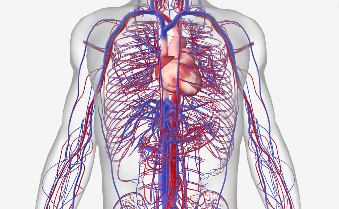
The blood circulatory system (cardiovascular system) provides nutrients and oxygen to all compartments in the body. It consists of the core and the blood crafts racing via the entire body. The arteries take blood away from the heart; the veins take it back to the heart. The system of blood vessels resembles a tree: The “trunk” – the main artery (aorta) – departments into large routes, which lead to smaller and smaller containers. The smallest arteries end in a grid of tiny vessels known as the capillary grid.
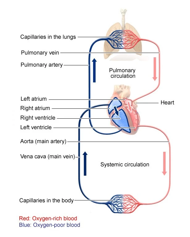
There isn’t only one blood circulatory procedure in the human body, but two, which are connected: Systemic circulation delivers organs, tissues, and cells with blood so that they get oxygen and different vital significance. Pulmonary circulation is where the fresh oxygen we live with joins the blood. At that exact time, carbon dioxide is dismissed from the blood.
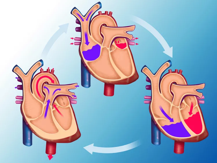
Blood circulation starts when the heart rests between two heartbeats: The blood streams from both atria (the upper two sections of the heart) into the ventricles (the lower two sections), which then expand. The subsequent phase is called the ejection period, which is when both ventricles pump blood into the significant streets.
In circulation, the left ventricle pumps oxygen-rich blood into the aorta. The blood crosses from the major artery to more extensive and smallish arteries and into the capillary grid. There the blood falls off oxygen, nutrients, and different important importance and picks up carbon dioxide and scrap products. The blood, which is now common in oxygen, is organized in veins and traverses to the right atrium and into the right ventricle.
This is where pulmonary circulation begins: The correct ventricle pumps low-oxygen blood into the pulmonary artery, which diverges off into smaller and smaller streets and veins. The veins form a fine grid around the pulmonary cysts (grape-like air sacs at the end of the airways). This is where carbon dioxide is discharged from the blood into the air inside the pulmonary cysts, and fresh oxygen penetrates the bloodstream. When we live out, carbon dioxide escapes our bodies. Oxygen-rich blood journeys via the pulmonary veins and the left atrium into the left ventricle. The next heartbeat creates a new rotation of systemic circulation.
Features of the Circulatory Procedure
The crucial elements of the human circulatory procedure are as follows:
- The human circulatory procedure consists of blood, the heart, blood vessels, and lymph.
- The human circulatory procedure circulates blood through two loops (double circulation) – One for oxygenated blood, and another for deoxygenated blood.
- The human heart consists of four compartments– two ventricles and two auricles.
- The human circulatory procedure possesses a body-wide grid of blood vessels. These comprise routes, veins, and capillaries.
- The primary role of blood vessels is to transport oxygenated blood and nutrients to all parts of the body. It is also charged with collecting metabolic wastes to be displaced from the body.
- Most circulatory system charts do not visually represent its sheer height. Theoretically, if the veins, arteries, and veins of a human were laid out, end to end, it would span a total distance of 1,00,000 kilometers (or roughly eight times the diameter of the Earth).
Structure of the Circulatory System
The circulatory procedure contains the heart, blood vessels, and blood. The cardiovascular procedure in all vertebrates consists of the heart and blood vessels. The circulatory procedure is further separated into two major courses– a pulmonary circulation, and a systemic circulation. Pulmonary circulation is a course loop from the right heart carrying deoxygenated blood to the lungs where it is oxygenated and returned to the left heart. Systemic circulation is a course loop that delivers oxygenated blood from the left heart to the remains of the body, and retrievals deoxygenated blood to the right heart via large veins known as the venae cavae. A macrocirculation and a microcirculation are two other ways to characterize the systemic circulation. Five to six quarts of blood, or around 4.7 to 5.7 liters, make up about 7% of the total body weight of an average adult. Red blood cells, white blood cells, platelets, and plasma all makeup blood. The circulatory system and the digestive system work together to supply the nutrients needed to keep the heart beating.
- Coronary circulation, cerebral circulation, renal circulation, and bronchial circulation—which travel to the bronchi in the lungs—are other circulatory channels that are connected.
The human circulatory procedure is closed, meaning that the blood is collected within the vascular grid. Nutrients travel via small blood crafts of the microcirculation to contact organs. The lymphatic system is an important subsystem of the circulatory method consisting of a grid of lymphatic vessels, lymph nodes, organs, tissues, and circulating lymph. This subsystem is an available system. A major position is to maintain the lymph, draining and returning interstitial liquid into the lymphatic ducts back to the core for recovery to the circulatory system. Another major position is working together with the immune method to supply defense against pathogens.
What Are the Parts of the Circulatory Method?
Two pathways come from the heart:
- Pulmonary circulation is a quick loop from the heart to the lungs and back too.
- Blood is circulated throughout the whole body, from the heart to every other part of it and back again.
In pulmonary circulation:
The pulmonary artery is a big street that arrives from the heart. It divides into two main components and carries blood from the heart to the lungs. In the lungs, the blood gathers up oxygen and drops off carbon dioxide. The blood then produces to the heart via the pulmonary veins.
In systemic circulation:
Next, blood that produces in the heart has gathered up lots of oxygen from the lungs. So it can now run released into the body. The aorta has branches that provide blood to every part of the body, including the heart’s own muscles. The aorta has branches that provide blood to every part of the body, including the heart’s own muscles. Like a tree, the branches get smaller and smaller as they get distant from the aorta.
A network of tiny blood arteries known as veins connects each bodily part’s very small artery departments to its very small veins. The capillaries have very light walls, and through them, nutrients and oxygen are provided to the cells. Waste developments are brought into the veins.
Veins then lead into small veins. Small veins lead to larger and larger veins as the blood comes from the heart. The veins’ valves maintain the proper blood flow. Two extensive veins that lead into the heart are the superior vena cava and inferior vena cava. (The terms superior and inferior indicate that veins are found above and below the heart, respectively, rather than that one vein is superior to the other.)
Once the blood is back in the heart, it must re-enter the pulmonary circulation and go around to the lungs to drop off the carbon dioxide and gather up more oxygen.
Organs of the Circulatory System
The human circulatory system includes 4 main organs that have distinct roles and functions. The vital circulatory system organs include:
- Heart
- Blood (technically, blood is thought a tissue and not an organ)
- Blood Vessels
- Lymphatic system
Heart
The heart pumps blood to all parts of the body delivering nutrients and oxygen to every cell, and releasing waste products. The left heart pumps oxygenated blood produced from the lungs to the rest of the body in the systemic circulation. The honorable heart pumps deoxygenated blood to the lungs in the pulmonary circulation. In the human heart, there is one atrium and one ventricle for each circulation, and with both systemic and pulmonary circulation there are four compartments in total: left atrium, left ventricle, right atrium, and right ventricle. The right atrium is the upper compartment of the right side of the heart. Deoxygenated (low in oxygen) blood from the right atrium enters the right ventricle and is then pushed through the pulmonary artery to the lungs. Blood that has just been oxygenated from the lungs and the pulmonary vein enters the left atrium before moving into the strong left ventricle and being pumped to the body’s other organs through the aorta.
Types Of Circulation
Pulmonary circulation
Pulmonary circulation is a region of the circulatory method in which oxygen-depleted blood is pumped out from the heart, via the pulmonary route, to the lungs and produced, oxygenated, to the heart through the pulmonary vein.
Oxygen-deprived blood from the superior and inferior vena cava joins the right atrium of the heart and courses through the tricuspid valve (right atrioventricular valve) into the right ventricle, from which it is then pumped via the pulmonary semilunar valve into the pulmonary artery to the lungs. Gas interaction occurs in the lungs, whereby CO2 is discharged from the blood, and oxygen is interested. The pulmonary vein produces the now oxygen-rich blood to the left atrium.
A different circuit from systemic circulation, bronchial circulation provides blood to the tissue of the bigger airways of the lung.
Systemic circulation
Systemic circulation is a course loop that supplies oxygenated blood from the left heart to the remains of the body via the aorta. The inferior vena cava and superior vena cava, two major veins, carry deoxygenated blood from the systemic circulation to the right heart, where it is pushed into the pulmonary circulation for oxygenation. Systemic circulation can also be represented as having two regions– a macrocirculation and a microcirculation.
Blood vessels
The blood vessels of the circulatory method are the arteries, veins, and veins. The large streets and veins that take blood to, and out from the heart are known as the extraordinary vessels.
Arteries
Oxygenated blood joins the systemic circulation when exiting the left ventricle, via the aortic semilunar valve. The first part of the systemic circulation is the aorta, an enormous and thick-walled artery. The aorta arches and offers branches providing the upper part of the body after giving through the aortic space of the diaphragm at the level of the thoracic ten vertebrae, it joins the abdomen. Later, it plunges down and supplies components to the abdomen, pelvis, perineum, and lower limbs.
The walls of the aorta are flexible. This elasticity helps to maintain blood stress throughout the body. The aorta recoils and is responsible for pulsing blood pressure when it absorbs nearly five liters of blood from the heart. As the aorta components into smaller streets, their elasticity goes on reducing and their submission goes on growing.
Capillaries
Streets branch into small passages named arterioles and then into the veins. The veins merge to bring blood into the venous system.
Veins
Capillaries connect into venules, which connect to veins. The venous system provides into two major veins: the superior vena cava – which mainly dehydrates tissues above the heart – and the insufficient vena cava – which mainly drains tissues below the heart. These two extensive veins empty into the right atrium of the heart.
Portal veins
The general rule is that routes from the heart branch out into veins, which collect into veins conducting back to the heart. Portal veins are a small exception to this. In humans the only considerable example is the hepatic portal vein which connects veins around the gastrointestinal tract where the blood sponges the various developments of digestion; rather than conducting directly back to the heart, the hepatic portal vein components into a second capillary method in the liver.
Coronary circulation
The heart itself is replenished with oxygen and nutrients through a small “loop” of the systemic circulation and emanates very little from the blood included within the four sections. The coronary circulation system delivers a blood supply to the core muscle itself. The coronary circulation begins near the root of the aorta through two coronary routes: the right coronary street and the left coronary route. After nourishing the heart muscle, blood returns via the coronary veins into the coronary sinus and from this one into the right atrium. Backflow of blood via its opening during atrial systole is discouraged by the Thebesian valve. Most miniature cardiac veins drain straight into the heart sections.
Cerebral circulation
The brain has a double blood supply, an anterior and a posterior circulation from arteries at its front and back. The anterior circulation occurs from the internal carotid arteries to give the front of the brain. The posterior circulation originates from the vertebral streets, to supply the rear of the brain and brainstem. The circulation from the show and the back join (anastomose) at the circle of Willis.
Renal circulation
Renal circulation is the blood supply to the kidneys, contains many technical blood vessels, and accepts around 20% of the cardiac output. It radiates from the abdominal aorta and produces blood to the ascending low vena cava.
Development
The development of the circulatory system begins with vasculogenesis in the embryo. The mortal arterial and venous systems originate from different places in the embryo. The arterial system originates mainly from the aortic arches, six pairs of angles that mold on the upper part of the embryo. The venous method stems from three bilateral veins during weeks 4 – 8 of embryogenesis. Fetal circulation begins within the 8th week of action. Fetal circulation does not contain the lungs, which are bypassed through the truncus arteriosus. Before birth, the fetus receives oxygen (and nutrients) from the mother via the placenta and the umbilical cord
Arteries
Beginning in week 4 of embryonic development, the dorsal aortae and the aortic arches give rise to the human vascular system. The first and second aortic angles regress and form only the maxillary streets and stapedial routes respectively. The arterial system itself appears from aortic arches 3, 4, and 6 (aortic arch 5 completely reverts).
The dorsal aortae, current on the dorsal side of the embryo, are initially current on both sides of the embryo. They subsequently fuse to form the foundation for the aorta itself. Approximately thirty fewer arteries branch from this at the back and sides. These branches form the intercostal streets, streets of the arms and legs, lumbar arteries, and lateral sacral arteries. Branches to the sides of the aorta will form the standard renal, suprarenal, and gonadal streets. Finally, branches at the show of the aorta consist of the vitelline streets and umbilical streets. The vitelline streets form the celiac, superior, and inferior mesenteric streets of the gastrointestinal tract. After birth, the umbilical streets will form the internal iliac streets.
Veins
The human venous system grows primarily from the vitelline veins, the umbilical veins, and the cardinal veins, all of which clear into the sinus venosus.
Function
In a model of arterial blood from a fit person breathing air at sea level pressure, around 98.5% of the oxygen is chemically linked with hemoglobin molecules. About 1.5% is physically disbanded in the other blood liquids and not related to hemoglobin. The hemoglobin molecule is the direct transporter of oxygen in vertebrates.
The other vital parts of the human circulatory method are as follows:
- It assists in sustaining all the organ methods.
- It thrills blood, nutrients, oxygen, carbon dioxide, and hormones throughout the body.
- It shields cells from pathogens.
- It performs as an interface for cell-to-cell interaction.
- The importance present in the blood help restores the injured tissue.
Clinical significance
Many disorders involve the circulatory system. These contain a number of cardiovascular disorders, involving the heart and blood vessels; hematologic conditions that involve the blood, such as anemia, and lymphatic disorders involving the lymphatic method. Cardiologists are medical experts who specialize in the heart, and cardiothoracic surgeons specialize in working on the heart and its surrounding areas. Vascular surgeons concentrate on the blood vessels.
Cardiovascular disease
Disorders affecting the cardiovascular procedure are called cardiovascular diseases.
Many of these disorders are called “lifestyle disorders” because they evolve over time and are connected to a person’s movement habits, diet, whether they smoke, and other lifestyle options a person drives. Atherosclerosis is the precursor to many of these disorders. It is where small atheromatous plaques make up the walls of medium and big arteries. This may eventually develop or rupture to block the arteries. It is also a risk element for acute coronary syndromes, which are disorders that are indicated by a rash deficiency of oxygenated blood to the heart tissue. Atherosclerosis is also associated with issues such as aneurysm appearance or splitting (“dissection”) of arteries.
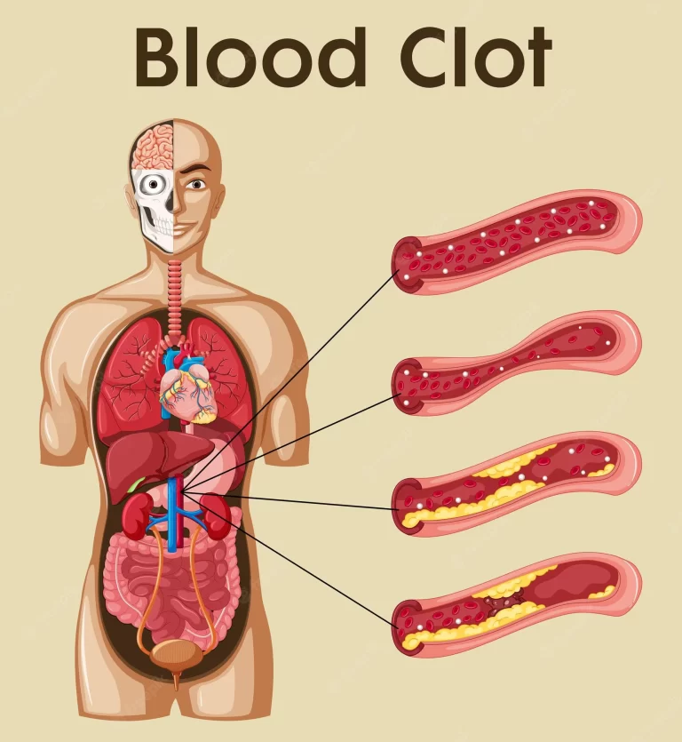
Another major cardiovascular disorder involves the result of a clot, called a “thrombus”. These can form in veins or arteries. Deep venous thrombosis, which mainly occurs in the legs, is one reason for clots in the veins of the legs, especially when an individual has been inactive for a prolonged time. These lumps may embolise, meaning journey to another place in the body. The effects of this may contain pulmonary embolus, transient ischaemic attacks, or stroke.
Cardiovascular diseases may also be hereditary in nature, such as heart imperfections or continuous fetal circulation, where the circulatory differences that are considered to happen after birth do not. Not all hereditary changes to the circulatory system are associated with disorders, a large number are anatomical interpretations.
Investigations
The procedure and health of the circulatory method and its pieces are calculated in a variety of manual and automatic ways. These contain easy procedures such as those that are elements of the cardiovascular assessment, including the taking of a person’s vibration as an indicator of a person’s heart rate, the taking of blood strain via a sphygmomanometer, or the benefit of a stethoscope to attend to the heart for buzzes which may display problems with the heart’s valves. An electrocardiogram can also be utilized to evaluate the way in which electricity is executed via the heart.
Other more invasive means can also be operated. A cannula or catheter inserted into an artery may be operated on to estimate pulse force and pulmonary wedge forces. Angiography, which affects injecting a dye into an artery to imagine an arterial tree, can be operated on in the heart (coronary angiography) or brain. At the exact time as the arteries are imagined, blockages or narrowings may be set through the insertion of stents, and active bleeds may be supervised by the insertion of coils. An MRI may be used to reproduce arteries, called an MRI angiogram. For evaluation of the blood supply to the lungs a CT pulmonary angiogram may be operated on. Vascular ultrasonography may be utilized to investigate vascular disorders involving the venous system and the arterial method including the diagnosis of stenosis, thrombosis, or venous deficiency. An intravascular ultrasound operating a catheter is also an opportunity.
FAQs
How does blood flow via the circulatory method step by step?
Blood reaches into the right atrium from the body, drives into the right ventricle, and is forced into the pulmonary arteries in the lungs. After gathering up oxygen, the blood spans back to the heart via the pulmonary veins into the left atrium, to the left ventricle, and out to the body’s tissues via the aorta.
How is blood pumped by the heart?
The pulmonary valve allows the right ventricle to flow oxygen-poor blood to the lungs. Blood that has been oxygenated by the lungs is taken from the left atrium and pumped via the mitral valve to the left ventricle. The aortic valve allows the left ventricle to send oxygen-rich blood to the body’s remaining tissues.
What allows blood circulation?
Improve cardiovascular exercise.
If you smoke, stop.
Debauch black or green tea.
If you are anemic, take iron accessories or eat iron-rich food.
Dry scrub your body.
Relieve stress.
Increase your intake of omega-3 fatty acids.
Sport condensation socks and elevate your legs.How does the bloodstream from the heart to the organs?
The arteries (red) carry oxygen and nutrients out from your heart to your body’s tissues. The veins (blue) carry oxygen-poor blood about to the heart. The aorta, a large artery that leaves the heart, is where arteries are born. They deliver blood that is rich in oxygen from the heart to every tissue in the body.
How is blood pumped via veins?
Blood primarily advances in the veins by the rhythmic motion of smooth muscle in the vessel wall and by the movement of the skeletal muscle as the body proceeds. Because most veins must carry blood against the pull of seriousness, blood is controlled from flowing rearward in the veins by one-way valves.