Tibial tuberosity
Table of Contents
What is Tibial tuberosity?
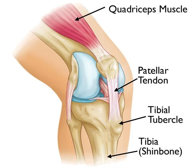
The tibial tuberosity is an elevation of the anterior aspect of the tibia bone. At full skeletal maturity, it is around 2 to 3cm distal to the proximal tibial articulating surface. It serves as an attachment for the patella tendon, intervening as a lever to extend the knee joint.
Attachment
The tibial tuberosity gives attachment to the patellar ligament, which attaches to the patella from where the suprapatellar ligament forms the quadriceps femoris muscles tendon. The quadriceps muscle group consist of four muscle, which is rectus femoris, vastus lateralis, vastus medialis, and vastus intermedius. The quadriceps muscle nerve supply is the femoral nerve.
Palpation
The two ligaments, the patella, and the tuberosity of the tibia are all located in superficial, easily palpable structures.
The function of The tibial tuberosity
It helps mainly by following functions such as:
- It makes the terminal part of the large structure that acts as a lever that helps to extend the knee joint.
- It also helps to prevent the knee from collapsing when the foot strikes the ground.
Clinical significance
Osgood-Schlatter Disease (OSD), or osteochondrosis, tibial tubercle apophysitis (painful inflammation of a bony outgrowth), or traction apophysitis of the tibial tubercle.
Osgood Schlatter disease is a common cause of anterior knee pain in the skeletally immature athletic population. Clinically, it presents as the atraumatic, gradual increase in pain anterior side of the knee, with tenderness at the patellar tendon insertion site at the tibial tuberosity. The condition occurs secondary to repetitive extensor mechanism stress activities such as jumping, sitting to stand, and sprinting as well as during sporting activities like Basketball, Volleyball, gymnastics, and Football.
Fracture of Tibial tuberosity :
Tuberosity of tibia fractures is an infrequent fracture, most common in teenage. In some specific activities like running and jumping motion, extreme contraction of the knee extensors can result in avulsion fractures of the tuberosity apophysis. A cast is all that is required if the fragment is not dislocated from its normal place on the tibia. However, if the tibial tuberosity fracture fragment is displaced at site of place, then surgery is necessary to allow for normal work.