
Neck muscles
Table of Contents
What are the neck muscles?
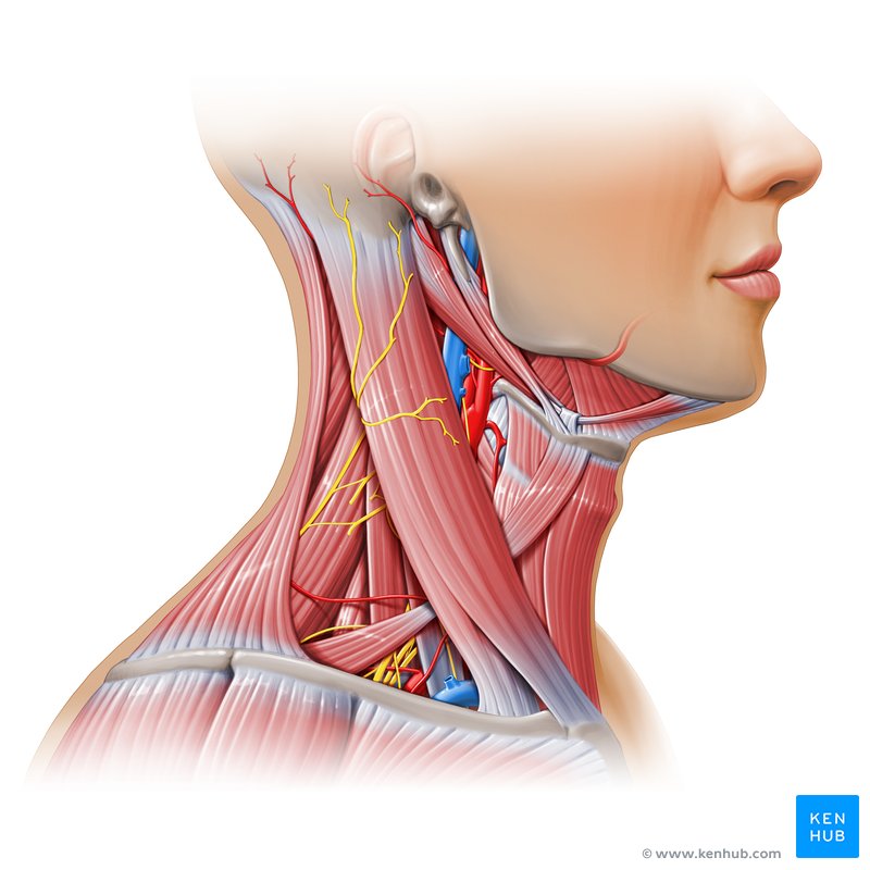
- The neck muscles are part of a complex musculoskeletal system (soft tissues & bones) that connects the base of the skull to the torso. Muscles contain fibers that contract (get smaller), allowing you to perform many of different movements. The neck muscles help you do everything from chewing & swallowing to nodding the head. You have more than 20 neck muscles. The muscles in the neck are skeletal muscles, meaning they are attached to bones by tendons. They are voluntary muscles, so you control how they move & work. Muscles of the neck are mainly responsible for the motion of the head in all directions. Like all other skeletal muscles in the body, neck muscles have lots of tiny, elastic fibers that allow the muscles to contract. Sheaths of tough connective tissue clasp the fibers together. Skeletal muscle fibers are red & white, so the muscles look striated (striped or streaked).
- Neck muscles consist of 3 main groups of muscles: anterior, lateral, & posterior muscle groups, based on their position in the neck. The muscles of the neck are further sub divided into more specific groups based on a number of determinants; including depth, precise location, & function. The location of a muscle or group of muscles in the neck generally relates to the function of the muscles in the neck. For example, the muscles in the posterior compartment of the neck are used for the extension of the neck. The muscles of the neck are closely related to a number of important structures that pass between the thorax & the head, which include major blood vessels, nerves, & elements of the respiratory & gastrointestinal systems.
Function of neck muscles
- Elevating the upper ribs so you can inhale.
- Helping with chewing, swallowing & speaking.
- Make certain facial expressions.
- Moving the head, neck & upper back, including the shoulder blades.
- Stabilizing & supporting the head, neck & spine.
Anterior neck muscles include:
- Platysma muscles
- Sternocleidomastoid muscles
- Subclavius muscles
- Scalenus muscles
- Hyoid muscles
Platysma muscle:
- Origin:
- Skin/fascia of infra- & supraclavicular regions
- Insertion:
- The lower border of the mandible, the skin of the buccal/cheek region, lower lip, modiolus, orbicularis oris muscle
- Nerve supply:
- Cervical branch of the facial nerve (CN VII)
- Blood supply:
- submental artery (facial artery), suprascapular artery (thyrocervical trunk)
- Actions:
- Depresses mandible & angle of mouth, tenses skin of lower face & anterior neck
Sternocleidomastoid muscle:
- Origins:
- Sternal head: superior part of the anterior surface of manubrium sterni
- Clavicular head: the superior surface of medial 3 of the clavicle
- Insertion:
- Lateral surface of the mastoid process of the temporal bone, Lateral half of the superior nuchal line of the occipital bone
- Nerve supply:
- Accessory nerve (CN XI), branches of cervical plexus (C2 to C3)
- Actions:
- Unilateral contraction: cervical spine: neck contralateral rotation, neck ipsilateral flexion
- Bilateral contraction: atlantooccipital joint/ superior cervical spine: head and neck extension; Inferior cervical vertebrae: neck flexion; sternoclavicular joint: elevation of clavicle & manubrium of the sternum
Subclavius muscle:
- Origin:
- Costal cartilage, sternal end of rib 1
- Insertion:
- The anteroinferior surface of the middle 3 of the clavicle
- Nerve supply:
- Subclavian nerve (C5-C6)
- Blood supply:
- Clavicular branch of thoracoacromial artery, suprascapular artery
- Action:
- Sternoclavicular joint: Anchors & depresses clavicle
Scalenes muscles:
Anterior scalene muscle
- Origin:
- anterior tubercle of transverse processes of vertebrae C3 to C6
- Insertion:
- anterior scalene tubercle of rib 1, superior border of rib 1 (anterior to subclavian groove)
- Nerve supply:
- anterior rami of spinal nerves C4 to C6
- Blood supply:
- ascending cervical branch of the inferior thyroid artery.
- Action:
- bilateral contraction – neck flexion
- unilateral contraction – neck rotation (contralateral), neck lateral flexion (ipsilateral), elevates rib
Middle scalene muscle
- Origin:
- posterior tubercles of transverse processes of vertebrae C2 to C7
- Insertion:
- superior border of rib one (posterior to subclavian groove)
- Nerve supply:
- anterior rami of spinal nerves C3 to C8
- Blood supply:
- ascending cervical branch of the inferior thyroid artery
- Action:
- neck lateral flexion, elevates rib 1st
Posterior scalene muscle
- Origin:
- posterior tubercles of transverse processes of vertebrae C5 to C7
- Insertion:
- external surface of rib 2nd
- Nerve supply:
- anterior rami of spinal nerves C6 to C8
- Blood supply:
- lower cervical branch of the inferior thyroid artery; superficial cervical artery
- Action:
- neck lateral flexion, Elevates rib 2nd
Hyoid muscles:
- The hyoid muscles are superficial muscles in the neck which attach to the hyoid bone. They are combined according to their position to the hyoid bone, the suprahyoid muscles lie above the hyoid bone & the infrahyoid muscles lie under it. These muscles act to stabilise the trachea & play a vital part in swallowing & speech.
- When the infrahyoid muscles contract, they depress the hyoid bone, allowing the suprahyoid muscles to contract & depress the mandible. If the suprahyoid muscles contract when the infrahyoid muscles are relaxed, the hyoid bone is elevated.
- The suprahyoid muscles consist of four muscles: digastric, stylohyoid, mylohyoid & geniohyoid.
Digastric muscle
- Origin:
- The anterior belly originates from the digastric fossa of the mandible
- The posterior belly originates from the mastoid notch of the temporal bone
- Insertion:
- both bellies join via the intermediate tendon which runs through the stylohyoid muscle and then inserts onto the body and greater cornu of the hyoid bone via a fibrous sling.
- Nerve supply:
- the anterior belly is derived from the first pharyngeal arch and is therefore innervated by the mylohyoid nerve, a branch of the mandibular nerve, which itself is a branch of the trigeminal nerve.
- The posterior belly is derived from the second pharyngeal arch and is therefore supplied by the digastric nerve, a branch of the facial nerve.
- Blood supply:
- Anterior belly from facial artery (submental branch) & the posterior belly by occipital artery & posterior auricular artery
- Action:
- elevation of the hyoid bone during swallowing
- mandible depression
Stylohyoid muscle
- Origin:
- the styloid process of the temporal bone
- Insertion :
- the body of the hyoid bone
- Nerve supply:
- the stylohyoid branch of the facial nerve which arises posterior to the parotid gland
- Blood supply:
- facial, posterior auricular & occipital arteries (branches of the external carotid artery)
- Action:
- elevation of the hyoid bone superiorly and posteriorly during swallowing
- mandible movement in depression
Mylohyoid muscle
- Origin:
- the mylohyoid line of the mandible
- Insertion :
- both parts of the muscles meet at a median tendon, known as the mylohyoid raphe, which then inserts onto the body of the hyoid bone
- Nerve supply:
- the mylohyoid nerve, a branch of the mandibular nerve
- Blood supply:
- Sublingual, inferior alveolar and submental arteries
- Action:
- forms the oral diaphragm
- elevation of the floor of the mouth
- elevation of the hyoid bone
- depression of the mandible
- tounge elevation
Geniohyoid muscle
- Origin:
- the inferior mental spine on the internal surface of the mandible
- Insertion:
- the body of the hyoid bone
- Nerve supply:
- the C1 nerve roots from the cervical plexus that run within the hypoglossal nerve
- Blood supply:
- Sublingual branch of the lingual artery
- Action:
- elevation of the hyoid bone
- depression of the mandible
- supports the lateral movement of the mandible.
Infrahyoid muscle
- There are four infrahyoid muscles: omohyoid, sternohyoid, sternothyroid and thyrohyoid.
Omohyoid muscle
- Origin:
- the inferior belly of the omohyoid muscle originates from the scapula and runs supromedially deep to the sternocleidomastoid
- Insertion:
- the superior belly of the omohyoid muscle inserts onto the inferior belly via an intermediate tendon
- the intermediate tendon is anchored to the clavicle via the deep cervical fascia
- the superior belly then ascends and attaches on the lateral border of the body of the hyoid bone.
- Nerve supply:
- the anterior rami of C1-C3 which is carried by a branch of the ansa cervicalis, part of the cervical plexus.
- Blood supply:
- superior thyroid artery
- Action:
- depression of the hyoid bone.
Sternohyoid muscle
- Origin:
- the sternum and sternoclavicular joint
- Insertion:
- the body of the hyoid bone
- Nerve supply:
- the anterior rami of C1-C3 which is carried by a branch of the ansa cervicalis
- Blood supply:
- lingual & superior thyroid arteries
- Action:
- depression of the hyoid bone.
Sternothyroid muscle
- Origin:
- manubrium of the sternum
- Insertion:
- thyroid cartilage
- Nerve supply:
- the anterior rami of C1-C3 which is carried by a branch of the ansa cervicalis
- Blood supply:
- lingual & superior thyroid arteries
- Action:
- depression of the thyroid cartilage and larynx.
Thyrohyoid muscle
- Origin:
- the thyroid cartilage
- Insertion:
- the body and greater cornu of the hyoid bone
- Nerve supply:
- anterior ramus of C1 which is carried within the hypoglossal nerve
- Blood supply:
- superior thyroid artery
- Action:
- depression of the hyoid bone
- elevation of the thyroid cartilage and larynx when the hyoid bone is fixed
Posterior neck muscles include:
- Superficial layer: Trapezius, splenius capitis, splenius cervicis
- Deep layer: Cervical transversospinales muscles
- Semispinalis capitis,
- Semispinalis cervicis
- Deepest layer: Suboccipital muscles:
- Rectus capitis posterior major,
- Rectus capitis posterior minor,
- Obliquus capitis superior,
- Obliquus capitis inferior
The superficial layer of muscles on the back of the neck
The Splenius Capitis muscle
- Origin:
- The splenius Capitis originates from the spinous processes of vertebrae C7 to T3 and the lower half of the nuchal ligament.
- Insertion :
- The splenius Capitis was inserted over the mastoid process and the lateral third of the superior nuchal line of the occipital bone.
- Blood supply :
- Blood supplies The splenius Capitis are the vertebral, occipital, superior intercostal, deep cervical, and transverse cervical arteries.
- Nerve supply :
- Nerve supply of splenius Capitis is posterior rami of the middle cervical spinal nerves and lower cervical spinal nerves
- Action :
- The splenius Capitis does lateral flexion of the neck and rotation of the neck to the same side.
The Splenius Cervices muscle
- Origin :
- The Splenius Cervices originate from the spinous processes of vertebrae T3 to T6
- Insertion :
- The Splenius Cervices are inserted over the transverse processes of vertebrae C1 to C3 or C4.
- Blood supply :
- the blood supply of splenius Cervicis is the vertebral, occipital, superior intercostal, deep cervical, and transverse cervical arteries.
- Nerve supply :
- The splenius cervicis muscles nerve supply are posterior rami of the middle cervical spinal nerves and lower cervical spinal nerves
- Action :
- The Splenius Cervices do lateral flexion of the neck and rotation of the neck to the same side.
Trapezius muscle
- Origin :
- The Trapezius muscle originates from the medial third of the superior nuchal line; external occipital protuberance, nuchal ligament, and spinous processes of C7 to T12 vertebrae.
- Insertion :
- The trapezius muscle inserts on the lateral third of the clavicle, acromion, and spine of the scapula.
- Nerve Supply :
- The Trapezius muscles nerve supply is the spinal root of the accessory nerve (CN XI) (motor) C3 and C4 Cervical nerves (pain and proprioception)
- Blood Supply :
- the Trapezius muscle’s arterial supply is the Transverse cervical artery (cervicodorsal trunk)
- Action :
- The function of the trapezius muscles is to stabilize and movement of the scapula. The upper fibers of the trapezius muscles elevate and upwardly rotate the scapula and extend the neck. The middle fibers of the trapezius muscles adduct (retract) the scapula. The lower fibers of the trapezius muscles depress and aid the upper fibers in upwardly rotating the scapula.
The deep layer of the muscles on the back of the neck
Semispinalis Capitus muscle
- Semispinalis Capitus belongs to the semispinalis group of the muscle, which in turn is part of the Transversospinal group of muscles (It is formed of muscles between a spinous process and the transverse process of the vertebrae below).
The semispinalis muscles have the longest fascicles of the transversospinalis group of the muscles, spanning six segments. The muscles in this group are the semispinalis capitis, semispinalis cervicis, and semispinalis thoracic muscles.
The semispinalis capitis is a long slender muscle that provided a long moment arm to provide an efficient extension of the neck.
- Origin:
- The Semispinalis capitis muscle originates from the articular processes of the C 5, C6, C7, and C8 as well as the transverse processes of T 1, T2, T3, T4, T5, T6.
- Insertion:
- The semispinalis capitis muscles insert onto the occiput in between the superior and inferior nuchal line.
- Nerve supply:
- The nerve supply of the semispinalis capitis muscle is greater occipital nerve, which additionally innervates the scalp
- Blood supply:
- The blood supply of the Semispinalis capitis muscles is descending branches of the occipital artery and the superior intercostal artery, via the dorsal rami of the upper two posterior intercostal arteries
- Action:
- Acting bilaterally: Extension of the head & neck.
- Acting unilaterally: Rotation of head & neck to the opposite side.
Semispinalis Cervicis muscle
- Semispinalis Cervicis belongs to the semispinalis group of the muscle, which in turn is part of the Transversospinal group of muscles (It is formed of muscles between a spinous process and the vertebrates transverse process below).
- The semispinalis muscles have the longest fascicles of the transversospinalis group of the muscles, spanning six segments. The muscles in this group include the semispinalis capitis, semispinalis cervicis, and the semispinalis thoracic muscles.
- Origin:
- The Semispinalis Cervicis muscles originate from the transverse processes of T1 to T6, articular processes of the 4th to 7th cervical vertebrae
- Insertion:
- The Semispinalis Cervicis muscles insert into the Spinous processes of C2 to C5
- Nerve supply:
- The nerve supply of the Semispinalis Cervicis muscles is the dorsal rami of cervical spinal nerves.
- Blood supply:
- The blood supply of the Semispinalis Cervicis muscles is the deep cervical artery
- Action:
- Acting bilaterally: extension of the cervical spine
- Acting unilaterally: lateral flexion of the neck and rotation to the neck’s opposite side.
The Deepest layer of the muscles on the back of the neck
Rectus capitis posterior major muscle
- Rectus capitis posterior major muscle is 1 of 4 small suboccipital muscles; with rectus capitis posterior minor muscle, obliquus capitis inferior muscle, & obliquus capitis superior muscle being the other 3.
- When both muscles contract bilaterally they perform the extension of the head on the neck, while the unilateral contraction rotates the head ipsilaterally at the atlantoaxial joint.
- Origin:
- The Rectus capitis posterior major muscle arise from the Tip of the Spinous process of the axis (C2)
- Insertion:
- The Rectus capitis posterior major muscle insert into The lateral part of an inferior nuchal line of the occipital bone
- Nerve supply:
- The muscle innervation of the Rectus capitis posterior major muscle is the Suboccipital nerve (posterior ramus of spinal nerve C1)
- Blood supply:
- The blood supply of The Rectus capitis posterior major muscle is the Vertebral artery & descending branches of the occipital artery
- Action:
- Bilateral contraction at the atlantooccipital joint: extension of the Head
- Unilateral contraction at the atlantoaxial joint: rotation of the Head (ipsilateral- Same side)
Rectus capitis posterior minor muscle
- Rectus capitis posterior minor muscle is a paired muscle that is located in the suboccipital compartment of the neck. It is part of the suboccipital group of the muscle which comprises four muscles in total, the other three being rectus capitis posterior major, obliquus capitis inferior, & obliquus capitis superior muscle.
- Origin:
- The Rectus capitis posterior minor muscle originates from the tubercle on the posterior arch of the atlas (C1)
- Insertion:
- The Rectus capitis posterior minor muscle is inserted on the Medial portion of the inferior nuchal line of the occipital bone.
- Nerve Supply:
- The nerve supply of the Rectus capitis posterior minor muscle is the Suboccipital nerve or dorsal ramus of the cervical spinal nerve (C1)
- Blood Supply:
- The blood supply of the Rectus capitis posterior minor muscle is the vertebral artery & the deep descending branch of the artery occipital .
- Action:
- Extension of the head
Obliquus capitis superior muscle
- Obliquus capitis superior/superior oblique muscle, is a small paired muscle that is situated deep in the upper cervical region, at the base of the occipital bone. It is one of four muscles that comprise the suboccipital group of the muscles along with rectus capitis posterior major, rectus capitis posterior minor, and obliquus capitis inferior muscles.
- Obliquus capitis superior muscles contribute to the extension of the head. However, Obliquus capitis superior muscles’ role as a postural muscle is more significant than its role as a prime mover.
- Origin:
- The Obliquus capitis superior muscle originates from the Superior surface of the transverse process of the atlas (C1)
- Insertion:
- The Obliquus capitis superior muscle inserted Between the superior and inferior nuchal lines of the occipital bone
- Nerve Supply:
- The nerve supply of The Obliquus capitis superior muscle is the Suboccipital nerve or dorsal ramus of the cervical spinal nerve (C1)
- Blood Supply:
- The blood supply of The Obliquus capitis superior muscle is the vertebral artery and the deep descending branch of the artery occipital.
- Action:
- Bilateral contraction – Atlantooccipital joint: extension of the Head
- Unilateral contraction – Atlantoaxial joint: lateral flexion of the head(ipsilateral-Same side)
Obliquus capitis inferior muscle
- Obliquus Capitis Inferior muscle (also known as the Inferior Oblique) is a small muscle that runs posteriorly and anteromedially from C1 to C2. Obliquus Capitis Inferior muscle is situated under the deep cervical vein and comprises the inferior border of the suboccipital triangle. Obliquus Capitis Inferior muscle is the only suboccipital muscle that does not attach to the skull.
- Obliquus capitis inferior is one of the four suboccipital muscles. Being grouped with rectus capitis posterior major muscle, rectus capitis posterior minor muscle, and obliquus capitis superior muscles; obliquus capitis inferior muscle is the largest muscle of the four. The functions of obliquus capitis inferior muscle are to facilitate the movements of the head and neck and maintain posture by supporting the atlantoaxial joint.
- Origin:
- The Obliquus capitis inferior muscle originates from the Base of the spinous process and adjoining lamina of the axis.
- Insertion:
- The Obliquus capitis inferior muscle inserts Along the inferior aspect of the tip of the transverse process of the atlas.
- Nerve Supply:
- The nerve supply of The Obliquus capitis inferior muscle is the Suboccipital nerve or dorsal ramus of the cervical spinal nerve (C1).
- Blood Supply:
- The blood supply of The Obliquus capitis inferior muscle is the Vertebral artery and the deep descending branch of the occipital artery.
- Action:
- Bilateral contraction – Atlantooccipital joint: extension of the Head
- Unilateral contraction – Atlantoaxial joint: rotation of the Head (ipsilateral-Same side)
Lateral neck muscles include:
Rectus capitis anterior muscle
- Origin:
- Anterior surface of the lateral mass and transverse process of atlas
- Insertion:
- Inferior surface of basilar part of the occipital bone
- Nerve supply:
- Anterior rami of spinal nerves C1, C2
- Blood supply:
- Vertebral artery, ascending pharyngeal artery
- Action:
- Atlantooccipital joint: Head flexion
Rectus capitis lateralis muscle
- Origin:
- Superior surface of the transverse process of atlas
- Insertion:
- Inferior surface of the jugular process of the occipital bone
- Nerve supply:
- Anterior rami of C1-C2 spinal nerves
- Blood supply:
- Branches of the occipital, vertebral, and ascending pharyngeal arteries
- Action:
- Stabilizes the atlantooccipital joint; head lateral flexion (ipsilateral)
Longus capitis muscle
- Origin:
- Anterior tubercles of transverse processes of C3-C6
- Insertion:
- Basilar part of occipital bone
- Nerve supply:
- Anterior rami of spinal nerves C1-C3
- Blood supply:
- Ascending cervical artery and the inferior thyroid artery
- Action:
- Bilateral contraction – head flexion;
- Unilateral contraction – head rotation (ipsilateral)
Longus Colli muscle
- Origin:
- Superior part: Anterior tubercles of transverse processes of vertebrae C3-C5
- Intermediate part: Anterior surface of bodies of vertebrae C5-T3
- Inferior part: Anterior surface of bodies of vertebrae T1-T3
- Insertion:
- Superior part: Anterior tubercle of vertebra C1
- Intermediate part: Anterior surface of bodies of vertebrae C2-C4
- Inferior part: Anterior tubercles of transverse processes of vertebrae C5-C6
- Nerve supply:
- Anterior rami of spinal nerves C2-C6
- Blood supply:
- Branches of the vertebral, ascending pharyngeal, and inferior thyroid arteries
- Action:
- Bilateral contraction: Neck flexion
- Unilateral contraction: Neck contralateral rotation, neck lateral flexion (ipsilateral)
Clinical significance of neck muscles:
- Neck muscles Spasms: Spasm also known as muscle cramps, muscle spasms occur when a muscle contract and can’t relax. Most spasms are short, lasting only for a few seconds. But you might have a sore or stiff neck afterward.
- Neck muscles Strains: A muscle strain is an injury to the muscle or the tendon. It occurs due to overstretching or tearing the muscle fibers.
- Torticollis: There is a shortening of one side of the sternocleidomastoid (SCM) muscle which leads to shortening of one side of the neck. It is congenital or due to secondary cause, for ex- SCM spasm, tumor in SCM, etc.
- Whiplash injury: If your head suddenly moves forward and then whips backward, you can injure the soft tissue in your neck. Whiplash usually involves the muscles, ligaments, and tendons.
Causes of neck pain/injury:
- Aging: Degenerative conditions such as osteoarthritis (the degenerative changes in joint cartilage) & spinal stenosis (narrowing of the spaces in the spine) can lead to neck pain as you age. Over time, stress & motion can lead to spinal disc degeneration, causing a herniated disc or pinched nerve.
- Injury: Trauma from the sudden forced movement of the neck or head & rebound in the opposite direction (whiplash) can cause pain & soreness. The muscles, ligaments, discs, vertebral joints & nerve roots in the spinal cord in the neck will be affected by trauma injuries.
- Mental stress: Tightening the neck muscles due to tension commonly causes neck pain & stiffness.
- Physical strain: Overusing the neck muscles during repetitive actions or strenuous activities can lead to stiffness and pain.
- Conditions that affect spinal balance: Poor posture (sitting for long periods of time; poor computer/keyboard/chair positioning), being overweight, and weak abdominal muscles can all affect spine posture & contribute to neck pain.
- Growths: In rare cases, masses including tumors, cysts & bone spurs can cause neck pain.
- Other health conditions: Meningitis, rheumatoid arthritis, and cancer.
Neck Muscles Exercises:
Neck muscles exercise mainly of two types:
- Neck muscles strengthening exercise
- Neck muscles stretching exercise
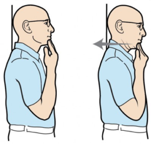
Chin tuck Exercise:
- Sit straight and look straight ahead with the ears directly over the shoulders.
- Put a finger on the chin.
- Without moving the finger, pull the chin and head straight backward until a good stretch is felt at the base of the head and top of the neck.
- Hold for 5 secs if possible.
Neck flexion Exercise (Neck tilt)
- In Relax sitting position flex your head down to rest your chin on your chest (Flex the neck fully).
- Gently tense your neck muscles and hold for 4 to 8 seconds.
- Return to a neutral position and repeat 10 times.
Neck flexion Exercise (side to side)
- Flex your head down towards your shoulder, and try to touch your shoulder with your ear (Without elevating your shoulder).
- Gently tense your neck muscles and hold for 8 seconds.
- Return your head to a neutral position and repeat in the opposite direction.
- Repeat 10 times on both side.
Neck Rotation exercise
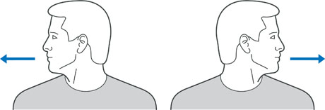
- Rotate your head towards one side, keeping your chin at the same height and moving within comfortable limits.
- Gently stretch your neck muscles and hold for 8 seconds.
- Return your head to the neutral position and repeat in the opposite direction.
- Repeat 10 times on both side.
Prone Cobra Exercise
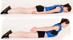
- This workout is done lying face down on the ground & uses gravity as resistance in the reinforcing process.
- Lying face down, put the forehead on a rolled-up hand towel for comfort.
- Place the arms at the side, palms downwards on the ground.
- Place the tongue on the roof of the mouth
- Pinch the shoulder blades together & lift the hands off the ground.
- Roll the elbows in, palms out, & thumbs upward.
- Gently lift the forehead about an inch off the towel keeping the eyes looking straight at the ground
- Hold the position for 10 secs.
- Perform 10 repetitions.
Isometric Neck Exercise
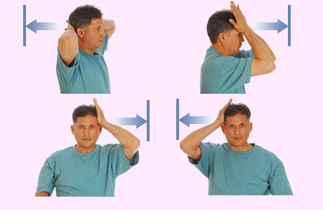
- Isometric neck exercise helps to strengthen your neck muscles. This exercise is mostly recommended by Physiotherapists from day one to relieve Neck pain.
Isometric Neck Exercise
How to perform this exercise In a sitting Position, - keep your Body straight, Put your both hands behind the neck (as seen in the images), and try your neck to push pressure on your hands and the same time Resist with your neck muscles, both hands maintain align position for 4 to 5 seconds and then relax.
The first day does 8 repetitions and the second day 10 repetitions. Do some exercise on your forehead and each side of the neck.
Do the exercise by pressing on the side of your head. Repeat 8 times, then alter sides.

Prone Rows Exercise
- Lie on your stomach with your arms dangling off the side of the bed (try angling your body so your head is facing the corner of your bed).
- Use a pillow under your stomach for comfort. Begin by pulling arms back while bending elbows and squeezing shoulders blades together then slowly return to starting position.
- Do not lift your head up while pulling your arms back.
- Repeat 20 times. Perform 2 time in a day.
Stretching Exercise:
Splenius capitis muscle stretch
- The left-side splenius capitis is stretched by flexing, right laterally flexing, and right rotating the head and neck at the spinal joints, while the left-the side shoulder girdle is allowed to elevate.
- You can hold for 30 seconds and 3 repetitionYou can hold for 30 seconds and 3 repetition
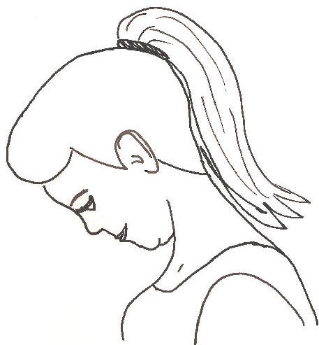
Splenius cervicis muscle stretch
- Stand or sit upright
- Keep your head up facing straight ahead
- Push your head forward side by sticking out your chin
- Note: Keep your head up during this stretch. Do not let your chin fall towards the floor.
- You can hold for 30 seconds and 3 repetition.
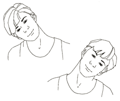
Trapezius muscle stretch
- Slowly take your left ear toward your left shoulder. It’s natural for your right shoulder to lift as you do this. If that happens, ease your head back toward the center until you can relax your right shoulder back down.
- Gently release this side, and then ease your right ear toward your right shoulder and complete the stretch on the other side, breathing deeply through it.
- You can hold for 30 seconds and 3 repetition.
Semispinalis capitis muscle stretch
- The semispinalis capitis muscle(of the transversospinalis group) is stretched by flexing the head and neck at the spinal joints. Adding in lateral
- flexion (not seen in the accompanying illustration) will increase the efficacy of the stretch for the opposite-side semispinalis capitis muscle.
- You can hold for 30 seconds and 3 repetition.
Splenius cervicis muscle stretch
- The left-side splenius cervicis muscle is stretched by flexing, right laterally flexing, and right rotating the neck at the spinal joints, while the left-side
- the shoulder girdle is allowed to elevate.
- You can hold for 30 seconds and 3 repetition.
Suboccipital Muscle Stretch
Manual Stretching
Patient position and procedure: Sitting
- How to do:
- Identify the spinous process of the 2nd cervical vertebra and stabilize it with your thumb or with the second metacarpophalangeal joint (and the
- thumb and index finger around the transverse processes). Asked the patient slowly nodding, doing only a tipping motion of the head on the upper spine. Guide the movement by placing the other hand across the forehead of the patient. You can hold for 30 seconds and 3 repetition.
Patient position and procedure: Supine
- How to do:
- Sit on a stool at the head of the treatment table with the forearms resting on the table. One hand stabilizes the C2 vertebra by grasping the transverse processes between the proximal part of the thumb & index finger; the other hand supports the occiput. Nod the head of the patient with the hand under the occiput to take up the slack of the suboccipital muscles; then ask the patient to roll the eyes upward.
- This causes a gentle isometric contraction of the muscles of the suboccipital. After holding 6 secs , ask the patient to roll the eyes downward.
- As the suboccipital muscles relax, take up the slack by passively nodding the head through a new range. The only motion should occur between the occiput & C2.
- The contraction is gentle in order to not cause overflow into the multi segmental erector spinal muscles & upper trapezius muscles.
- You can hold for 30 secs and 3 repetition.
Self-Stretching
- How to do:
- Patient position and procedure: Supine or sitting. Instruct the patient to nod the head, bringing the chin toward the larynx until a stretch is felt in the suboccipital region. Putting light pressure under the occipital area with the palm of your hand while tipping the patient’s head forward reinforces the motion.
- You can hold for 30 seconds and 3 repetition.

11 Comments