
Adductor muscle
Table of Contents
Introduction of Adductor muscle
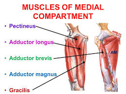
- The muscles in the medial compartment of the thigh are together called the hip adductor muscle. There are 4 muscles in this group; adductor longus, adductor brevis, adductor Magnus, & gracillis. The hip adductor compartments’ function is to move the thigh & lower extremity closer to the body’s central axis.
- All the hip adductor muscle are supplied by the obturator nerve, which arises from the lumbar plexus.
- In closed chain activation, the hip adductors help stabilize the pelvis and lower extremity during the stance phase of gait, & assist in postural control.
- They further secondary roles including hip flexion & rotation.
- The Obturator Externus, pectineus, Gemelli (superior & inferior ) & quadratus femoris may also be included by some authorities.
Adductor longus muscle
Introduction
- The Adductor longus is one of the adductor muscles of the medial thigh. Together with adductor brevis, adductor Magnus, gracilis & obturator externus, it makes up the hip adductors.
- This large fan-shaped muscle is located most anteriorly of this group & covers the middle part of the adductor Magnus & the anterior part of adductor Brevis.
- The muscle forms the medial border of the femoral triangle.
- The muscles in this compartment are believed to be evolved from both hip extensor & flexor columns.
Origin:
- The strong tendon from the anterior aspect of the pubic body is inferior to the pubic crest.
Insertion:
- Expands into a fan shape, attaching widely to the linea aspera on the middle third of the femur.
- This insertion point is between the insertion of the adductor Magnus & the origin of the vastus medialis muscle, & inferior to the adductor brevis insertion.
Nerve supply
- Hip adductors & others
- Anterior devision of obturator nerve (L2, L3, & L4).
Blood supply
- Deep femoral artery (Arteria profunda femoris)
- Deep femoral artery
- Arteria profunda femoris
- The blood supply to the adductor longus comes from two arteries, the profunda femoris artery (a branch of the femoral artery) & obturator artery (a branch of the internal iliac artery). The proximal portion of the muscle is supplied by the medial circumflex artery (branch of the profunda femoris artery). The Profunda femoris vein receives tributaries that correspond to the branches of the artery.
Function
- The main function of the adductor group of muscles is to adduct the thigh at the hip joint.
- The adductor longus muscle also plays a role in external or lateral rotation& flexion of the thigh.
- Along with the other hip adductors it helps to stabilize the pelvis in standing & aiding in balancing the body on the lower limb during walking.
Relations
- The adductor longus muscle lies in front of the adductor Magnus, adductor brevis muscle, the anterior branch of the obturator nerve, & profunda femoris vessels. The pectineus muscle is lateral to it while the gracilis lies medially. Anterior relations to its upper part are the spermatic cord & fascia lata, while the femoral artery & vein are present anterior to it in the lower part near its attachment.
Assessment
- It is important to do a thorough assessment of the pelvis & core as well as the thigh muscles. Designing a strengthening program to address which muscles are weak & which muscles are tight is vital.
Adductor Brevis Muscle
Introduction
- The Adductor brevis is the flat, triangular muscle that is found in the inner thigh. This muscle runs from the pubis to the medial aspect of the femur. Together with the adductor longus, adductor Magnus, gracilis & pectineus muscles, it comprises a group of muscles called the adductors of the thigh.
- These inner thigh muscles produce movements of the hip joint; primarily thigh adduction, but they also participate in flexion, internal & external rotation & stabilization of the pelvis while standing or walking. The Adductor brevis, being one of the shortest muscles in this group, is a weak adductor of the thigh.
Origin:
- Adductor brevis originates from the body of the pubis & inferior pubic rami.
Insertion
- It attaches to the linea aspera on the posterior part of the femur, proximal to the adductor longus.
Structure
- It is somewhat triangular in form, & arises by a narrow origin from the outer surfaces of the body of the pubis & inferior ramus of the pubis, between the gracilis & obturator externus.
- The Adductor brevis muscle widens in a triangular fashion to be inserted into the upper part of the linea aspera immediately lateral to the insertion of the pectineus & above that of the adductor longus.
Nerve supply
- Like the majority of the thigh adductors, the adductor brevis is supplied by the obturator nerve. The Obturator nerve is derived from the lumbar plexus (anterior branches of spinal nerves L2-L4).
Blood supply
- The blood supply for the adductor brevis muscle typically comes from the deep femoral artery (profunda femoris) & from its branch called the artery for the adductors. It can also be supplied by partially from the medial circumflex femoral & obturator artery. Venous blood from this region is drained by the deep femoral vein, whose path follows that of its artery prior to emptying into the femoral vein.
Function
- Hip adduction & Hip flexion. Whether it acts to rotate the femur laterally or medially is dependent on location.
- All adductor muscles in the thighs pull the legs toward the middle when walking, in order to keep balance. Overall adductor brevis play an important role in balancing the pelvis during standing & walking. This happens in the closed chain activation.
Relations
- Lying in the middle of the medial compartment of the thigh, the adductor brevis muscle is found posterior to the adductor longus & anterior to the adductor Magnus. Superiorly situated at the obturator externus muscle alongside the medial circumflex femoral artery. The inferior aspect of the adductor brevis muscle runs along with the gracilis & adductor Magnus. Near adductor brevis insertion on the femur, the middle perforating artery pierces the muscle.
Assessment
- It is necessary to do a thorough assessment of the pelvis & core as well as the thigh muscles. Designing a strengthening program to address which muscles are weak & which muscles are tight is vital.
Adductor Magnus muscle
Introduction
- The adductor Magnus’s muscle is a large triangular muscle of the lower limb, with its apex located on the hip bone, & it’s base on the linea aspera of the femur. It is located both in the posterior & medial fascial compartments of the thigh. The distribution of this muscle in 2 compartments is reflected in the fact that it receives a dual nerve supply. Regardless of its, the adductor Magnus= muscle is classified as a muscle of the medial compartment of the thigh.
- The adductor Magnus is the largest & strongest muscle in the medial compartment of the thigh, which also consists of the adductor longus, adductor brevis, pectineus, & gracilis muscles. With respect to their function, these 5 muscles are collectively called the adductors of the thigh, even though their actions are a bit more complex than that.
- Aside from thigh adduction, these muscles also participate in movements such as thigh flexion, extension, external & internal rotation, as well as pelvis stabilization during walking.
Origin
- The part of the muscle that is considered the adductor part has its proximal attachment to the inferior ramus of the pubis & the ramus of the ischium.
- The part of the muscle considered the hamstring portion has its proximal attachment to the ischial tuberosity.
- Every portion of the muscle has separate & distinct distal attachment points.
Insertion
- Adductor part: posterior surface of the proximal femur, middle of linea aspera, medial supracondylar line
- Hamstring part: adductor tubercle of medial condyle of femur & supracondylar line.
Structure
Pubofemoral (adductor) portion
- Those fibers which arise from the ramus of the pubis are short, horizontal in direction, & are inserted into the rough line of the femur leading from the greater trochanter to the linea aspera, and medial to the gluteus maximus.
- Those fibers from the ramus of the ischium are directed downward & laterally with different degrees of obliquity, to be inserted, by means of a broad aponeurosis, into the linea aspera & the upper part of its medial prolongation below.
Ischiocondylar (hamstring) portion
- The medial portion of the muscle, composed principally of the fibers arising from the tuberosity of the ischium, forms a thick fleshy mass consisting of coarse bundles which descend almost vertically, & end about the lower third of the thigh in a rounded tendon that is inserted into the adductor tubercle on the medial condyle of the femur, & is connected by the fibrous expansion to the line leading upward from the tubercle to the linea aspera.
Nerve supply:
- The nerve supply of the adductor Magnus’s muscle is reflected by its position in both the medial & the posterior compartment. Like other muscles of the medial compartment, the adductor part is supplied by the posterior division of the obturator nerve. (L2, L4). The hamstring part, sometimes considered a portion of the hamstring group of muscles, is also supplied accordingly by the tibial component of the sciatic nerve (L4).
Variation
- The upper, lateral part of the adductor Magnus is an incompletely separated division often considered a separate muscle — the adductor minimus. These 2muscles are frequently separated by a branch of the superior perforating branch of the profunda femoris artery.
Blood supply
- The adductor Magnus’s muscle gets its important arterial blood supply from the perforating branches of the deep femoral artery, passing through its use-aponeurotic openings. In addition, the superior portion of the muscle is supplied by the medial femoral circumflex artery, & the inferior portion receives blood from the femoral, popliteal & genicular arteries.
Function
- The adductor Magnus’s muscle is a powerful adductor of the thigh, working in a coordinated fashion with the adductor longus & brevis & pectineus muscles. In addition to thigh adduction, all of these muscles serve as the main stabilizers of the pelvis on the lower limb during walking.
- The 2 parts of the adductor Magnus muscle, the adductor & hamstring part, have some similarities but also some different functions. Besides adducting the thigh, the adductor portion also contributes to the flexion of the thigh, especially the superior horizontal portion of the adductor part. The hamstring part adducts the thigh as well but also cooperates with the hamstring group of muscles & assists the extension of the thigh.
- The adductor Magnus does not partake in the adduction of the abducted thigh when standing because gravity alone is enough for that function. The adductor Magnus is most active during adduction of the flexed thigh when standing, for example when kicking with the medial side of the foot in soccer, or during the supine positions. The fibers of the adductor part of the muscle that attaches to the linea aspera can also act as lateral rotators due to their oblique attachment. It is also believed that, together with the adductor longus, this muscle acts as a medial rotator of the thigh, however, these actions of medial & lateral rotation depend on the position of the thigh & the mechanical axis of the femur.
Relations
- The adductor Magnus is the occupy majority of the medial portion of the thigh. Anteriorly to the adductor, Magnus’s muscles are the pectineus, adductor longus & adductor brevis. Neurovascular structures in relation to the anterior surface of the adductor Magnus are the femoral artery & vein, the deep femoral artery & vein, &the posterior branches of the obturator artery, nerve & vein.
- On the posterior aspect, the adductor Magnus’s muscle is in relation to the gluteus maximus, semimembranosus, semitendinosus & the sciatic nerve. The medial border of the adductor Magnus’s muscle is related to the gracilis & sartorius muscles, whereas its superior border is related to the obturator externus and quadratus femoris muscle. The adductor portion of the muscle runs parallel with the quadratus femoris muscle, leaving a gap through which the femoral circumflex artery passes. At times, adductor Magnus muscles can be fused with the no-gap present.
- The adductor Magnus’s muscle takes part in creating the boundaries of the adductor canal also known as Hunter’s canal. The adductor canal is a narrow passageway in the middle third of the thigh, transmitting the descending genicular & muscular branches of the femoral artery & their corresponding veins, the saphenous nerve, & the nerve to the vastus medialis. The adductor canal ends by going through the adductor hiatus & into the popliteal fossa. The adductor canal is bounded by:
- Anteriolaterally: Vastus medialis muscle
- Posterimedially: Adductor Magnus & longus muscles
- Anteriomedially (also called roof): Subsartorial fascia & the sartorius muscle
- The adductor canal has a lot of clinical significance, one of which is to provide access to anesthesia for leg & foot procedures. This can be achieved by the saphenous nerve block at the level of the adductor canal.
Assessment
Palpation
- The tendon of the Adductor Longus is the most proximal tendon amongst the adductors of the hip joint, Gracilis is medial to the Adductor Longus. Adductor Magnus is located posterior to the Gracilis muscle. Adductor Magnus is palpated on the medial aspect of the thigh while resisting the hip adduction against resistance & feel for the engagement of the musculature.
Power
- Position: Side-lying.
- Test: Adduction of the underneath extremity from the table without rotation, flexion, an extension of the hip, or tilting of the pelvis. Strength is graded by the pressure applied over the medial aspect of the thigh in the direction of abduction i.e downward towards the thigh.
Length
- Insufficient length of muscles results in a contracture of the hip adductors or hip adduction deformity.
- In standing, the pelvis is laterally tilted, it is high on the side of contracture; making it necessary to plantarflex the foot on the same side so that the toes touch the floor. As an alternative, if the foot is flat on the floor, the opposite extremity is either flexed at the hip joint or abducted in order to compensate for the apparent shortness of the adducted side.
Gracilis muscle
Introduction
- The Gracilis muscle is a long & slender muscle situated in the medial (adductor) compartment of the thigh. It forms part of the adductor muscle group together with the adductor longus, adductor brevis, adductor Magnus & pectineus muscles. The Gracilis muscle is the most superficial hip adductor. It is also the weakest member but the only hip adductor that crosses & acts on two joints; the hip & knee.
- Gracilis extends from the coxal bone to the tibia and, you are capable of thigh adduction & flexion, as well as leg flexion & medial (internal) rotation. These actions have several important roles, for instance, balancing the trunk during walking.
Origin
- The gracilis muscle originates from the inferior ischiopubic ramus, & body of the pubis.
Insertion
- The gracilis muscle descends almost vertically down the leg & inserts on the medial tibia at the pes anserinus. The pes anserinus is further the attachment site of the Sartorius & the Semitendinosus.
Structure
- It arises by the thin aponeurosis from the anterior margins of the lower half of the symphysis pubis & the upper half of the pubic arch.
- The muscle fibers run vertically downward and end in a rounded tendon. This tendon passes behind the medial condyle of the femur, curves around the medial condyle of the tibia where it becomes flattened, & inserts into the upper portion of the medial surface of a body of the tibia, below the condyle. For this cause, the muscle is a lower limb adductor. At its insertion, the tendon is located immediately above that of the semitendinosus muscle, & its upper edge is overlapped by the tendon of the sartorius muscle, which it joins to form the pes anserinus. Pes anserinus is divided from the medial collateral ligament of the knee joint by a bursa.
- A few of the fibers of the lower part of the tendon extend into the deep fascia of the leg.
Nerve supply
- The gracilis muscles are supplied by the anterior branch of the obturator nerve (L2-L4). The anterior branch of the obturator nerve is supplied by the adductor longus, & sometimes the adductor brevis.
- There are 5 -7 muscle fiber bundle compartments in the gracilis, with the nerve branches coursing along with each compartment, indicating possible independent neuromuscular compartment functioning.
Blood supply
- Gracilis muscle receives the majority of its vascular supply from the ‘artery to the adductors’, which is a branch of the deep femoral artery. The ‘artery to the adductors’ enters the gracilis via its lateral surface, approximately one-third away from its origin.
- The proximal part of the muscle also receives a small proportion of blood supply from the medial circumflex femoral artery and the distal third of the gracilis muscle is also to be supplied by a minor branch of the femoral artery.
Relations
- Gracilis is the most superficial muscle of the medial compartment of the thigh. It is overlaid superficially by the skin & subcutaneous tissue, while its medial part is also covered by the deep layer of fascia lata. The part of the fascia lata between the sartorius & gracilis tendons is pierced by the saphenous nerve, which exits the adductor canal to become subcutaneous. The saphenous branch of the descending genicular artery also courses between sartorius & gracilis.
- Adductor brevis & adductor Magnus muscles are located deep in the gracilis. However, the spiral trajectory of this muscle can result in some complex & changing relations. Therefore, the gracilis is also neighboring the medial border of adductor Magnus, the lower border of adductor brevis, & it is located medial to adductor longus. The tibial collateral ligament is located deep in the gracilis tendon, being separated from it by the anserine bursa.
Function
- Gracilis acts on the hip & knee joints, resulting in several movements:
- Strong leg flexion & the medial (internal) rotation around the knee joint when the knee is in a semi-flexed position.
- Weak thigh flexion & the adduction around the hip joint, simply aid the other, more powerful thigh adductors.
- The main function of the gracilis is to help the hamstring muscles flex the knee, for example during the initial swing phase in walking, or during boat rowing. The medial rotation of the leg also becomes evident during walking, when the foot is solidly planted on the floor. When the lower extremity is fixed, the gracilis muscle laterally rotates the femur & pelvis around the tibia, which acts as a fulcrum. This function is important to balance the trunk. However, all movements of the gracilis become evident during horse riding, when the muscle helps the rider to grip the horse (thigh adduction) & control the flexed knee.
Assessment
- Adductor tendinopathy is usually felt as groin pain on palpation of the adductor tendons, adduction of the legs &/or of the
- affected leg. Pain can develop gradually or appear as acute, sharp pain.
- A swelling or a lump may be also felt affected in the adductor muscle(s), stiffness in the groin site, or an inability to contract or stretch the adductors. In severe cases, physical activities will be restricted as the tendon can no longer sustain replicate tensile loading.
Pectineus muscle
Introduction
- The pectineus is a flat muscle found in the superomedial part of the anterior thigh. The fascial part of the thigh muscles is specific in that each of them is innervated by a particular nerve. Due to having a dual nerve supply, the pectineus is one of a few muscles classified into 2 compartments simultaneously: anterior & medial. The others are adductor longus & adductor Magnus.
- For didactic purposes, the pectineus is usually described together with the 5 muscles of the medial compartment of the thigh. Namely, these muscles are the gracilis, adductor longus, adductor brevis & adductor Magnus. This muscle comprises the functional group of thigh adductors.
- Besides adducting the thigh, the functions of the pectineus consist of the additional movements of thigh flexion, external (lateral) rotation & internal (medial) rotation. Apart from being a prime mover, the pectineus is also a postural muscle as it stabilizes the pelvis & balances the trunk on the lower extremity during walking.
Origin
- pectineal line & adjacent bone of the pelvis
Insertion
- the oblique line extending from the base of the lesser trochanter to linea aspera on the posterior surface of the femur.
Nerve supply :
- The pectineus is predominately supplied by the femoral nerve (L2, L3). However, in some people pectineus may receive innervation from 2 separate nerves of the lumbar plexus.
- This composite innervation reflects the dual compartmentalization of the pectineus into both the anterior & medial compartments of the thigh. In these cases, the anterior part of the muscle sits is supplied by the femoral nerve (L2, L3), a feature of muscles of the anterior thigh. While the posterior, smaller part of the muscle is innervated by a branch of the obturator nerve (L3, L4), the accessory obturator nerve.
Blood supply
- The superficial portion of the muscle is supplied by the medial circumflex femoral artery, a branch of the femoral artery. The deep part of the muscle is vascularised by the anterior branch of the obturator artery, itself a branch of the internal iliac artery.
Function
- Due to the course of its fibers, the pectineus both flexes & adducts the thigh at the hip joint when it contracts. When the lower limb is in the anatomical position, contraction of the muscle first causes flexion to happen at the hip joint. This flexion can go as far as the thigh at a 45-degree angle to the hip joint.
- At that point, the angulation of the fibers is such that the contracted muscle fibers pull the thigh towards the midline, producing thigh adduction. An example of a sequential movement that involves both actions of the pectineus muscle is crossing your legs at the knee or ankle. Hip flexion solely is an action that, in synergy with the psoas major, iliacus, rectus femoris & sartorius, enables the carry-through phase of the gait cycle.
- Some sources also state that the pectineus muscle contributes to external & internal rotation of the thigh. This is still heavily debated, although it is noted that the insertion of the pectineus is lateral to the midline, beyond the mechanical axis of rotation of the femur, suggesting that there might be some merit to the argument.
- Besides being the prime mover, this muscle is also important to stabilizer the pelvis. In fact, it is suggested that all the muscles attaching between the pelvis & proximal femur act as adaptable ligaments of the hip joint, preserving its integrity during body movements.
Structure
- The pectineus muscle arises from the pectineal line of the pubis & to a slight extent from the surface of the bone in front of it, between the iliopectineal eminence & pubic tubercle, and from the fascia covering the anterior surface of the muscle; the fibers pass downward, backward, & lateral, to be inserted into the pectineal line of the femur which conducts from the lesser trochanter to the linea aspera.
Relations
- The muscle lies in the same plane as, & medially to, the adductor longus. While laterally, it is related to the psoas major muscle & the medial circumflex femoral artery & vein.
- The anterior surface of the pectineus forms the medial part of the floor of the femoral triangle together with the adductor longus, while the iliacus & psoas major complete the lateral side of the ground. This surface of the pectineus is covered with the deep layer of fascia lata that separates it from the femoral artery, femoral vein & great saphenous vein that course through the femoral triangle.
- Posterior to pectineus are the adductor magnus, adductor brevis & obturator externus muscles, & the anterior branch of obturator nerve. The fascia lata peripherally bounds the anterior & posterior thigh compartments. These 2 are internally separated by medial & lateral intermuscular septa that attach to the femur. Although the adductor (medial) compartment to which the pectineus belongs does not have its own fascial border to separate it from the anterior & posterior compartments, it is still described separately due to the common function & muscular innervation of its constituents.
Clinical importance of Hip Adductor muscle
- The adductor longus muscle forms the medial border of the femoral triangle. The superior border is formed by the inguinal ligament & the lateral border is by sartorius. The femoral nerve, artery & vein are located in this triangular region. The nerve runs most laterally, close to the anterior superior iliac spine (ASIS), & runs under the inguinal ligament. The nerve supplies the iliacus in this region, & descends to supply the anterior (extensor) compartment of the thigh.
- Adductor Tendinopathy is a common cause of medial leg & groin pain, especially among athletes. It is a general injury in such sports as eg ice skating, horse riding, soccer, football, karate, and running. Sudden changes in direction cause quick adduction against a large abduction force thereby stressing the tendon, most commonly at its origin. It is caused by a disproportional strain of the muscles, often in combination with a poor warm-up & a lack of stretching.
- Risk factors- previous hip or groin injury as well as age, weak adductors, muscle fatigue, decreased range of motion, & inadequate stretching of the adductor muscle complex. Anatomical variances also play important role eg excessive pronation or leg-length discrepancy.
- Symptoms – pain extending to the inguinal & knee region when stretching & straining the muscles.
Joint pain
- Runners are at risk of tight adductors
- Tight adductors muscle can cause knee pain, especially seen in runners. The function of the adductor muscles is to pull the thighs together & rotate the upper leg inwards, as well as stabilize the hip.
- The adductor Magnus present to display a relatively mixed muscle fiber type proportion, albeit with a greater proportion of type I muscle fibers.
- Postural (type 1) has the tendency to shorten when chronically stressed.
- Adductor muscles may be torn at their origin from the pelvis or in their bulk on the inside of the thigh.
Adductor Canal Compression Syndrome
- Adductor canal syndrome is an unusual cause of acute arterial occlusion in younger men. It is the result of arterial compression by an abnormal musculotendinous band arising from the adductor Magnus muscle & lying adjacent & superior to the adductor tendon.
- Baseball: potential Gracilis injury
- Groin strain/adductor tendinopathy commonly occurs in high-impact sports that involve ballistic movements or stretching eg soccer, hockey, and baseball. Tearing of the muscles usually happens at a proximal region near bony attachments at the pelvis
- In women, the muscular volume of the medial vastus is greater than in men, while the volume of the gracilis muscle (& the sartorius muscle) is smaller. This predisposes females to Anterior Cruciate Ligament (ACL) injury compared to males.
- Surgeons may use the gracilis tendon in reconstruction surgery of the Anterior Cruciate Ligament (ACL)
- Pes anserine bursitis is an inflammatory condition of the bursa of the conjoined insertion of the sartorius, gracilis & semitendinosus. Pes anserine bursitis is commonly seen in patients who have obese, have osteoarthritis, and are female.
- The pectineus muscle can be injured by overstretching; specifically, by stretching a leg or legs too far out to the side or front of the body. Pectineus injuries can also be caused by rapid movements like kicking or sprinting, changing directions too fastly while running, or even sitting with a leg crossed for too long.
- The most common symptom of an injured pectineus muscle is pain. Other may include bruising, swelling, tenderness & stiffness.
Hip adductor muscle stretching exercise
- The adductors involve the inner leg muscles, called the groin. The adductor muscle stretch will include the group of muscles that has a significantly large muscle mass. The key action of these muscles is to pull the leg inward. There is more useful for them in sports such as soccer, where these muscles are used in kicking a soccer ball with the inside of the foot. Finally, they are utilized in flexion & extension of the thigh when against resistance or running.
- Adductor Lunge Stretch
- Standing Lateral Lunge Adductor Stretch
- Butterfly Stretch
- Adductor AIS Release
- Supine Wall Stretch
- Standing Adductor Stretch
- Kneeling adductor stretch
- Half-kneeling adductor dips
- Frogger stretch
- Lateral squat
- Crossover stretch
- Seated Adductor Stretch
- Reclining angle bound pose
- Standing banded adduction
- Hip Opener and Groin Stretch
- Squatting Groin Stretch
- Frog Squat With Arm Raise
- Runner’s Lunge
1. Adductor Lunge Stretch

How to perform stretch:
- Begin kneeling on the ground in a lunge position.
- Bend your torso forward to bring the outside of the shoulder towards the interior of the lead knee.
- Then, lunge forward so the hips slide forward.
- You can feel a stretch along the inside of the forwarding leg.
- Hold this adductor stretch for 5- 10 seconds then release.
- Perform 10-12 repetitions.
2. Standing Lateral Lunge Adductor Stretch
How to perform stretch:

- Stand in a broad stance position.
- Now bend your & knee & move your hips to that side.
- maintain posterior pelvic tilt during the stretch
- You can feel stretch inside of the opposite thigh.
- Hold for 5-10 seconds & release.
- You can enhance holding time in progression.
- Do this adductor stretch 4-5 times.
3. Butterfly Stretch
How to perform stretch:
- Sit down on the ground with your legs in front of you.
- Reach forward & grab your left foot.
- You can bend the knee to help the hand & foot join.
- Slowly pull your left foot up towards the groin bending until it is at a comfortable place & the sole is facing your right thigh.
- Bend your right knee to bring your right foot toward the groin so that it is sole touches the sole of your left foot.
- Hold both feet with your hands & place your elbows on the knees.
- While maintaining the back straight, allow your knees to fall towards the ground.
- You can give gentle pressure to the inner thigh by pressing gradually on the knees with the elbows.
- You might feel pulling & tension in your groin.
- Hold the butterfly stretch for 20-30 seconds.
- Release & repeat 2-3 times.
4. Adductor AIS Release
How to perform stretch:
- Begin in a supine position with a resistance strap on around one foot.
- The opposite end of the strap is grasped in the hand.
- Then, actively slide the foot sideways.
- make sure to slide the foot, don’t raise it or the hip flexors can contract.
- At the end of the movement gradually pull the foot further outwards with the resistance strap.
- You can feel a stretch in the adductors.
- Hold the stretch for 5 to 10 seconds then slide to return.
- Perform 8-10 repetitions.
5. Supine Wall Stretch

How to perform stretch:
- In the supine position in front of a wall with the leg up on the wall.
- move towards the wall as you can, and maintain the comfortable stretch in the hamstrings.
- Maintain the leg straight while they slowly split open until you feel a stretch inner side of the legs.
- Hold the stretch for 15-30 seconds.
- Relax & repeat 3 times.
6. Standing Adductor Stretch
How to perform stretch:
- Start standing with the feet around 3 feet apart.
- Transfer the weight to one side & bend your knee.
- Keep the other knee straight to feel a stretch on the inside of the thigh.
- Hold the stretch for 15-30 seconds.
- Relax & repeat on the other side.
7. Kneeling adductor stretch

How to perform stretch:
- Begin with your knees & hands on the ground.
- Straighten out a left leg to the side. Try to keep the leg on the floor.
- Move your butt back to the heel of your right bent knee.
- You can feel the stretch on the inner thigh of the left leg.
- Pause for 8-10 seconds, then release the stretch & back in.
- Repeat 8-10 times.
8. Half-kneeling adductor dips
How to perform stretch:
- Start in a half-kneeling position with the right knee on the floor & the left leg bent with the foot on the floor.
- Move the left leg out to the side as much as you can.
- Your foot might be at a right angle to your knee.
- With your hands on your hips, dip the body toward the left leg.
- You can feel the stretch on the inner thigh—especially on the right side.
- Repeat 8-10 times.
9. Frogger stretch
How to perform stretch:

- Start with your knees & forearms on the ground with your knees & feet wider.
- Try to keep the inner part of your feet on the floor.
- Sit the butt back to the heels, feeling the stretch on the inner thighs.
- Pause for 10-15 seconds, then release the stretch & back in.
- Repeat 8-10 times.
10. Lateral squat
How to perform stretch:

- Stand tall & put the feet double shoulder-width apart.
- Transfer your weight to your left leg, bend your left knee, & push your hips back as if you are taking a sitting position.
- Drop as low as possible while keeping your right leg straight.
- Keep the chest up & your weight on the left leg.
- Breathe & hold for 15 to 20 seconds prior to returning to the beginning position.
- Repeat 2-3 times, then move to the other side.
11. Crossover stretch
If you love dancing, this move might come naturally as it is the same as the “grapevine” dance move.
How to perform stretch:
- Start with the feet together, then step to the right with the right foot.
- Cross your both foot.
- Step to the right again with your right foot, & bring your left foot to join your right foot.
- Once both the feet are together and repeat on the opposite side.
- You can start gradually, but pick up the stride as you get used to the move.
- Do it 5-10 times.
12. Seated Adductor Stretch

How to perform stretch:
- Straight the legs out to the side, and make a “V” shape.
- To prevent joint strain, do not overdo this pose.
- For many people, simply sitting like this is enough to give an inner thigh stretch.
- If you want to feel more stretch, keep your back straight, and lean towards the floor from your hip joints.
- Stay there for around 10-15 seconds. breathe normally.
13. Reclining angle bound pose
This is the best stretch if you spend most of the day in a sitting position.
How to perform stretch:
- Take a supine position on the ground.
- Bend both knees & draw your soles inward so that their borders are touching each other.
- Move the knees down toward the floor so that you can feel the groin muscles stretching.
- Breathe & hold this position for 15- 30 seconds.
- Repeat 3-4 times. Try to draw the feet closer to the buttocks with every stretch.
14. Squatting Groin Stretch
How to perform stretch:
- Take a standing position with the feet wide apart, toes pointing outwards.
- Slowly start squatting down until your knees are directly over the ankles & bend to 90 degrees.
- Put your hands on top of your inner thighs & gently push outward to open the hips.
- You will feel a stretch in the inner side of each leg.
- Hold for 20 – 30 seconds, relax & repeat 3 times.
15. Hip Opener and Groin Stretch
How to perform stretch:
- Begin in a forward lunge position & drop your right knee to the floor.
- Place the left elbow on the inside of your left knee as pictured.
- Press the left elbow gently into your left knee & twist your torso to the right.
- Reach the right arm behind you until you feel a gentle stretch in the lower back & left groin.
- Hold the stretch for about 20-30 seconds, release & repeat on the right leg.
16. Squatting Groin Stretch
How to perform stretch:
- Take a standing position with the feet wide apart, toes pointing outwards.
- Slowly start squatting down until your knees are directly over the ankles & bend to 90 degrees.
- Put your hands on top of your inner thighs & gently push outward to open the hips.
- You will feel the stretch in the inner side of each leg.
- Hold for 20 – 30 seconds, relax & repeat 3 times.
17. Frog Squat With Arm Raise
How to perform stretch:
- Moving directly from the first stretch, place the right hand on the ground.
- Continue to gradually push your inner thigh outward, as you reach the left hand directly up to the roof, fingers pointed upward.
- With each breath, twist the upper body slightly further, reaching as high as you can.
- Your right heel might lift slightly.
- Then, switch the other sides.
18. Runner’s Lunge
How to perform stretch:
- Get on all fours, facing the front of the mat.
- Plant the fingertips gently into the floor as you extend your right leg behind you, maintaining the knee rested or lifted slightly.
- Press the right heel toward the back of the room.
- Bring the left foot forward so it is in line with your left hand.
- Maintain your head upwards.
- Inhale & exhale, driving the hips further into the ground with each breath.
- Then, move to the other sides.
Health Benefits Of Hip Adductor Muscle Exercises.
- The hip adductors are frequently overlooked when it comes to strengthening exercises but this is a mistake. By not strengthening these muscles you are leaving yourself suffering from groin injuries.
- Below we explain some benefits of doing hip adductor exercises.
Better Balance: Better Balance exercise helps you to better balance as they keep our bodies upright when doing a sudden lateral motion. Some hip adductor exercises are also encouraged unilateral movement which catches the small muscles & helps to improve balance.
Enhanced Rotational Power: Strong hip adductors give our bodies produce more rotational power. The internal rotation of the hips required in some games like baseball or tennis requires rotational power & torque which is partly supported by these adductor muscles.
Boost Hip Extension: Hip extension plays the important role in some of the big compound lifts such as squatting & deadlifts. doing hip adductor exercises in your normal workout routine can improve your lifts, boost athletic performance and make daily living activities easier.
Reduce Risk of Injury: Groin pulls are a common injury sustained in sportsmen & even in daily life. The cause of groin strain happens due to tight or weak hip adductors. By strengthening & stretching the adductor muscles you can decrease the chances of experiencing this injury.
Athletes, in particular, depend on this muscle group to aid in explosive movements such as jumping, running, & quickly moving from side to side.
- The gracilis muscle is generally used as a flap in microsurgery. According to the classification of Mathes & Nahai, it presents a type II blood supply, allowing it to be transferred to its artery derived from the medial circumflex femoral artery. This artery arrives at the muscle about 10 cm from the pubic symphysis. At this point, the nerve also enters.
- As a pedicled function flap, the gracilis muscle can be transferred to treat anal incontinence. This technique is called graciloplasty. . The gracilis microsurgical free flap is generally used in the reconstruction of upper & lower limbs, in breast reconstruction & – as a free function flap – to restore forearm function, or in the dynamic reconstruction of facial paralysis.
- The pectineus muscle can become injured by overstretching; & specifically, by stretching a leg or legs too far out to the side or front of the body. Pectineus injuries can also be caused by rapid movements like kicking or sprinting, changing directions too fastly while running, or even sitting with a leg crossed for too long.
- The most common symptom of an injured pectineus muscle is pain. Other may include bruising,swelling,tenderness & stiffness.
Hip Adductor Muscle Strengthening Exercises.
- Hip adductor exercises utilize multiple small muscles in the inner thigh that are responsible for bringing the thighs together, providing balance & support and proper hip alignment. Most people should only consider doing hip adductor exercises whenever they walk past the hip adductor machine in the gym but we are here to change that Mentality.
- It is essential to strengthen the hip adductor muscles to increase mobility & flexibility, enhance stability & prevent leg injuries. Strong abductors are an integral part of sports performance, maintaining mobility, & injury prevention, as you age. This is the main muscle group that contributes to hip mobility & strength. In terms of strength training, the adductors are mostly overlooked because they can be difficult to train properly.
- Standing Leg Circles
- Side-Lying Hip Adduction
- Squat Side Kick
- Standing side Leg Raise
- Sumo Squat
- Cross Scissors
- Dumbbell Side Lunge
- . Cossack Squat
- Copenhagen Side Plank
- Cable Hip Adductor
- Seated Hip Adduction
- Adductor machine
- Wide stance squat
- Standing banded adduction
- Seated banded adduction
- Standing Leg Circles
How to do it?
- This is the dynamic warm-up exercise, this exercise improves blood circulation to the hip muscle & upper leg.
- for standing leg circle exercise, stand with feet hip-width apart.
- Raise the left leg off the floor. while balancing on the right leg, make a small circle with your left leg.
- Complete 10 to 20 repetitions on the left side then change to the right leg.
- Note: If you have a balance problem then stand close to a prop where you can help to maintain your body by holding something.
2. Side-Lying Hip Adduction
How to do it?

- This is the best exercise for the hip adductors that can be done on the ground while isolating a single leg at a time.
- All you need to do is to focus on contracting the hip adductor muscles to raise your leg off the floor.
- For this exercise, you have to lie down on your left side and take your arms in front of you with your elbows & forearm on the floor for support. raise the right leg over your lower leg placing your heels against the thigh of your bottom leg.
- maintain your leg extended, and raise it upward as much as possible.
- Slowly return to the starting position.
- Do 10-25 repetitions on the left side then move to the right leg.
- Note: You can add some provocation to your exercise by using a resistance band attached to an anchor or by strapping an ankle weight to the leg.
3. Squat Side Kick

How to do it?
- The squat side kick bodyweight exercise will utilize both the adductors & the abductors & is a great all-around lower limb exercise.
- This exercise combines 2 movements that enable both muscle strengthening & stretching which is important to decrease the risk of suffering from groin pain.
- For the Squat Side Kick exercise, you have to stand with a shoulder hip-width apart, and your hands are together in front of you.
- Flexed your knees until your thighs are parallel to the floor, then stand up & transfer your weight to your right leg while kicking out the left leg to the side.
- Back to squatting position then repeat it on another leg.
- Note: Your core muscles might be engaged throughout the movement with your back straight, chest up, & do not let your knees extend past your toes while squatting.
4. Standing side Leg Raise
How to do it?

This is the best bodyweight exercise that can hit the hip adductors of one leg while hitting the hip abductors of the other leg as
- you will be using an isometric hold on your other leg to maintain it in the air.
- For this exercise, you have to stand with shoulders hip-width apart. now on the left leg away from the body.
- Then Lift the right leg as far as comfortable, & hold it there for 4-6 seconds.
- Raise your left leg to your right leg until they touch then lower back to the starting position. Complete 10-18 repetitions of 2-3 sets.
- Note: To make this exercise more challenging strap ankle weights to both legs.
5. Sumo Squat
How to do it?
- This variation of the squatting will hit the muscle groups in the lower limb plus the inner thighs.
- Maintain your back straight & your chest up throughout the exercise.
- Even though the sumo squat has a smaller range of motion compared with a regular squat, it is still an effective exercise that can be incorporated into normal workouts.
- For this exercise, you have to stand wider than the shoulder-width stance with your pointing outside.
- Take a squatting position, drop your hip down & back while your back is straight & chest up until your thighs are parallel to the ground.
- Push off through the ground and return to the starting position.
- Note: If you want to add some provocation to your exercise then hold a kettlebell in your hand.
6. Cross Scissors
How to do it?
- This is the best exercise to work hip adductor muscles & the core muscle simultaneously.
- Cross scissors are challenging as you require to stay in a crunched position throughout the movement.
- Maintain this position by crossing your legs in front of you need all your stabilizing muscles to be contracted.
- For this exercise, you have to Sit down & brace yourself by putting your hands on the floor back to you. move your leg off the ground in front of you at a 30-40-degree angle with one leg crossed over the other.
- Your core muscle might be engaged throughout the movement in a semi-“V” position change your legs out to the sides then bring them back closed while crossing the other leg over.
- Do this with an Alternate leg until you complete 10-20 repetitions on each side.
- Note: You can enhance the difficulty of cross scissors by sitting in a “V” position without bracing the upper limb with your arms.
7. Dumbbell Side Lunge
How to do it?
- The side lunge is an excellent exercise to improve your balance, stability, & lower limb strength.
- The side-to-side movement is both a strengthening & stretching exercise. Dumbbell Side Lunge exercise utilizes your hip abductors & hip adductor muscles.
- For this exercise, you have to stand with shoulder hip-width apart & hold the dumbbells at your chest level.
- Take a large step to your side & drop your hip down & back until your thighs are parallel to the floor while your foot is planted on the ground.
- Push through your flexed foot, & bring your back to the initial position.
- Repeat it for 10-15 repetitions then move to the next leg.
- Note: To make this exercise easy you can do bodyweight side lunges. your back might be in a neutral position & chest up while trying not to lean forward.
8. Cossack Squat
How to do it?
- Cossack Squat exercise moves the body through the frontal plane of motion, going side to side.
- Working lower limb at this angle can improve the flexibility of hips, knees, & ankles.
- You will increase your stability by mastering this exercise by getting a good stretch & strengthening the hip adductors.
- For this exercise, you have to stand with feet in a wide stance with toes pointing outside by dropping your hips downward & back.
- Squat down to that single side and shift your weight while your other leg is extended out with your heels on the floor, toes pointing up.
- Push through the ground with your flexed leg and bring your back to starting position.
- Repeat this exercise on the other leg by shifting the weight & lowering it down into a squat position on the other side.
- Note: Cossack Squat exercise requires a high degree of mobility so if you can not go all the way down then go as far as you can while trying to improve each workout. your back might be maintained in a neutral position throughout the exercise.
- Copenhagen Side Plank
How to do it?
- Copenhagen Side Plank exercise is the most difficult variation of plank that not only hits the core muscles but also strengthens the hip adductors. Copenhagen Side Plank exercise will help to balance out the strength in the muscles on the outside of the hip.
- For this exercise, you have to lie down on the floor & perpendicular to the bench then brace yourself on your forearm & elbows.
- Lift with your flexed knee lift your top leg & place it on the bench.
- Hold this position.
- Complete 20-25 repetitions for 2 sets then do this on another side.
- Note: Increase the difficulty by doing this exercise with legs extended with only your ankle placed on the bench.
- Cable Hip Adductor
How to do it?
- You may notice women at the gym doing this exercise while the men neglect it.
- It is time to move the stigma that cable hip adductions are not mainly, everyone might be doing this exercise to strengthen the adductors to reduce the risk of injury.
- Make sure you are warmed up with some dynamic stretches before performing this exercise then try to start with less weight & maximum repetitions until you are comfortable enough to increase the weight.
- Find an attachment that you can use to strap onto the ankle closest to the pulley.
- Set up the pulley around calve level. Stand to the side of the pulley. Brace yourself using your hand against the machine in a safe place where your fingers won’t get pinched. Your active leg might be up off the floor towards the pulley.
- Pull your leg away from the pulley towards the middle of your body.
- Slowly let leg return to its starting position Completed the desired number of repetitions
- Note: exercise can also be done with this same technique by attaching a resistance band to a fixed anchor point.
- Seated Hip Adduction
- The seated hip adduction will isolate the hip adductor muscles when you are seated, only having to focus on bringing thighs together.
- Add Seated Hip Adduction exercise towards the end of your leg day after completing the bigger compound lifts such as squatting.
- Seat in the machine with back against the backrest
- Set the width of the knee pads to a manageable position that gives a stretch to the inner thighs but does not over-stretch the adductor muscles.
- Set up a lightweight for the 1st time set so that you do not overdo it.
- Squeeze thighs together while exhaling, until your knees meet in the center of the body.
- Slowly return to the initial position.
- Complete maximum repetitions.
- Adductor machine
How to do it?
- Most people think of isolating the adductors, they may think of the classic adductor machine established in gyms across the world.
- Though this machine can do the best job of training the adductor, it is not the only movement that can yield good results.
- Considering that you can modify the amount of weight & width of the pads, this exercise is great for beginners. It is best to begin super light to get a feel for the exercise & avoid getting injured.
- For this exercise, you have to sit on the machine begin with sitting on the machine with the pads positioned between your legs as wide as is comfortable, & select your ability-wise resistance.
- In a controlled manner, contract thighs together just until the pads touch, & feeling the muscles contract.
- gradually reverse the movement, returning your thighs to the initial position. Repeat this exercise number of sets & repetitions.
- If just beginning, try 2–3 sets of 10 reps.
- Wide stance squat
How to do it?
- Squatting is a king of leg exercises & this will stimulate the whole leg.
- There have different squat variations but here we discuss the wide stance squat also called sumo squat which will utilize inner thigh muscles.
- You can do this exercise with a variety of weighted equipment like a barbell, kettlebell, dumbbell, or sandbag — or with just only body weight.
- For the Wide stance squat exercise, you have to stand a little wider than the shoulder hip-width apart, and turn your toes outside.
- Shift your weight backward & slowly lower your hips until your thighs are parallel to the ground.
- Then return to the starting position by pushing through the ground, and your inner thigh muscles are contracted.
- Do 10-15 repetitions of 2-3 sets.
- Standing banded adduction
How to do it?
- For this exercise, you need a resistance band, and wrap a resistance band on a solid anchor. you have to stand on either side of the body facing the anchor point & the resistance band.
- Allow the resistance band to pull the leg to the side by resisting the movement.
- To start this exercise you have to stand tall & bring a banded leg towards the midline of the body, you feel the good contraction in your inner thighs.
- Gradually release your leg back to the side with control movement.
- Switch the legs & do 10 to 20 repetitions for 3 to 4 sets on each leg.
15. Seated banded adduction
How to do it?
- For the Seated banded adduction exercise you need a resistance band, and wrap a resistance band on a solid anchor.
- You have to sit on the bench on either side of your body facing the anchor point & the resistance band.
- Put the internal foot through the resistance band, the band is positioned just below the knee joint. allow the resistance band to pull the leg towards the anchor point.
- Move your leg towards the center of the body by contracting the adductor muscle.
- Repeat this on each leg.
- Do 2-3 sets of 12-15 repetitions.

- Lateral lunge
How to do it?
- For the Lateral lunge exercise, you have to stand with feet hip-width apart. step out of your left leg & start with sending your hip back as you flex your left knee.
- You can extend your arms forward for counterbalance if you like. during the descent, the knee should not move more than 2 inches beyond your toes, &your knee should be aligned between 2 & 3 toes.
- The toe turns outwards slightly. push-off through left to return to a starting position.
- Repeat on the right side leg to complete the 1st repetitions.
- Do 3 to4 sets of 8 to 10 repetitions on each leg.
When did you not do this exercise?
- If your health care provider advised to you take a rest.
- If you are already suffering from back & knee pain.
- If your lower limb bone recently fractured.
- If you feel pain during this exercise.
36 Comments