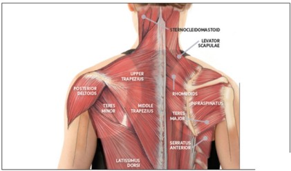
Shoulder to Neck Muscles
Table of Contents
What is the shoulder to neck muscles?
- The shoulder to neck muscles are the skeletal muscles. These muscles are interlinked muscles. They play a major role in performing a wide variety of movements. These muscles have their own importance while they work as a stabilizer of the neck and shoulder structures and also play a vital role in movements of the neck or shoulder. While the muscles of the neck are closely related to a number of important structures that pass between the thorax and the head, which include major blood vessels, nerves, and elements of the respiratory and gastrointestinal systems.
- The shoulder is a ball-&-socket joint that allows you to perform a wide range of movements. You use these muscles for actions from throwing a ball to reaching an item on a shelf. Also termed as glenohumeral joint, it has more range of motion than any other joint in your body. There are about 8 muscles in the shoulder that support this joint. They give it strength, stability & shape.
- The muscles on the Back of the neck are muscles that cover the neck area. The neck muscles are part of a complex musculoskeletal system that connects the base of the skull to the torso. muscles of the neck are mainly responsible for the movement of the head in all directions. They consist of 3 main groups of muscles: anterior, lateral, & posterior muscle groups, based on their position in the neck. The position of a muscle or group of muscles in the neck generally relates to the function of the muscles in the neck. For example, the muscles in the posterior compartment of the neck are responsible for the extension of the neck.
Shoulder muscles
Deltoid muscle
- The Deltoid muscle is a thick, triangular shoulder muscle. The deltoid muscle is name base on the shape of the Greek letter ‘delta’ (Δ). The Deltoid muscle is located at the rounded contour of your shoulder. It is also famous as the ‘common shoulder muscle’.The entire deltoid muscle starts from the spine of the scapula to the lateral portion of the clavicle. It travels inferiorly covering the glenohumeral joint on all sides and inserts onto the humerus bone. The Deltoid is formed of acromial, clavicular and scapular spinal parts. The acromial part (middle fibers) abducts the arm, while the clavicular and scapular spinal parts play a significant role in stabilization, ensuring a steady plane of abduction. Additionally, the clavicular part (anterior fibers) can act as a flexor and internal rotator of the arm, while the scapular spinal part (posterior fibers) can extend and externally rotate the arm.
- Origin :
- The Deltoid muscle originates from the anterior border & the adjoining surface of the lateral one-third of the clavicle,
- the lateral border of the acromion process where 4 septa of origin are attached & the lower lip of the crest of the spine of the scapula.
- Insertion :
- The Deltoid muscle is inserted into the deltoid tuberosity of the humerus.
- Nerve supply :
- The nerve supply of the deltoid muscle is the Axillary nerve (C5 and C6).
- Blood supply :
- The blood supply of the deltoid muscle is the thoracoacromial artery (acromial & deltoid branches), the circumflex humeral arteries, & the profunda brachial artery
- Action :
- The multipennate acromial fibers are powerful abductors of the upper limb at the shoulder joint from beginning to 90 degrees of range.
- The anterior fibers are flexors & Internal rotators of the shoulder.
- The posterior fibers are the extensors & external rotators of the shoulder.
Rotator cuff muscle
- The Rotator Cuff (RC) is a common name for the group of 4 distinct muscles and their tendons, which provide strength and stability during a motion to the shoulder complex. They are also referred to as the SITS muscle, with reference to the first letter of their names (Supraspinatus,
- Infraspinatus, Teres minor, and Subscapularis, respectfully). The muscles arise from the scapula and connect to the head of the humerus, forming a cuff around the glenohumeral (GH) joint.
- Rotator cuff muscles are formed from 4 muscles, they are,
Supraspinatus muscle
Infraspinatus muscle
Teres minor muscle
Subscapularis muscle
Supraspinatus muscle
- Origin:
- supraspinatous fossa of scapula.
- Insertion:
- greater tubercle of the humerus.
- Nerve supply:
- suprascapular nerve (C4, C6).
- Blood supply:
- Suprascapular artery.
- Action:
- initiation of abduction of arm to 15° at the glenohumeral joint; stabilization of humeral head in the glenoid cavity.
Infraspinatus muscle
- Origin:
- infraspinatous fossa of scapula.
- Insertion:
- greater tubercle of the humerus.
- Nerve supply:
- suprascapular nerve (C5, C6).
- Blood supply:
- Suprascapular artery, circumflex scapular arteries.
- Action:
- external rotation of the arm at the glenohumeral joint; stabilization of the humeral head in the glenoid cavity.
Teres minor muscle
- Origin:
- the lateral border of the scapula.
- Insertion:
- greater tubercle of the humerus.
- Nerve supply:
- axillary nerve (C5, C6).
- Blood supply:
- Suprascapular artery, dorsal scapular artery.
- Action:
- external rotation & adduction of the arm at the glenohumeral joint; stabilization of the humeral head in the glenoid cavity.
Subscapularis muscle
- Origin:
- medial 2/3 of the subscapular fossa.
- Insertion:
- lesser tubercle of the humerus.
- Nerve supply:
- upper & lower subscapular nerves (C5, C7).
- Blood supply:
- Suprascapular artery, axillary artery, subscapular artery.
- Action:
- internal rotation of arm; stabilization of humeral head in the glenoid cavity.
Teres major muscle
- Origin:
- Inferior angle & lower part of the lateral border of the scapula.
- Insertion:
- Intertubercular sulcus (medial lip) of the humerus.
- Nerve supply:
- Lower subscapular nerve (C5-C7).
- Blood supply:
- Thoracodorsal branch of subscapular artery & posterior circumflex humeral artery.
- Action:
- Extension & internal rotation of the humerus (arm).
Pectoralis major muscle
- The pectoralis major is a paired, superficial muscle located on the anterior surface of the thoracic cage. If you’re a gym lover, you’ll hear these muscles also being referred to as the pecs muscles. The pectoralis major has a broad origin, based on which it is divided into three parts: clavicular part, sternocostal part, and abdominal part. All three parts converge laterally and insert onto the greater tubercle of the humerus.
- Origin:
- The pectoralis major muscle consists of 3 parts:-
- The clavicular part origins from the anterior surface of the medial half of the clavicle.
- The sternocostal part originates from the anterior surface of the sternum & the anterior aspects of the costal cartilages of ribs one, and six.
- The smallest, abdominal part origins from the anterior layer of the rectus sheath.
- Insertion:
- The Pectoralis major muscle fibers from all 3 parts pass laterally, converging towards the proximal humerus. They give off a broad tendon that inserts along the crest of the greater tubercle of the humerus. The upper & lower fibers of the muscle are inserted at the crest of the greater tubercle of the humerus. Upper fibers are more anterior & caudal on the crest, while posterior fibers twist on themselves & are more posterior and cranial than the upper fibers.
- Nerve supply:
- The pectoralis major gets two engine innervation by the average pectoral nerve & the parallel pectoral nerve, otherwise called the horizontal from the thoracic nerve.
- The sternal head gets innervation from the C7, C8 & T1 nerve roots, through the lower trunk of the brachial plexus & the average pectoral nerve.
- The clavicular head gets innervation from the C5 & C6 nerve roots through the upper trunk & horizontal string of the brachial plexus, which radiates the sidelong pectoral nerve. The parallel pectoral nerve is circulated above the profound surface of the pectoralis major muscle.
- Blood supply:
- The pectoralis major muscle Blood supply is the pectoral branches of the thoracic acromial artery & the perforating branches of the internal thoracic artery.
- Action:
- Pectoralis major muscle does mainly are adduction of shoulder joint or depression of the arm & rotation of the arm forward about the axis of the body (internal rotation).
Pectoralis minor muscle
- The pectoralis minor muscle is triangular in shape & is located under the pectoralis major, & both formed the anterior wall of the axilla.
- Origin:
- It originates from the 3rd to 5th ribs, near the costochondral junction, the intervening fascia covering external intercostal muscles.
- Insertion :
- It inserts on the medial border & upper surface of the coracoid process.
- Nerve supply :
- It is supplied by the medial & lateral pectoral nerves.
- Blood supply :
- The blood supply to the pectoralis minor comes from several sources:
- Thoracoacromial artery (branch of the second part of an axillary artery) gives 2 supplying branches – pectoral & deltoid.
- Superior thoracic artery (branch of the 1st part of an axillary artery).
- Lateral thoracic artery (branch of the axillary artery).
- Action :
- The action of pectoralis minor is to,
- draws the scapula forward.
- depresses the point of the shoulder.
- also helps in forced inspiration.
Latissimus dorsi muscle
- This latissimus dorsi is one of the largest muscles in the back.
- It is sometimes referred to as lats & this muscle is known for its large & flat “V” shape.
- The function of this muscle is to span the width of the back & helps to control the movement of the shoulders joint.
- Origin:
- Vertebral part: Spinous processes of vertebrae T seven to T twelve, Thoracolumbar fascia
- Iliac part: Posterior 3rd of the crest of ilium
- Costal part: Ribs nine & twelve.
- Scapular part: Inferior angle of scapula
- Insertion:
- Intertubercular sulcus of the humerus, between the pectoralis major and teres major muscle.
- Nerve supply:
- Thoracodorsal nerve (C6-C8)
- Blood supply:
- Thoracodorsal artery, perforating arteries of the 9th-11th posterior intercostal arteries, & 1st-3rd lumbar arteries
- Action:
- Shoulder joint: Arm internal rotation, Arm adduction, Arm extension, Assists in respiration.
Biceps muscle
- The biceps brachii muscle is one of the chief muscles of the arm and is located in the front part of the upper arm. The biceps muscle has a “short head” and a “long head” that work as a single muscle. The Biceps muscle is named because of its two heads which are the Long head and Short head. The Biceps muscle is located at the front side of your upper arm starting from shoulder to end below the elbow joint (From shoulder to elbow). The biceps muscle is attached to the forearm bones by tough connective tissues called tendons. The biceps muscle lies superficial to the brachialis and coracobrachialis muscles and essentially forms the front side of the upper arm. While both its origin are covered by the deltoid muscle, its insertion tendon can be easily seen and palpated at the crook of the elbow joint.
- Origin:
- Short head originate from Apex of the Coracoid process of the scapula
- The long head originates from the Supraglenoid tubercle of the scapula
- Insertion:
- Radial tuberosity of the radius
- Deep fascia of the forearm (insertion of the bicipital aponeurosis)
- Nerve supply:
- Musculocutaneous nerve (C5- C6)
- Blood supply:
- Branches of brachial artery
- Action:
- Flexion & supination of the forearm at the elbow joint, fragile flexor of the arm at the glenohumeral joint.
Triceps brachii Muscles
- The triceps brachii is the muscle that runs down the back of the humerus, which is the long bone of the upper arm and ends at the top of the ulna, which is the long bone of the forearm. The triceps brachii muscle gets its name because it contains three muscle ‘heads’ or points of origin.
- These include the Medial head, Lateral head, and Long head. Triceps brachii is a three-headed muscle of the arm. It represents the only constituent of the posterior muscle group of the arm, spanning almost the entire length of the humerus. The triceps brachii muscle consists of a long, medial, and lateral head, that originates from their respective attachments on the humerus and scapula. The main function of the triceps brachii is the extension of the forearm at the elbow joint. Its long head contributes to the extension and adduction of the arm at the shoulder joint.
Long head
- Origin:
- infra glenoid tubercle of the scapula
- Insertion:
- the posterior surface of the olecranon process of the ulna, capsule of the elbow joint & antebrachial fascia.
- Nerve supply:
- radial nerve
- Blood supply:
- deep brachial artery
- Action:
- Because it attaches to the scapula, the long head not only extends the elbow yet it will also have a small action on the glenohumeral joint. With the arm adducted, the triceps muscle acts to clasp the head of the humerus in the glenoid cavity. This action assists to prevent any displacement of the humerus.
- The long head also assists with the extension & adduction of the arm at the shoulder joint.
- The lateral head is also working during the extension of the forearm at the elbow joint when the forearm is supinated/pronated.
Lateral Head
- Origin:
- the posterior surface of the humerus, superior to the radial groove
- Insertion:
- the posterior surface of the olecranon process of the ulna, capsule of the elbow joint & antebrachial fascia.
- Nerve supply:
- radial nerve
- Blood supply:
- deep brachial artery
- Action:
- Strongest head of the 3. It is active during the extension of the forearm at the elbow joint when the forearm is supinated/pronated.
Medial head
- Origin:
- posterior aspect of the humerus, inferior to the radial groove
- Insertion:
- the posterior surface of the olecranon process of the ulna, capsule of the elbow joint & antebrachial fascia.
- Nerve supply:
- radial nerve
- Blood supply:
- deep brachial artery
- Action:
- As the medial head does not attach to the scapula & therefore has no movement on the glenohumeral joint, whether with stabilization or movement. However, the medial head is working during the extension of the forearm at the elbow joint when the forearm is supinated or pronated.
Neck muscles
- Anterior neck muscles include the Platysma muscle, Sternocleidomastoid muscle, Subclavius muscle, Scalenes muscles, and Hyoid muscles.
Platysma muscle:
- This muscle covers a portion of a neck muscle known as the sternocleidomastoid. The platysma muscle is expansive in size, with a broad width that spans the collarbone, or clavicle, and the side of the neck. Its point of origination is the upper portions of the pectoral, or chest, and the deltoid, or shoulder.
- Origin:
- Skin/fascia of infra- & supraclavicular regions
- Insertion:
- The lower border of the mandible, the skin of the buccal/cheek region, lower lip, modiolus, orbicularis oris muscle
- Nerve supply:
- Cervical branch of the facial nerve (CN VII)
- Blood supply:
- submental artery (facial artery), suprascapular artery (thyrocervical trunk)
- Actions:
- Depresses mandible & angle of mouth, tenses skin of lower face & anterior neck
Sternocleidomastoid muscle
- The sternocleidomastoid muscle is a two-headed long, bilateral muscle of the neck, and is the most visible cervical muscle. The main functions of the muscle are rotation of the neck to the opposite side and flexion of the neck. The muscle nerve supply is the accessory nerve.
- The muscle name sternocleidomastoid is derived from the manubrium of the sternum (Sterno-) and the clavicle (cleido-) and has an insertion at the mastoid process of the temporal bone of the skull.
- Origin:
- Sternal head:
- superior part of the anterior surface of manubrium sterni
- Clavicular head:
- the superior surface of medial 3 of the clavicle
- Insertion:
- the lateral surface of the mastoid process of the temporal bone, the Lateral half of the superior nuchal line of the occipital bone
- Nerve supply:
- the accessory nerve (CN XI), branches of cervical plexus (C2-C3)
- Blood supply:
- superior thyroid artery & the external carotid artery
- Actions:
- Unilateral contraction: cervical spine: neck ipsilateral flexion, neck contralateral rotation
Bilateral contraction: atlantooccipital joint/ superior cervical spine: head and neck extension; Inferior cervical vertebrae: neck flexion. - sternoclavicular joint: elevation of clavicle & manubrium of the sternum
Subclavius muscle:
- The subclavius is a small triangular muscle, placed between the clavicle and the first rib. Along with the pectoralis minor and pectoralis major muscles, the subclavius muscle makes up the anterior wall of the axilla.
- Origin:
- Costal cartilage, sternal end of rib 1
- Insertion:
- The anteroinferior surface of the middle 3 of the clavicle
- Nerve supply:
- Subclavian nerve (C5-C6)
- Blood supply:
- Clavicular branch of thoracoacromial artery, suprascapular artery
- Action:
- Sternoclavicular joint: Anchors & depresses clavicle
Scalenes muscles:
- Scalene Muscle is a group of three pairs of muscles in the lateral neck, namely the anterior scalene. It is located deeply just behind the Sternocleidomastoid muscle. The brachial plexus and subclavian artery run between the anterior and middle scalene muscles. This provides an important anatomical location for anesthetics to do an inter scalene block. The subclavian vein & phrenic nerve run anteriorly to the anterior scalene while the subclavian vein passes horizontally across the muscle, while the phrenic nerve runs vertically down the muscle. The subclavian artery is situated posterior to the muscle.
Anterior scalene muscle
- Origin:
- anterior tubercle of transverse processes of vertebrae C3 to C6
- Insertion:
- anterior scalene tubercle of rib 1, superior border of rib 1 (anterior to subclavian groove)
- Nerve supply:
- anterior rami of spinal nerves C4 to C6
- Blood supply:
- ascending cervical branch of the inferior thyroid artery.
- Action:
- bilateral contraction – neck flexion
- unilateral contraction – neck rotation (contralateral), neck lateral flexion (ipsilateral), elevates rib
Middle scalene muscle
- Origin:
- posterior tubercles of transverse processes of vertebrae C2 to C7
- Insertion:
- the superior border of rib one (posterior to subclavian groove)
- Nerve supply:
- anterior rami of spinal nerves C3 to C8
- Blood supply:
- ascending cervical branch of the inferior thyroid artery
- Action:
- neck lateral flexion elevates rib 1st
- Posterior scalene muscle
Posterior scalene muscle
- Origin:
- posterior tubercles of transverse processes of vertebrae C5 to C7
- Insertion:
- the external surface of rib 2nd
- Nerve supply:
- anterior rami of spinal nerves C6 to C8
- Blood supply:
- lower cervical branch of the inferior thyroid artery; superficial cervical artery
- Action:
- neck lateral flexion, Elevates rib 2nd
Hyoid muscles:
- The hyoid muscles are superficial muscles in the neck that attach to the hyoid bone. They are combined according to their position on the hyoid bone, the suprahyoid muscles lie above the hyoid bone & the infrahyoid muscles lie under it. These muscles act to stabilize the trachea & play a vital part in swallowing & speech.
- When the infrahyoid muscles contract, they depress the hyoid bone, allowing the suprahyoid muscles to contract & depress the mandible. If the suprahyoid muscles contract when the infrahyoid muscles are relaxed, the hyoid bone is elevated.
- The suprahyoid muscles consist of four muscles: digastric, stylohyoid, mylohyoid & geniohyoid.
Digastric muscle
- Origin:
- The anterior belly originates from the digastric fossa of the mandible
- The posterior belly originates from the mastoid notch of the temporal bone
- Insertion:
- Both bellies join via the intermediate tendon which runs through the stylohyoid muscle and then inserts onto the body and greater cornu of the hyoid bone via a fibrous sling.
- Nerve supply:
- the anterior belly is derived from the first pharyngeal arch and is therefore innervated by the mylohyoid nerve, a branch of the mandibular nerve, which itself is a branch of the trigeminal nerve.
- The posterior belly is derived from the second pharyngeal arch and is therefore supplied by the digastric nerve, a branch of the facial nerve.
- Blood supply:
- Anterior belly from a facial artery (submental branch) & the posterior belly by occipital artery & posterior auricular artery
- Action:
- elevation of the hyoid bone during swallowing
- mandible depression
Stylohyoid muscle
- Origin:
- the styloid process of the temporal bone
- Insertion :
- the body of the hyoid bone
- Nerve supply:
- the stylohyoid branch of the facial nerve which arises posterior to the parotid gland
- Blood supply:
- facial, posterior auricular & occipital arteries (branches of the external carotid artery)
- Action:
- elevation of the hyoid bone superiorly and posteriorly during swallowing
- mandible movement in depression
Mylohyoid muscle
- Origin:
- the mylohyoid line of the mandible
- Insertion :
- both parts of the muscles meet at a median tendon, known as the mylohyoid raphe, which then inserts onto the body of the hyoid bone
- Nerve supply:
- the mylohyoid nerve, a branch of the mandibular nerve
- Blood supply:
- Sublingual, inferior alveolar, and submental arteries
- Action:
- forms the oral diaphragm
- elevation of the floor of the mouth
- elevation of the hyoid bone
- depression of the mandible
- tongue elevation
Geniohyoid muscle
- Origin:
- the inferior mental spine on the internal surface of the mandible
- Insertion:
- the body of the hyoid bone
- Nerve supply:
- the C1 nerve roots from the cervical plexus that run within the hypoglossal nerve
- Blood supply:
- Sublingual branch of the lingual artery
- Action:
- elevation of the hyoid bone
- depression of the mandible
- supports the lateral movement of the mandible.
Infrahyoid muscle
- There are four infrahyoid muscles: omohyoid, sternohyoid, sternothyroid and thyrohyoid.
- Omohyoid muscle
- Origin:
- the inferior belly of the omohyoid muscle originates from the scapula and runs superomedial deep to the sternocleidomastoid
- Insertion:
- the superior belly of the omohyoid muscle inserts onto the inferior belly via an intermediate tendon
- the intermediate tendon is anchored to the clavicle via the deep cervical fascia
- the superior belly then ascends and attaches to the lateral border of the body of the hyoid bone.
- Nerve supply:
- the anterior rami of C1-C3 which is carried by a branch of the ansa cervicalis, part of the cervical plexus.
- Blood supply:
- superior thyroid artery
- Action:
- depression of the hyoid bone.
Sternohyoid muscle
- Origin:
- the sternum and sternoclavicular joint
- Insertion:
- the body of the hyoid bone
- Nerve supply:
- the anterior rami of C1-C3 which is carried by a branch of the ansa cervicalis
- Blood supply:
- lingual & superior thyroid arteries
- Action:
- depression of the hyoid bone.
Sternothyroid muscle
- Origin:
- manubrium of the sternum
- Insertion:
- thyroid cartilage
- Nerve supply:
- the anterior rami of C1-C3 which is carried by a branch of the ansa cervicalis
- Blood supply:
- lingual & superior thyroid arteries
- Action:
- depression of the thyroid cartilage and larynx.
Thyrohyoid muscle
- Origin:
- the thyroid cartilage
- Insertion:
- the body and greater cornu of the hyoid bone
- Nerve supply:
- anterior ramus of C1 which is carried within the hypoglossal nerve
- Blood supply:
- superior thyroid artery
- Action:
- depression of the hyoid bone
- elevation of the thyroid cartilage and larynx when the hyoid bone is fixed
Posterior neck muscles include:
- Superficial layer: Trapezius muscle, splenius capitis muscle, splenius cervicis muscle.
- Deep layer: Cervical transversospinales muscles
- Semispinalis capitis muscle,
- Semispinalis cervicis muscle
- Deepest layer: Suboccipital muscles:
- Rectus capitis posterior major muscle,
- Rectus capitis posterior minor muscle,
- Obliquus capitis superior muscle,
- Obliquus capitis inferior muscle,
- Rhomboids major muscle,
- Rhomboids minor muscle.
The superficial layer of muscles on the back of the neck
The Splenius Capitis muscle
- Splenius capitis muscle is a broad, straplike muscle in the back of the neck. It pulls on the base of the skull from the vertebrae in the neck and upper thorax. It is involved in movements such as shaking the head.
- Origin:
- The splenius Capitis originates from the spinous processes of vertebrae C7 to T3 and the lower half of the nuchal ligament.
- Insertion :
- The splenius Capitis was inserted over the mastoid process and the lateral third of the superior nuchal line of the occipital bone.
- Blood supply :
- Blood supplies The splenius Capitis are the vertebral, occipital, superior intercostal, deep cervical, and transverse cervical arteries.
- Nerve supply :
- Nerve supply of splenius Capitis is posterior rami of the middle cervical spinal nerves and lower cervical spinal nerves
- Action :
- The splenius Capitis does lateral flexion of the neck and rotation of the neck to the same side.
The Splenius Cervices muscle
- Origin :
- The Splenius Cervices originate from the spinous processes of vertebrae T3 to T6
- Insertion :
- The Splenius Cervices are inserted over the transverse processes of vertebrae C1 to C3 or C4.
- Blood supply :
- the blood supply of splenius Cervicis is the vertebral, occipital, superior intercostal, deep cervical, and transverse cervical arteries.
- Nerve supply :
- The splenius cervicis muscles nerve supply are posterior rami of the middle cervical spinal nerves and lower cervical spinal nerves
- Action :
- The Splenius Cervices do lateral flexion of the neck and rotation of the neck to the same side.
Trapezius muscle
- The trapezius (or trapezoid) is a large paired surface muscle that extends longitudinally from the occipital bone to the lower thoracic vertebrae of the spine and laterally to the spine of the scapula. It moves the scapula and supports the arm.
- Origin :
- The Trapezius muscle originates from the medial third of the superior nuchal line; external occipital protuberance, nuchal ligament, and spinous processes of C7 to T12 vertebrae.
- Insertion :
- The trapezius muscle inserts on the lateral third of the clavicle, acromion, and spine of the scapula.
- Nerve Supply :
- The Trapezius muscles nerve supply is the spinal root of the accessory nerve (CN XI) (motor) C3 and C4 Cervical nerves (pain and proprioception)
- Blood Supply :
- The Trapezius muscle’s arterial supply is the Transverse cervical artery (cervicodorsal trunk)
- Action :
- The function of the trapezius muscles is to stabilize and movement of the scapula. The upper fibers of the trapezius muscles elevate and upwardly rotate the scapula and extend the neck. The middle fibers of the trapezius muscles adduct (retract) the scapula. The lower fibers of the trapezius muscles depress and aid the upper fibers in upwardly rotating the scapula.
- The deep layer of the muscles on the back of the neck
The deep layer of the muscles on the back of the neck
Semispinalis Capitus muscle
- Semispinalis Capitus belongs to the semispinalis group of the muscle, which in turn is part of the Transversospinal group of muscles (It is formed of muscles between a spinous process and the transverse process of the vertebrae below).
- The semispinalis muscles have the longest fascicles of the transversospinalis group of the muscles, spanning six segments. The muscles in this group are the semispinalis capitis, semispinalis cervicis, and semispinalis thoracic muscles.
- The semispinalis capitis is a long slender muscle that provided a long moment arm to provide an efficient extension of the neck.
- Origin:
- The Semispinalis capitis muscle originates from the articular processes of the C 5, C6, C7, and C8 as well as the transverse processes of T 1, T2, T3, T4, T5, & T6.
- Insertion:
- The semispinalis capitis muscles insert onto the occiput in between the superior and inferior nuchal line.
- Nerve supply:
- The nerve supply of the semispinalis capitis muscle is greater occipital nerve, which additionally innervates the scalp
- Blood supply:
- The blood supply of the Semispinalis capitis muscles is descending branches of the occipital artery and the superior intercostal artery, via the dorsal rami of the upper two posterior intercostal arteries
- Action:
- Acting bilaterally: Extension of the head & neck.
- Acting unilaterally: Rotation of head & neck to the opposite side.
Semispinalis Cervicis muscle
- Semispinalis Cervicis belongs to the semispinalis group of the muscle, which in turn is part of the Transversospinal group of muscles (It is formed of muscles between a spinous process and the vertebrates transverse process below).
- The semispinalis muscles have the longest fascicles of the transversospinalis group of the muscles, spanning six segments. The muscles in this group include the semispinalis capitis, semispinalis cervicis, and semispinalis thoracic muscles.
- Origin:
- The Semispinalis Cervicis muscles originate from the transverse processes of T1 to T6, articular processes of the 4th to 7th cervical vertebrae
- Insertion:
- The Semispinalis Cervicis muscles insert into the Spinous processes of C2 to C5
- Nerve supply:
- The nerve supply of the Semispinalis Cervicis muscles is the dorsal rami of cervical spinal nerves.
- Blood supply:
- The blood supply of the Semispinalis Cervicis muscles is the deep cervical artery
- Action:
- Acting bilaterally: extension of the cervical spine
- Acting unilaterally: lateral flexion of the neck and rotation to the neck’s opposite side.
- The Deepest layer of the muscles on the back of the neck
The Deepest layer of the muscles on the back of the neck
Rectus capitis posterior major muscle
- Rectus capitis posterior major muscle is 1 of 4 small suboccipital muscles; with rectus capitis posterior minor muscle, obliquus capitis inferior muscle, & obliquus capitis superior muscle being the other 3.
- When both muscles contract bilaterally they perform the extension of the head on the neck, while the unilateral contraction rotates the head ipsilaterally at the atlantoaxial joint.
- Origin:
- The Rectus capitis posterior major muscle arises from the Tip of the Spinous process of the axis (C2)
- Insertion:
- The Rectus capitis posterior major muscle inserts into The lateral part of an inferior nuchal line of the occipital bone
- Nerve supply:
- The muscle innervation of the Rectus capitis posterior major muscle is the Suboccipital nerve (posterior ramus of spinal nerve C1)
- Blood supply:
- The blood supply of The Rectus capitis posterior major muscle is the Vertebral artery & descending branches of the occipital artery
- Action:
- Bilateral contraction at the atlantooccipital joint: extension of the Head
- Unilateral contraction at the atlantoaxial joint: rotation of the Head (ipsilateral- Same side)
Rectus capitis posterior minor muscle
- Rectus capitis posterior minor muscle is a paired muscle that is located in the suboccipital compartment of the neck. It is part of the suboccipital group of the muscle which comprises four muscles in total, the other three being rectus capitis posterior major, obliquus capitis inferior, & obliquus capitis superior muscle.
- Origin:
- The Rectus capitis posterior minor muscle originates from the tubercle on the posterior arch of the atlas (C1)
- Insertion:
- The Rectus capitis posterior minor muscle is inserted on the Medial portion of the inferior nuchal line of the occipital bone.
- Nerve Supply:
- The nerve supply of the Rectus capitis posterior minor muscle is the Suboccipital nerve or dorsal ramus of the cervical spinal nerve (C1)
- Blood Supply:
- The blood supply of the Rectus capitis posterior minor muscle is the vertebral artery & the deep descending branch of the artery occipital.
- Action:
- Extension of the head
Obliquus capitis superior muscle
- Obliquus capitis superior/superior oblique muscle is a small paired muscle that is situated deep in the upper cervical region, at the base of the occipital bone. It is one of four muscles that comprise the suboccipital group of the muscles along with rectus capitis posterior major, rectus capitis posterior minor, and obliquus capitis inferior muscles.
- Obliquus capitis superior muscles contribute to the extension of the head. However, Obliquus capitis superior muscles’ role as a postural muscle is more significant than its role as a prime mover.
- Origin:
- The Obliquus capitis superior muscle originates from the Superior surface of the transverse process of the atlas (C1)
- Insertion:
- The Obliquus capitis superior muscle inserted Between the superior and inferior nuchal lines of the occipital bone
- Nerve Supply:
- The nerve supply of The Obliquus capitis superior muscle is the Suboccipital nerve or dorsal ramus of the cervical spinal nerve (C1)
- Blood Supply:
- The blood supply of The Obliquus capitis superior muscle is the vertebral artery and the deep descending branch of the artery occipital.
- Action:
- Bilateral contraction – Atlantooccipital joint: extension of the Head
- Unilateral contraction – Atlantoaxial joint: lateral flexion of the head(ipsilateral-Same side)
Obliquus capitis inferior muscle
- Obliquus Capitis Inferior muscle (also known as the Inferior Oblique) is a small muscle that runs posteriorly and anteromedially from C1 to C2. Obliquus Capitis Inferior muscle is situated under the deep cervical vein and comprises the inferior border of the suboccipital triangle. Obliquus Capitis Inferior muscle is the only suboccipital muscle that does not attach to the skull.
- Obliquus capitis inferior is one of the four suboccipital muscles. Being grouped with rectus capitis posterior major muscle, rectus capitis posterior minor muscle, and obliquus capitis superior muscles; obliquus capitis inferior muscle is the largest muscle of the four. The functions of obliquus capitis inferior muscle are to facilitate the movements of the head and neck and maintain posture by supporting the atlantoaxial joint.
- Origin:
- The Obliquus capitis inferior muscle originates from the Base of the spinous process and adjoining lamina of the axis.
- Insertion:
- The Obliquus capitis inferior muscle inserts Along the inferior aspect of the tip of the transverse process of the atlas.
- Nerve Supply:
- The nerve supply of The Obliquus capitis inferior muscle is the Suboccipital nerve or dorsal ramus of the cervical spinal nerve (C1).
- Blood Supply:
- The blood supply of The Obliquus capitis inferior muscle is the Vertebral artery and the deep descending branch of the occipital artery.
- Action:
- Bilateral contraction – Atlantooccipital joint: extension of the Head
- Unilateral contraction – Atlantoaxial joint: rotation of the Head (ipsilateral-Same side)
- Lateral neck muscles include:
Rhmboids major muscle
- The Rhomboid major muscle is a skeletal muscle on the back that connects the scapula with the vertebrae of the spinal column. In human anatomy, it acts together with the rhomboid minor to keep the scapula pressed against the thoracic wall and to retract the scapula toward the vertebral column.
- Origin :
- It originates from the spinous processes of the thoracic vertebrae T2 to T5 as well as the supraspinous ligament.
- Insertion :
- It inserts into the medial border of the scapula, from about the level of the scapular spine to the scapula’s inferior angle.
- Nerve supply :
- The dorsal scapular nerve (c5) supplies the muscle.
- Blood supply :
- Rhomboid minor and major receive arterial blood from three sources:
- Dorsal scapular artery and deep branch of the transverse cervical artery. Both of these blood vessels stem from the thyrocervical trunk.
- Dorsal branch of upper five or six posterior intercostal arteries, branches of the thoracic aorta.
- Action :
- It pulls and rotates the scapula medially. It also holds the scapulae into the thorax wall.
- In addition, the contraction of the rhomboids fixes and stabilizes the scapula into position. This provides an anchor point in space from which various muscles of the upper limb can act and a fulcrum around which the upper limb can move.
Rhomboid minor muscle
- The Rhomboid minor is a small skeletal muscle on the back that connects the scapula with the vertebrae of the spinal column. Along with the Rhomboid major, it keeps the scapulae pressed against the thoracic wall.
- Origin :
- It originates from,
- the inferior border of the nuchal ligament,
- the spinous processes of the seventh cervical and first thoracic vertebrae,
- the intervening supraspinous ligaments.
- Insertion :
- It inserts into a small area of the medial border of the scapula at the level of the scapular spine.
- Nerve supply :
- The dorsal scapular nerve (c5) supplies the muscle.
- Blood supply :
- Rhomboid minor and major receive arterial blood from three sources:-
- Dorsal scapular artery and deep branch of the transverse cervical artery. Both of these blood vessels stem from the thyrocervical trunk.
- Dorsal branch of upper five or six posterior intercostal arteries, branches of the thoracic aorta.
- Action :
- It acts as a retractor and rotator of scapulae.
Lateral neck muscles
- Lateral neck muscles include: Rectus capitis anterior muscle, Rectus capitis lateralis muscle, Longus capitis muscle, and Longus Colli muscle.
Rectus capitis anterior muscle
- The rectus capitis anterior (also rectus capitis anterior muscle, anterior rectus capitis, Latin: musculus rectus capitis anterior) is a short muscle located anterior to the vertebral column, stretching between the atlas and the base of the skull. It is involved in the flexion of the neck. Along with the rectus capitis lateralis it belongs to the anterior suboccipital muscles. A short, flat muscle, situated immediately behind the upper part of the Longus capitis.
- Origin:
- The anterior surface of the lateral mass and transverse process of the atlas
- Insertion:
- The inferior surface of the basilar part of the occipital bone
- Nerve supply:
- Anterior rami of spinal nerves C1, C2
- Blood supply:
- Vertebral artery, ascending pharyngeal artery
- Action:
- Atlantooccipital joint: Head flexion
Rectus capitis lateralis muscle
- The rectus capitis lateralis muscle is a short muscle situated anterior to the vertebral column that extends between the atlas &the base of the skull & is involved in lateral flexion of the neck, rectus capitis anterior also belongs to the anterior suboccipital muscles.
- Origin:
- The superior surface of the transverse process of the atlas
- Insertion:
- The inferior surface of the jugular process of the occipital bone
- Nerve supply:
- Anterior rami of C1-C2 spinal nerves
- Blood supply:
- Branches of the occipital, vertebral, and ascending pharyngeal arteries
- Action:
- Stabilizes the atlantooccipital joint; head lateral flexion (ipsilateral)
Longus capitis muscle
- The longus capitis is a prevertebral muscle of the neck. Situated anterior to the vertebral column it is involved in flexion and rotation of the head and cervical vertebrae. It is broad and thick above, narrow below, and arises by four tendinous slips.
- Origin:
- Anterior tubercles of transverse processes of C3-C6
- Insertion:
- Basilar part of occipital bone
- Nerve supply:
- Anterior rami of spinal nerves C1-C3
- Blood supply:
- Ascending cervical artery and the inferior thyroid artery
- Action:
- Bilateral contraction – head flexion;
- Unilateral contraction – head rotation (ipsilateral)
Longus Colli muscle
- longus colli muscle situated on the anterior surface of the vertebral column, between the atlas and the third thoracic vertebra.
- Origin:
- Superior part: Anterior tubercles of transverse processes of vertebrae C3-C5
- Intermediate part: Anterior surface of bodies of vertebrae C5-T3
- Inferior part: Anterior surface of bodies of vertebrae T1-T3
- Insertion:
- Superior part: Anterior tubercle of vertebra C1
- Intermediate part: Anterior surface of bodies of vertebrae C2-C4
- Inferior part: Anterior tubercles of transverse processes of vertebrae C5-C6
- Nerve supply:
- Anterior rami of spinal nerves C2-C6
- Blood supply:
- Branches of the vertebral, ascending pharyngeal, and inferior thyroid arteries
- Action:
- Bilateral contraction: Neck flexion
- Unilateral contraction: Neck contralateral rotation, neck lateral flexion (ipsilateral)
Clinical significance of shoulder and neck muscles:
- Spasms: Spasm also known as muscle cramps, muscle spasms occur when a muscle contract and can’t relax. Most spasms are short, lasting only for a few seconds. But you might have a sore or stiff neck afterward.
- Strains: A muscle strain is an injury to the muscle or the tendon. It occurs due to overstretching or tearing the muscle fibers.
- Torticollis: There is a shortening of one side of the sternocleidomastoid SCM muscle which leads to shortening of one side of the neck. It is congenital or due to secondary cause, for ex- SCM spasm, tumor in SCM, etc.
- Whiplash: If your head suddenly moves forward and then whips backward, you can injure the soft tissue in your neck. Whiplash usually involves the muscles, ligaments, and tendons.
- Trapezitis: Trapezitis is an inflammatory pain arising from the trapezius muscle which causes a severe neck spasm. It can occur due to muscle strain, bad neck posture, or overuse of a muscle.
- Rotator cuff injuries: Rotator cuff muscle injuries occur due to sprain, strain, and accidental cause to the cuff muscles.
Stretching exercise:
Across-the-chest stretch:
- Perform this stretching in a sitting or standing position.
- First, bring your right arm across your chest.
- Then place the arm in the crease of your left elbow joint.
- You are using your left hand to support your arm.
- Hold this stretching position for up to 1 minute & repeat on the opposite side.
- Do this stretching each side 3–5 times.
- To deepen the stretch, lift your arm at shoulder height.
Seated twist stretch:
- You are sitting in a chair with your ankle joint directly under your knee joint.
- Twist your upper body to the right & bringing the back of your left hand to your thigh.
- Then place your right hand down wherever you feel too comfortable.
- Hold this stretching position for up to 30 seconds & repeat on the left side.
- Do this stretching each side 3–5 times.
Doorway shoulder stretch:
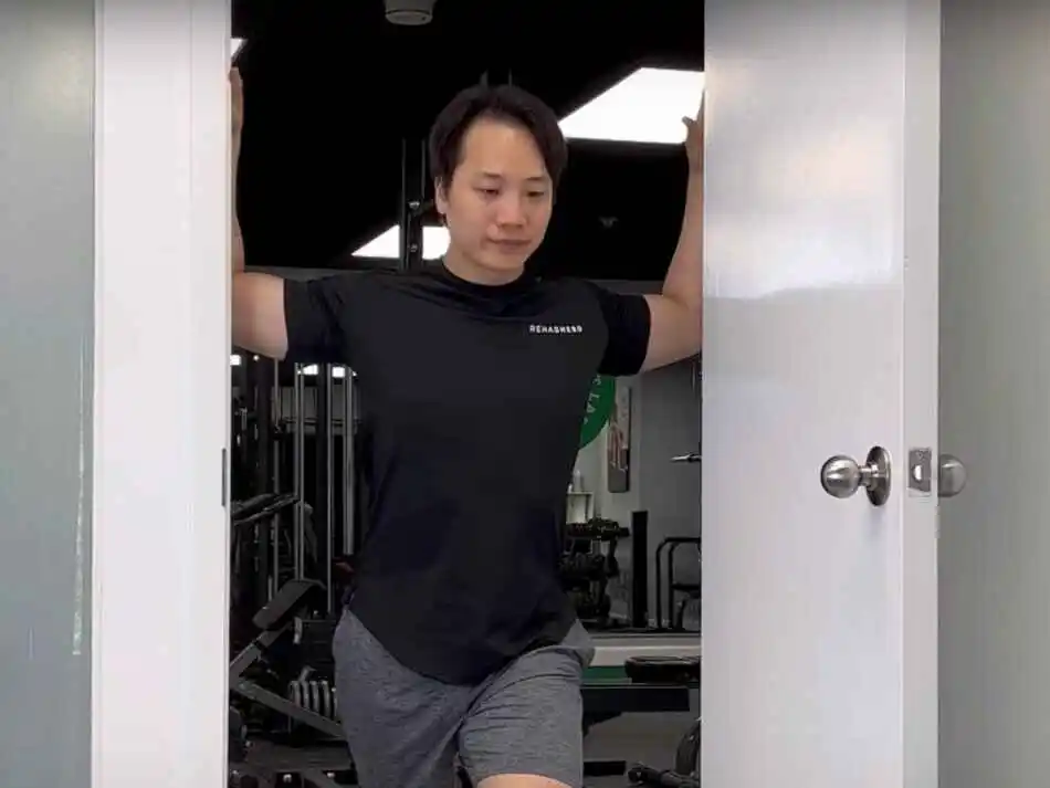
- You are standing near a doorway with your elbow joint & arms forming a 90-degree angle.
- First, step your right foot forward as you press your palms into the sides of the door frame.
- Try to Lean forward & engage your core muscle.
- Hold this stretching position for up to 30 seconds.
- Then repeat the stretching with your left foot forward.
- Do this stretching exercise on each side 2–3 times.
Neck release stretch:
- This exercise is a gentle way to loosen tension in your neck & shoulder joints.
- Perform this stretching in a sitting or standing position.
- Then lower your chin toward your chest.
- You feel a stretch along the back of your neck.
- Try to gently tilt your head to the left side to stretch your right shoulder joint.
- Hold this stretching position for up to 1 minute & repeat on the opposite side.
- Do this stretching each side 3–5 times.
For to deepen in this stretch:
- First place 1 hand on your shoulder joint & 1 hand above your ear to gently guide the movement.
- Then lower your chin toward your chest.
- You feel a stretch along the back of your neck.
- Try to gently tilt your head to the left to stretch your right shoulder joint.
- Hold this stretching position for up to 1 minute & repeat on the opposite side.
- Do this stretching each side 3–5 times.
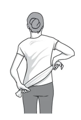
Towel stretch:
- First, hold one end of a three-foot-long towel behind your back & grab the opposite end with your other hand.
- You are holding the towel in a horizontal position.
- You must use your good arm to pull the affected arm upward to stretch it.
- You are also performing an advanced version of this exercise with your towel draped over your good shoulder joint.
- Then hold the bottom of the towel with the affected arm.
- Try to pull the arm toward the lower back with the use of the unaffected arm.
- Do this 10 to 20 times a day.
Bent Arm Scaption Plane Stretch:
- You are kneeling position next to an elevated surface.
- Then put one palm down on the surface with your elbow joint pointed up.
- Try to Roll the shoulder joint capsule in & out.
- You feel the best stretch in your pec during this stretching pose.
- Then move in & out of the stretch 10 times & hold this exercise for 15-30 seconds.
- Do the 1 set of 3 repetitions & 3 times per day.
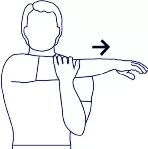
Shoulder stretch:
- You are in a standing position.
- Try to raise your shoulder joint.
- Hold this exercise for 5 seconds.
- Then squeeze your shoulder blades back & together.
- Hold this exercise for 5 seconds.
- Try to pull your shoulder blades downward.
- Hold this exercise for 5 seconds.
- Relax & repeat this exercise 10 times.
Armpit stretch:
- Use the good arm, and lift with the affected arm about breast-high.
- Gently bend the knees and open up the armpit.
- Dee pend the knee bend slightly, gently stretch armpit.
- With each knee bend and stretch a little further, but not force it.
- Do this exercise 10 to 20 times each day.
Splenius capitis muscle stretch
- The left-side splenius capitis is stretched by flexing, right laterally flexing, and right rotating the head and neck at the spinal joints, while the left-the-side shoulder girdle is allowed to elevate.
- You can hold for 30 seconds and 3 repetitions you can hold for 30 seconds and 3 repetition
Splenius cervicis muscle stretch
- Stand or sit upright
- Keep your head up facing straight ahead
- Push your head forward side by sticking out your chin
- Note: Keep your head up during this stretch. Do not let your chin fall towards the floor.
- You can hold for 30 seconds and 3 repetitions.
Trapezius muscle stretch
- Slowly take your left ear toward your left shoulder. It’s natural for your right shoulder to lift as you do this. If that happens, ease your head back toward the center until you can relax your right shoulder back down.
- Gently release this side, and then ease your right ear toward your right shoulder and complete the stretch on the other side, breathing deeply through it.
- You can hold for 30 seconds and 3 repetitions.
Semispinalis capitis muscle stretch
- The semispinalis capitis muscle(of the transversospinalis group) is stretched by flexing the head and neck at the spinal joints. Adding in lateral flexion (not seen in the accompanying illustration) will increase the efficacy of the stretch for the opposite-side semispinalis capitis muscle.
- You can hold for 30 seconds and 3 repetitions.
Splenius cervicis muscle stretch
- The left-side splenius cervicis muscle is stretched by flexing, right laterally flexing, and right rotating the neck at the spinal joints, while the left-side shoulder girdle is allowed to elevate.
- You can hold for 30 seconds and 3 repetitions.
Strengthening exercise:
Shoulder circles:
- You are standing with your left hand on the back of a chair.
- Then allow your right hand to hang down.
- Try to make a circle on your right hand 5 times in each direction.
- Repeat this exercise on the opposite side.
- Do this exercise 2–3 times per day.
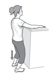
Pendulum exercise:
- The patient is in a standing position beside a table with the hand of the unaffected shoulder on the table & feet are slightly wider than shoulder-width apart.
- Then Bend the hip joint approximately to 75 to 90 degrees & let the affected arm hang down toward the floor.
- Shift the weight from side to side, letting the arms swing freely from side to side.
- Shift the weight forward & backward, letting the arms swing freely front to back.
- Once they feel comfortable with these movements, move the body so that the arm swings in a circle.
- Must be Keep the circle small, less than 8 inches.
- Continue for 30 seconds.
- Every day, increase the time to 3 to 5 minutes.
- Repeat this exercise 10 times per day.
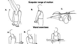
Wand exercise:
- This exercise is helpful to you for increasing ROM exercise.
- The patient is holding the wand with the help of both hands.
- Then perform the shoulder & elbow joint movement.
- Shoulder flexion, abduction, adduction, external & internal rotation & elbow flexion, and extension movement perform with the wand.
- Every exercise does the 2–3 times per day.
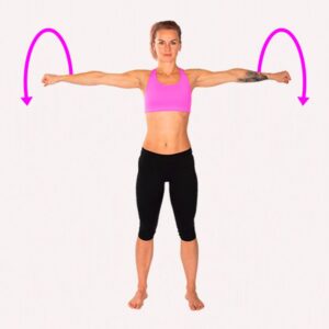
Lateral raises:
- First, hold a pair of light dumbbells for this exercise.
- You are standing with the feet slightly wider than the hip joint width apart.
- Try to raise the weights to the sides till are at shoulder level.
- Must be remembered to engage the core muscles & slowly lower the weights down to the sides.
- Repeat this exercise for 2 sets, in every set consisting of 12–15 reps & 3 to 4 times per week.
Trapezius Strengthening exercise:
- First, place your knee joint on a bench or chair.
- Try to lean forward so that your hand is reached the bench & also help to support your weight.
- Your other hand is placed at your side & palm facing your body.
- Try to raise your arm & rotating your hand to the thumbs-up position.
- Stop this exercise when your hand is at shoulder height & your arm is parallel to the floor.
- Slowly lower your arm to the original position for a count of 5.
- Do this exercise 2–3 times per day.
Shoulder Flexion isometric exercise:
- To perform the isometric shoulder flexion exercise you are standing to face a wall.
- Try to flex your elbow joint on the side of the shoulder then make a fist.
- You Place the folded napkin in between your fist & the wall & slowly press your hand into the wall.
- Hold this pressing for 7 to 10 seconds then slowly release the pressing.
- You do not need to try to push the wall over.
- Just you are gently pressing the wall to activate your shoulder flexor muscles.
- Do the 12 to 15 repetitions.
Shoulder abduction isometric exercise:
This exercise mostly targets your rotator cuff muscles.
- To perform shoulder abduction you stand six inches away from the wall.
- Then turn your body perpendicular to the wall & close to the wall at the side which is affected by the shoulder joint side.
- Make a fist on that side & press the towel into it.
- You are also using a folded towel for extra support.
- You are only gently pressed into the wall & try to lift your arm outside.
- Hold this pressure for 10 seconds & slowly release pressure from the wall.
- Do the 10 to 15 repetitions.
Shoulder joint external rotation isometric exercise:
This exercise helps to strengthen your rotator cuff muscles, mostly teres minor & infraspinatus.
- To perform shoulder external rotation you are stand to perpendicular to a wall about 6 inches from the wall.
- The affected shoulder joint is close to the wall.
- First flexed your elbow joint up to 90 degrees then make a fist.
- Try to press the towel from the back of your hand into the wall as you rotate your arm in an outward direction.
- Then gently press the towel into the wall.
- Hold this pressing for 5 to 10 seconds & slowly release the pressure from the wall.
- Complete 10 to 15 repetitions.
Shoulder joint internal rotation isometric exercise:
- To perform shoulder internal rotation you are standing facing a door frame & and outside the wall corner.
- The affected shoulder joint is near the door corner.
- Then flexed your elbow joint up to 90 degrees & make a fist.
- Try to gently press the towel into the corner wall & door jamb like as you rotate your arm inward towards your belly.
- Hold this pressing for 5 to 10 seconds & slowly release.
- Do the 10 to 15 repetitions.
Shoulder Extension isometric exercise:
- To perform shoulder extension you are standing to 6 inches away from the wall & your back is facing the wall.
- Your elbow joint is straight so your hand stays just near your buttocks & makes a fist.
- Try to gently press the towel into the wall behind you.
- Hold this pressing for 5 to 10 seconds & slowly release.
- Complete the 10 to 20 repetitions.
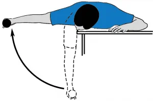
Scapular Retraction/Protraction movement:
- You are lying on your stomach means in a prone position on a table & bed with your injured arm hanging over the side.
- Must keep your elbow joint straight & lift the weight slowly.
- Try to squeeze your shoulder blade toward the opposite side as you can possible.
- Then return slowly to the starting position & repeat this exercise.
- Do this exercise 2–3 times per day.
Bent-Over Horizontal Abduction:
- You are lying on your stomach means in a prone position on a table & bed with your injured arm hanging over the side.
- Must keep your arm straight & slowly raise it to eye level.
- Then slowly lower it back to the starting position & repeat this exercise.
- Do this exercise 2–3 times per day.
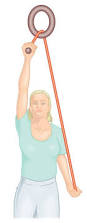
Pully exercise:
- For the shoulder active Assisted Exercise does the pully exercise.
- First, hold a rope–pully on both hands then perform the movement of the shoulder.
- Flexion,abduction, external & internal rotation.
- This exercise is performed 10 times in 1 session & 3 sessions per day.
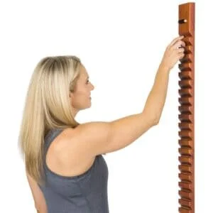
Finger ladder Exercises:
- The patient is in a standing position & facing a ladder that is hanging over a wall.
- Ask them to patient place the affected hands on the ladder at a low level.
- Then slowly start an upward climb on the finger ladder till it reached the top & slowly down back to the starting.
- This exercise is performed 10 times in 1 session & 3 sessions per day.
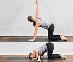
Thread the needle:
- Start this exercise on your hands & knees.
- First, lift your right hand toward the ceiling with your palm facing away from your body.
- Then lower your arm to bring the arm under your chest & over to the left side of your body with your palm facing up.
- It activates your right shoulder joint & arm to avoid collapsing into this area.
- Must keep your left hand on the floor for support.
- Then lift the arm toward the ceiling & bring the arm around to the inside of your right thigh.
- Hold this exercise position for up to 30 seconds & relax in a Child’s Pose.
- Then repeat this stretching exercise on the left side.
- Do this exercise 2–3 times per day.
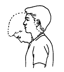
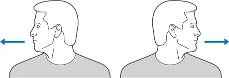
Chin tuck Exercise:
- Sit straight and look straight ahead with the ears directly over the shoulders.
- Put a finger on the chin.
- Without moving the finger, pull the chin and head straight backward until a good stretch is felt at the base of the head and top of the neck.
- Hold for 5 secs if possible.
Neck flexion Exercise (Neck tilt)
- In Relax sitting position flex your head down to rest your chin on your chest (Flex the neck fully).
- Gently tense your neck muscles and hold for 4 to 8 seconds.
- Return to a neutral position and repeat 10 times.
Neck flexion Exercise (side to side)
- Flex your head down towards your shoulder, and try to touch your shoulder with your ear (Without elevating your shoulder).
- Gently tense your neck muscles and hold for 8 seconds.
- Return your head to a neutral position and repeat in the opposite direction.
- Repeat 10 times on both sides.
Neck Rotation exercise
- Rotate your head towards one side, keeping your chin at the same height and moving within comfortable limits.
- Gently stretch your neck muscles and hold for 8 seconds.
- Return your head to the neutral position and repeat in the opposite direction.
- Repeat 10 times on both sides.
Prone Cobra Exercise
- This workout is done lying face down on the ground & uses gravity as resistance in the reinforcing process.
- Lying face down, put the forehead on a rolled-up hand towel for comfort.
- Place the arms at the side, palms downwards on the ground.
- Place the tongue on the roof of the mouth
- Pinch the shoulder blades together & lift the hands off the ground.
- Roll the elbows in, palms out, & thumbs upward.
- Gently lift the forehead about an inch off the towel keeping the eyes looking straight at the ground
- Hold the position for 10 secs.
- Perform 10 repetitions.
Isometric Neck Exercise
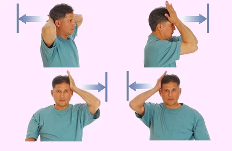
- Isometric neck exercise helps to strengthen your neck muscles. This exercise is mostly recommended by Physiotherapists from day one to relieve Neck pain.
Isometric Neck Exercise
How to perform this exercise In a sitting Position, - keep your Body straight, Put your both hands behind the neck (as seen in the images), and try your neck to push pressure on your hands and the same time Resist with your neck muscles, both hands maintain align position for 4 to 5 seconds and then relax.
The first day does 8 repetitions and the second day 10 repetitions. Do some exercise on your forehead and each side of the neck.
Do the exercise by pressing on the side of your head. Repeat 8 times, then alter sides.
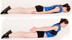
Prone Rows Exercise
- Lie on your stomach with your arms dangling off the side of the bed (try angling your body so your head is facing the corner of your bed).
- Use a pillow under your stomach for comfort. Begin by pulling arms back while bending elbows and squeezing shoulders blades together then slowly return to starting position.
- Do not lift your head up while pulling your arms back.
- Repeat 20 times. Perform 2 times a day.