
Ankle Pain: Cause, Symptoms, Diagnosis, Physiotherapy Treatment and Exercise
Table of Contents
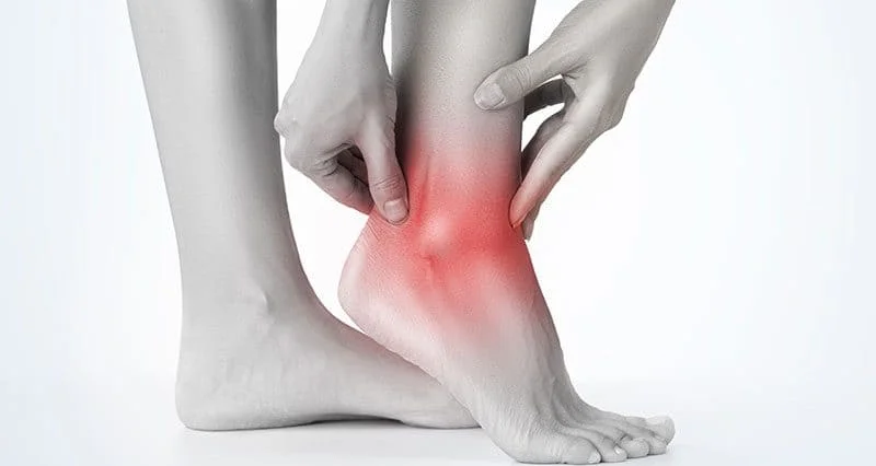
What is ankle Pain?
- Ankle pain means any kind of pain or discomfort affecting any part of the ankle.
- Ankle pain can happen for many reasons. The most reliable causes are injury, arthritis, and normal wear and tear. Depending on the cause, people may feel pain or stiffness anywhere around the ankle and the ankle may also swell, and individuals may not be able to put any weight on it.
What is an ankle joint?
- The ankle joint is also famous as a talocrural joint, which is a synovial joint, located in the lower limb.
- This ankle joint is formed by the bones of the lower limbs like as tibia and fibula & talus.
- Functionally, the ankle joint is a hinge type joint, which allows dorsiflexion and plantarflexion of the foot. Ligaments hold these bones together.
- A complex structure of tendons, muscles &other soft tissues of the ankle allows the foot for joint movement.
What are the considerable causes of ankle pain?
- An ankle pain happens when the ankle joint is twisted more than its normal position.Most ankle injuries happen whether during sports activities or while walking on an uneven surface that forces the foot &ankle into a twisted position. The unnatural position of the ankle in high-wedged shoes or walking on rough surfaces or sandals is also, the factor that may contribute to ankle injuries.
- In addition,
- Bursitis: Fluid-filled sacs called bursae to cushion your bones when they move. Bursitis occurs when sacs become irritated and inflamed.
- Fractures: An accident/injury may cause bones to break (fracture). Ankle fractures range from mild to severe. A broken ankle causes ankle swelling and pain, for example, stress fractures.
- Sprains: Sprained ankle is the favorite cause of ankle pain. An ankle sprain occurs when any ligaments stretch or tear. Sprained/ twisted ankles occur when the ankle rolls forcefully out of its normal position.
- Tendonitis: Irritated, inflamed tendons are soft-tissue injuries called tendonitis. Tendons connect muscles to bones. Sometimes, a tendon may tear (such as an Achilles tendon rupture). A torn tendon may need surgical repair.
- Several diseases, disorders, and conditions may also lead to ankle pain. These include:
- Arthritis: Pain and stiffness in the ankle joint can result from ankle arthritis. Arthritis occurs if cartilage (tissue in joints that cushions bones) breaks down. The breakdown causes bones to rub together. Arthritis happens because of overuse, injuries, and obesity, and it’s mostly seen in people over 65. Many types of arthritis can affect the ankles. Common types include rheumatoid arthritis, osteoarthritis, and Septic arthritis.
- Flatfoot: A very low arch (or no arch at all) can cause pain and swell in the ankles and feet. Sometimes, childrens’ arches do not develop normally as they grow up, resulting in the condition.
- Gout: A type of arthritis, gout results from a buildup of uric acid throughout the body. Generally, uric acid leaves the body in urine. Over the amount of uric acid creates crystals that are settled in the joints. Gout in the ankle is very painful.
- Infection: Several types of infection, including cellulitis, can cause swelling and pain in the ankle joint. A bone infection is called osteomyelitis.
What are the signs and symptoms of ankle injury?
- Ankle swelling
- Bruising
- Redness
- Numbness or tingling
- Instability
- Joint Pain
- Inability to bear weight on the affected ankle
- Ankle joint Stiffness
- Weakness
- Discoloration
- Warmth
- Tenderness
Diagnosis:
- During a physical examination, the doctor will examine the patient’s ankle, foot, and lower limb. The doctor will touch the area of skin around the injury to check for proper points of tenderness and move the patient’s foot to check the range of motion and to know what positions cause discomfort or pain. If the injury is severe, a doctor must recommend 1 or more of the following imaging scans to find a broken bone or to evaluate in more detail the extent of ligament damage
- X-ray:-While an X-ray, a very small amount of radiation passes through the patient’s body to produce images of the bones of the whole ankle. This test is the best for finding out bone fractures.
- Magnetic resonance imaging (MRI):- MRI uses radio waves & a strong magnetic field to produce detailed cross-sectional or 3-D images of the soft internal tissue of the ankle, including ligaments.
- CT scan:– Through CT scans doctor can get more detail regarding the bones of the joint. CT scan takes X-rays from many variant angles and mixes them to make cross-sectional or 3-D images.
- Ultrasound:- An ultrasound uses sound waves to produce images. These images may help doctors find the condition of a ligament/tendon when the foot is in a different position.
Some special test for ankle pain:-
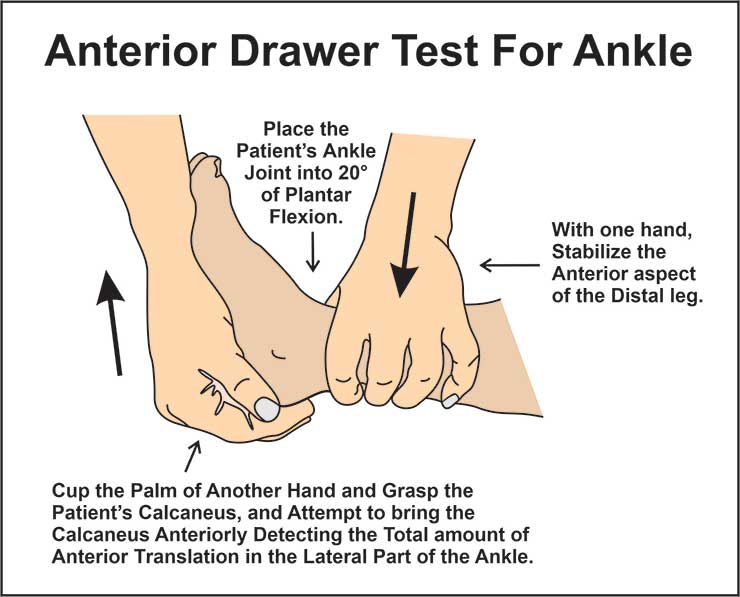
Anterior Drawer test – To find out any injury in anterior talofibular ligament:
The patient is in the supine position, the ankle joint is 20° flexed, then the heel is resting on the examiner’s palm which is resting on the table. After stabilizing the calcaneus bone. The examiner after that stabilizes the tibia & fibula while drawing the calcaneus bone anteriorly observing the amount of anterior translation at the lateral aspect of the ankle & the change in end feel. The amount of anterior translation & the eventual weakening of the end feel, changing from hard ligamentous to weak elastic, is observed. An anterior translation greater than 1 cm as compared to the healthy opposite ankle and an evident weakening of the end feel is most indicative of a partial rupture or complete rupture of the anterior talofibular ligament.
Talar Tilt test – To find out any injury in the Anterior Talofibular ligament & the Calcaneofibular ligament.
The patient is seated with an unsupported foot and ankle. The position of the foot is 10-20 degrees of plantarflexion. The distal lower limb is stabilized with one hand just proximal to the mallei & the hindfoot is inverted with the opposite hand. The lateral side of the talus is palpated to determine if tilting occurs. The laxity is compared to the opposite side.
Squeeze test
The squeeze test compresses the proximal fibula opposite to the tibia to assess the integrity of the bones, interosseous membrane, and syndesmotic ligaments. Pain starts with fracture or diastasis and is considered positive.
Kleiger’s test or external rotation
The patient is in a seated position, with knee hanging in 90 degrees, ankle relaxed. Examiner should sit at the level of the ankle to be tested with one hand stabilizing the leg from behind, the other hand grasping the ankle in a neutral position then externally rotating the foot, If there is pain at the site of the interosseous membrane, or medially. Pain may radiate upward to the leg depending on the severity of the injury then the test is positive.
Impingement sign ankle
The clinician faces the patient in a seated position. The clinician holds the calcaneus with the hand on the lateral side of the foot with fingers around the calcaneal tuberosity and thumb over the anterolateral portion of the ankle. The medial hand grasps the forefoot. Initially, the foot is held in plantarflexion. While pressure is applied with the examining thumb, the foot is brought from a plantarflexed position to full dorsiflexion. If Pain is reproduced from combined movement with thumb pressure and pain is greater in dorsiflexion than in plantarflexion then the test is positive. If Pain is not increased with the combined movement then the test is negative.
Treatment
Medical treatment
- In most cases, pain relievers used — such as ibuprofen (Advil, Motrin IB, others) or naproxen sodium (Aleve, others) or acetaminophen (Tylenol, others) — are enough to manage the ankle pain.
Physiotherapy treatment
Electrotherapy treatment
- There are many types of electrocal modalities available, but especially in ankle pain three modalities would be beneficial which are describe below:
- Ultrasound: Ultrasound is usually apply on tender point for reduction of pain.
- TENS (Transcutaneous electrical nerve stimulation): TENS modality is mainly for reduction of pain, swelling.
- IFT (Interferential Therapy ): If the patient has any type of swelling, pain, or inflammation, IFT would be the most effective modality.
Exercises
- Regular exercise must help the patient to relieve pain and strengthen muscles.
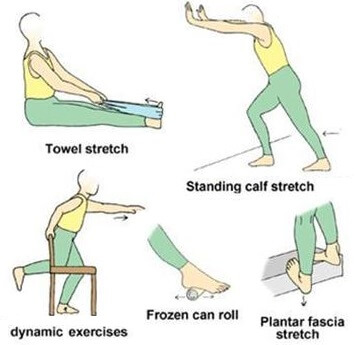
Stretching exercise:
- Achilles tendon & plantar fascia stretch:
- In the starting, loop a towel around the ball of the foot & pull it back towards your body with a straight knee. Hold for 30 seconds for each foot and do it 3 times.
- Sitting plantar fascia stretch:
- Cross the foot over the contralateral knee. The base of the toes is put on the ground & gently pull back until you feel a stretch along the underside of your foot. Hold for thirty seconds per repetition. Repeat 3 times on each foot.
- Plantar fascia stretch:
- Put a towel on the floor in front of your chair, with the heels which are on the ground, try to pick up the towel by grabbing it with your toes, this exercise Repeat 10-20 times.
- Standing calf stretch:
- The patient must stand facing a wall then place both hands on the wall which is in front of the patient. Put one foot anterior to the other with the front foot about 30cm away from the wall, keep the posterior knee straight with your back heel flat on the floor. Slowly bend the front knee until the patient can feel a stretch in the calf of the back lower leg. Relax and repeat ten times. This exercise may also be done with the back knee slightly bent for a milder stretch.
- Deep calf stretch:
- A patient must stand on a step with the heel hanging off the edge. Patients may allow holding onto something for support. Allow patient heels to drop towards the floor and feel a stretch in the calf muscles & Achilles tendon. Hold this for 30 seconds. Repeat this exercise 3 times.
Range of motion exercise:
- Flexion:
- Tell the patient to bend the foot toward himself/herself as long as he/she can towards the face side.
- Repeat 10 times, twice a day.
- Extension:
- Bend foot away from the body.Repeat 10 times, twice a day.
- inversion:
- Turn your foot inward as long as the patient can. Repeat 10 times, twice a day.
- eversion:
- The foot turns outward as long as you can. Repeat 10 times, twice a day.
- Alphabet:
- Firstly, draw the letters of the alphabet in the air by imaging with your foot. Do lowercase and uppercase letters, and make sure to move from the ankle, not from the hips.
- Circles:
- Move your ankle in circles: five to ten circles in one direction, then do the same in the other. Repeat minimally 3 times a day.
Strengthening exercise:
- Seated foot & heel raise:
- Embark with patient’s feet flat on the floor. Raise both heels off the floor then hold for a few seconds before placing heels back on the ground. After that raise the toes off the floor and hold for a few seconds before coming back to the floor. Repeat 10 times.
- Toe bend:
- Embark with the feet flat on the floor. Raise both heels off the floor after that hold for a few seconds before placing heels back on the ground. After, raise toes off the floor & hold for a few seconds before putting them on the floor. Repeat 10 times.
- Big toe lift and hold:
- With your feet on the ground, lift merely a big toe while keeping the other toes on the ground. Hold this exercise for a few (around10) seconds and relax. Repeat 10 times.
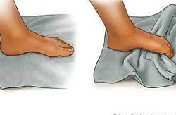
- Towel pickup:
- Place a towel on the ground in front of the chair. With your heel which is on the ground, try to pick up the towel by grabbing it with your toes. Repeat this exercise 10-20 times. After that, this exercise must be performed by adding a small weight to the towel.
- Flexion:
- Sit on a chair and tie the theraband around something that won’t move, like a heavy table. Extend your lower leg and wrap the other end of the band around the top of your foot. Do flexion of your foot, with the band resisting the motion. Hold for 10 seconds. Repeat 10 times.
- Extend:
- Sit on a chair. Take one side of the theraband in each hand, wrap it around the ball of your injured foot, and stretch your leg out in front of you. Extend your foot away from you. Hold for 10 seconds. Repeat 10 times.
- Inversion:
- Tie the theraband around the leg of the chair on the same side of the injured foot. Wrap the other end of theraband around the inside of the injured foot. Then, try to turn your foot inside. Do this exercise 10 times, and hold it for 10 seconds.
- eversion:
- Tie the theraband on the leg of your chair opposite to your injured foot. Wrap the other end around the outside of the injured foot. Try to turn your foot outside, and hold for 10 seconds, repeat this exercise 10 times.
- Step-ups:
- Embark with the injured foot on the bottom stair and your good foot on the ground. Straighten your knee so he/she can lift yourself up on the injured leg, then put it down. Repeat this exercise 5 times, minimally 3 times a day.
- Heel raises:
- Sit on the chair with flat feet flat on the ground. Although raise your heels up slowly, keep your toes on the ground. Lower your heels back down. Repeat this exercise 10 times, minimally 2 to 3 times a day. As muscles start to gain strength, do this exercise in a standing position. When you are stronger still, do this standing on just one (you’re injured) foot.
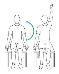
Balance and Coordination Exercises:
The last step is to work on balance and coordination. You must try these exercises:
- Balance:
- Do this balance exercise near a chair, table, or in a doorway so the patient will have support if the patient needs it. Stand on a chair or table and balance on just your injured leg for 30 seconds. Repeat this exercise 3 times a day. Try to do this for up to 3 minutes. In the next stage, do this exercise in the same but with closed eyes.
- Heel walk:
- First, walk forward and backward as long as the patient can on his/her heels.
- Tippy toe walk:
- Walk forward and backward as long as the patient can on his/her toes.
- Single-legged balance:
- Lift one foot off the ground. Try to balance on one foot for as long as you can, up to 30 seconds. Repeat on the other side. Repeat it 5 times a day.
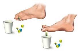
- Marble pickup:
- Sit up straight on a chair, with feet flat on the ground. Place an empty bowl and a bowl of 20 marbles on the ground in front of the patient’s feet. Using only the toes of one foot, try to pick up each marble and place marble in the empty bowl. Repeat marble picks up exercise using the other foot.
The most considerable five home treatments for ankle pain are:
- Rest: If a person has an injury namely a sprain, a person should stay off his/her feet for a while. Crutches or walking boots can help a person get around without putting weight on a person’s ankle.
- Ice: For reduction of swelling, apply ice or a cold compress to the injured area for 15 to 20 minutes every 2-3 hours.
- Compression: Try to wrap an elastic bandage around the patient’s ankle to reduce inflammation. Be aware not to wrap it too tight.
- Elevation: Resting with the injured ankle elevated above the patient’s heart reduces swelling. The patient can also try to sleep with his/her foot elevated at night.

4 Comments