Superior tarsal muscle Origin, Insertion, Function, Exercise
Table of Contents
Introduction
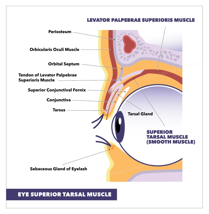
- The superior tarsal muscle is a smooth muscle bordering the levator palpebrae superioris muscle that allows the raising of the upper eyelid. Not to be mistaken with the orbitalis muscle or the circular fibers of the ciliary muscle, both of which are also called Müller’s muscle. The superior tarsal (Muller’s muscle) is a smallish muscle located within the superior eyelid. It is a smooth muscle, but it is viewed as a structural portion of the more extensive skeletal muscle of the eyelid; levator palpebrae superioris. It rises from the deep side of the levator palpebrae superioris to the upper margin of the superior tarsal plate of the eyelid.
- The smooth muscle, the superior tarsal is controlled by the autonomic nervous system, specifically its sympathetic division. The important action of this muscle is to elevate the upper eyelid in a form of sympathetic predominances, such as excitement, fear,& amazement. Moreover, the fixed tone of the muscle also gives to keeping the eyes open & maintaining the size of the palpebral fissure constant in the state of awakeness.
- Injury to this muscle, or the nerves which supply it, will outcomes in ptosis of the affected eye as seen in Horner syndrome, a condition in which there is damage to the cervical sympathetic chain. Surgeries that involve repair of ptosis, or upper eyelid correction procedures, will generally face Muller’s muscle & might be very careful in adjustments when resecting portions of the upper eyelid.
Origin
The superior tarsal muscle originates from the undersurface of the levator palpebrae superioris muscle.
Insertion
- The superior tarsal muscle is inserted in the upper margin of the superior tarsal plate.
Structure
- The superior tarsal muscle arises from the underside of the levator palpebrae superioris & inserts at the superior tarsal plate of the eyelid. The superior tarsal be as a smooth muscle of the upper eyelid. It arises from the deep surface of the levator palpebrae superioris & inserts inferiorly, to the superior tarsal plate of the upper eyelid. It is observable with the naked eye & is about 15 mm wide by 10 mm long.
- The superior tarsal muscle acquired its innervation from the sympathetic nervous system. More specifically, the postganglionic sympathetic fibers stem from the superior cervical ganglion & form the internal carotid plexus around the cervical segment of the internal carotid artery. Following the petrous & cavernous segments of the internal carotid artery, these nerve fibers enter the skull & traverse the cavernous sinus. Finally, the nerve fibers for the superior tarsal muscle allow the orbit in a form of a tight nervous plexus bound around the ophthalmic artery, which is a branch of the internal carotid artery. Consider that the ophthalmic artery innervated the blood supply for the superior tarsal muscle.
- The main motion of the superior tarsal muscle is to elevate the upper eyelid in a state of sympathetic main. The function of this action is to permit a wider visual field in states of acute life-threatening situations (fight or flight response). In extra, the muscle has a static tone, which includes up to the tone of the levator palpebrae superioris muscle. The tones of these 2 muscles are in balance with the adversarial tone of orbicularis oculi, thereby keeping the eyes open & defining the size of the palpebral fissure.
Function
- Its action is not fully clear but may be an accessory muscle to raise the upper eyelid. The superior tarsal muscle has a remarkable action in that it assists the levator palpebrae superioris by sustaining the elevation of the upper eyelid after the levator palpebrae superioris has raised it. It also acts to raise the upper eyelids a further 2 mm after the levator palpebrae superioris has already originally raised them as a sympathetic nervous system response.
- The superior tarsal muscle was discovered to have a much more main role in levator action when compared to the levator palpebrae superioris than earlier thought. Research has set that Muller’s muscle shares a large amount of power with the levator palpebrae superioris when elevating the eyelid
Nerve supply
- The superior tarsal muscle gets its suppliers from the sympathetic nervous system. Postganglionic sympathetic fibers get in the superior cervical ganglion & transit via the internal carotid plexus, where small branches spread with the oculomotor nerve as it hands through the cavernous sinus. The sympathetic fibers continue to the superior branch of the oculomotor nerve, where they go to Muller’s muscle on its inferior portion.
Blood supply and lymphatics
- Oxygenated blood proceeds to the superior tarsal muscle via the lateral muscular branch of the ophthalmic artery. This branch as well as gives blood to the lateral rectus, superior rectus, superior oblique, & levator palpebrae muscles. The ophthalmic artery holds a medial muscular branch supplying blood to the remaining extraocular muscles.
- The superior tarsal muscle drains deoxygenated blood into the precise veins as the remaining extraocular muscles. These veins, called vortex veins – originating from the posterior portion of the eye, are where the blood exiting Muller’s muscle reaches first. This blood then unloads into the superior & inferior orbital veins which eventually pass to the cavernous sinus.
History
- The muscle emerges its name from the Greek ταρσός ‘flat surface’, typically used for drying. The term Müller’s muscle is sometimes utilized as a synonym. Yet, the same term is also used for the circular fibers of the ciliary muscle, & also for the orbitalis muscle that preserves the inferior orbital fissure. Given the possible confusion, the use of the term Müller’s muscle might be discouraged unless the context clears any mysteriousness.
Embryology
- The superior tarsal muscle evolves in a way, not unlike the other extraocular muscles. These subtypes of muscles are distinct in that; when contrast to periocular tissues, these muscles came from the paraxial mesoderm in the prechordal plate, which is unusual in that connective tissue tendons are arid from neural crest cells instead. Specifically, around week 5 is when Muller’s muscle 1st seems in development.
Muscle fibers
- The definite muscle subtype of the superior tarsal muscle is different from the further extraocular muscles. Muller’s muscle is made of smooth muscles, especially thin fibers of smooth muscular tissue. This muscle arises at the bottom of the levator palpebrae superioris & attaches to the superior tarsal plate.
- Precise differences which are seen in this muscle contrast to other smooth muscles exist. 1 such distinction is the point that the superior tarsal muscle is invited not solely of smooth muscle but instead is mixed tissues. These tissues include connective tissue, blood vessels, & fat. Also, one fundamental difference between the superior tarsal muscle & others is that the smooth muscle cells of this muscle are distributed sporadically & are not connected. Lastly, another difference indicates that the arising of Muller’s muscle may have fibers of striated muscle connected with fibers of the smooth muscle.
Physiologic Variants
The superior tarsal muscle has typical physiologic variants when comparing different individuals, & these center near the attachment of the muscle to the superior tarsal plate. Four variations live, & they are below:
- Muller’s muscle binding to the upper border of the superior tarsal plate
- 2M: The superior tarsal muscle binds to the medial side of the plate
- 2L: The superior tarsal muscle binding to the lateral side of the plate
- The Muller’s muscle attaches along the whole part of the superior tarsal plate.
- The 3rd variation was the most repeatedly observed.
Clinical importance
- Horner’s syndrome happens when the sympathetic nerve supply to the face & eye is damaged. This condition frequently occurs due to trauma of the neck and shoulder regions & consequential injury of the cervical sympathetic ganglia &/or nerves. Injuries of this kind will result in a loss of effective innervation of the smooth muscles & glands in the head.
- Horner’s syndrome is characterized by a triad of symptoms; partial ptosis (a drop of the upper eyelid), miosis (constricted pupil) & loss of hemifacial sweating (anhidrosis). certain times, it can also be followed by the posterior displacement of the eyeball within the orbit (enophthalmos).
FAQ
The superior tarsal muscle, called Muller’s muscle, is a structural muscle that acts to maintain the elevation of the upper eyelid. It collects innervation from the sympathetic nervous system & is unique in that it consists of thin fibers of the smooth muscle.
The oculomotor or 3rd cranial nerve innervates the levator palpebrae superioris to elevate the upper eyelid. The levator palpebrae superioris becomes a tendinous aponeurosis, which fuses with the anterior superior part of the superior tarsal plate & possibly the pretarsal skin.
Müller’s muscle is a smooth muscle in the upper eyelid that is supplied by the sympathetic nervous system. When the muscle is contracted, it elevates the lid roughly 2.5mm.
The superior tarsal muscle arises on the underside of the levator palpebrae superioris & inserts on the superior tarsal plate of the eyelid.
superior eyelid
The superior tarsal (Muller’s muscle) is a small muscle located within the superior eyelid. It is a smooth muscle, but it is considered a structural part of the larger skeletal muscle of the eyelid; levator palpebrae superioris.
The inferior tarsal muscle which composes part of the lower eyelid retractor a function to retract the lower eyelid & innervates sympathetic nerves like the Müller muscle, an organ homologous with the inferior tarsal muscle.
The superior tarsal (Muller’s muscle) is a small muscle situated within the superior eyelid. It is a smooth muscle, but it is considered a structural part of the larger skeletal muscle of the eyelid; levator palpebrae superioris.
Structure. The superior tarsal muscle arises on the underside of the levator palpebrae superioris & inserts on the superior tarsal plate of the eyelid.
The tarsal plates serve as the main structural site of the eyelids. They are constructed of dense connective tissue & contain the Meibomian glands & eyelash follicles. There are approximately 30 meibomian glands in the upper eyelid & 20 meibomian glands in the lower eyelid.