
Median Nerve Anatomy, Origin, Course, Function
The median nerve is the main peripheral nerve of the upper limb. It controls most of the hand’s movement so it’s called the laborer’s nerve. it arises from the lateral and medial cord of the brachial plexus, originating in the sp[inal cord and runs down the anterior forearm to the lateral part of the hand and digits.
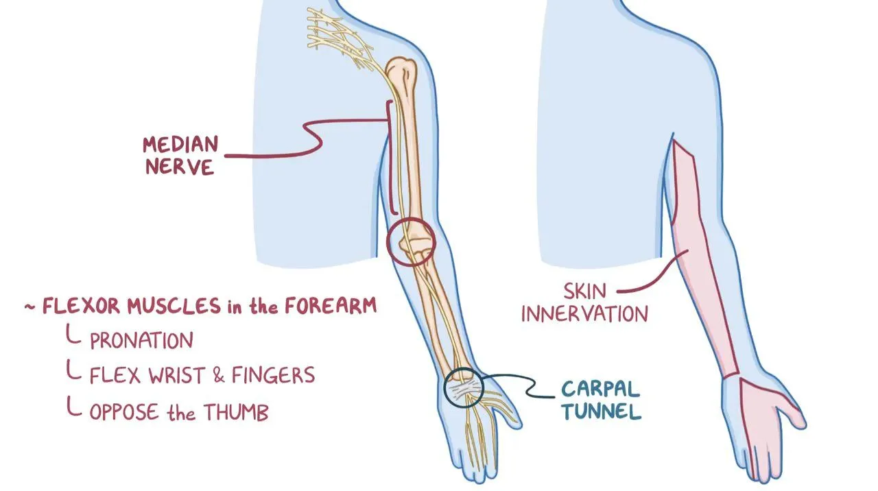
The median nerve gives the motor and sensory functions to the muscles. Motor functions like pronation movement of forearm, wrist flexion movement, flexion of digits of fingers. The sensory function like pain and temperature gives to the lateral 3 and 1/2 fingers on the palmer surface and on dorsum aspect gives to the thumb and radial half of 2nd to 4th digits of fingers in hand.
Table of Contents
Median Nerve Origin:
The median nerve is one nerve of the five terminal branches of the brachial plexus. The junction of the lateral and the medial cords form this nerve, and it has contributions from all anterior rami of C5-T1. The median nerve is the sensory and motor nerve of the arm.
It supplies most of the superficial and deep flexors muscles in the forearm, thenar, and lumbrical muscles. It also gives the sensation to the thumb, index finger, middle finger, and half of the hand’s ring finger.
Nerve Roots of Median Nerve:
C5 to C7 Roots – Lateral Cord of Brachial Plexus.
C8 and T1 Roots – Medial Cord of Brachial Plexus.
Course Of Median Nerve:
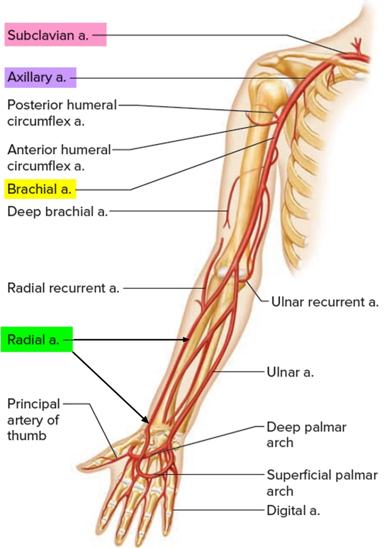
It starts from the lateral and medial cords of the brachial plexus, originating in the spinal cord, and runs through the anterior portion of the arm and forearm. Then arising from the lateral and medial cords of the brachial plexus, the median nerve enters the arm at the axilla. then it travels with the brachial artery down the shaft of the humerus bone and enters into the cubital fossa, which is at the surface of the elbow joint.
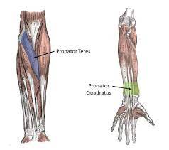
At the level of the elbow, like the median nerve courses under the bicipital aponeurosis, it provides innervation to many proximal muscles of the forearm, including the pronator teres, flexor carpi radialis, flexor digitorum sublimis, and palmaris longus muscles. As the nerve together from the bicipital aponeurosis, it passes through the superficial and deep heads of the pronator teres.
From the cubital fossa, the median nerve runs down the anterior forearm, and then it passes through the carpal tunnel, it is a narrow pathway on the palm-side of the wrist. After reaching the hand, the median nerve supplied different muscles of the hand.
As the median nerve down the forearm, it travels deep to the flexor digitorum superficialis and superficial to the flexor digitorum profundus (FDP). In the forearm, the median nerve mainly supplies to the FDP, however, the anterior interosseous nerve(AIN) gives innervation to the FDP, flexor pollicis longus, and pronator quadratus.
At the end of its course in the forearm, the median nerve and all its branches supplied all of the flexor muscles of the forearm except the ulnar half of the flexor carpi ulnaris and the flexor digitorum profundus.
Median nerve’s Branches and its innervation:-
The median nerve gives branches in the forearm and hand regions. The branches in the forearm include:
The muscular branches to pronator teres muscle, palmaris longus muscle, flexor digitorum superficialis muscle, and flexor carpi radialis muscle, these branches innervate the corresponding muscles.
The anterior interosseus nerve(AIN), supplies the flexor pollicis longus and radial part of flexor digitorum profundus. The anterior interosseous nerve runs on the interosseous membrane with the anterior interosseous artery and passes deep to the pronator quadratus to supply the pronator quadratus muscle. Its articular branches end in the distal radioulnar, radiocarpal, and carpal joints.
The major branches in the hand include:
The cutaneous nerve of the palm supplies the proximal part of the palm. This branch does not enter the carpal tunnel but is spared in carpal tunnel syndrome.
The two common palmar digital nerves, the first of which supplies the radial two lumbricals. The second nerve goes through the ring and middle finger and is divided into digital nerves that give the sensation to certain areas of the hand.
The recurrent branch to the thenar eminence muscle (flexor pollicis brevis, abductor pollicis brevis, opponens pollicis). It is also known as the ‘million dollar nerve’ to signify its importance for basic hand function.
The median nerve gives the motor supply to the flexor muscles in the forearm, apart from flexor carpi ulnaris and the ulnar head of flexor digitorum profundus (which is supplied by the ulnar nerve). It also supplies the thenar muscles as well as the radial two lumbricals.
List of Muscles innervated by Median nerve:
Flexor muscles in the forearm:

- Pronator teres muscle
- Palmaris longus muscle
- Pronator quadratus muscle
- Flexor digitorum superficialis
- Flexor carpi radialis
- Flexor digitorum profundus
Thenar eminence muscles:
- Flexor pollicis brevis
- Abductor pollicis
- Opponens pollicis
Radial two lumbricals.
The Function of the Median nerve:–
Motor function:

The median nerve gives the function of most of the muscles in the anterior forearm and some intrinsic hand muscles.
Anterior Forearm: In the forearm, the median nerve directly innervates muscles in the superficial and intermediate layers:
Superficial layer muscles: Pronator teres, flexor carpi radialis and palmaris longus.
Intermediate layer muscle: Flexor digitorum superficialis.
These all muscles perform pronation of the forearm.
The anterior interosseous nerve, which supplies the deep flexors:
Deep layer: Flexor pollicis longus, pronator quadratus, and the lateral half of the flexor digitorum profundus (the medial half of the muscle is innervated by the ulnar nerve).
These muscles perform the flexion of the wrist and flexion of the digits of the hand.
In Hand
The median nerve gives 2 branches in the hand.
The recurrent branch of the median nerve gives function of the thenar muscles for the movements of the thumb.
The palmar digital branch of the lateral two lumbricals – these muscles function of flexion at the metacarpophalangeal joints and extension at the interphalangeal joints of the first(Index) and second(middle) fingers.
Sensory function:
The median nerve is responsible for the cutaneous innervation of part of the hand. mainly 2 branches are present:
Palmar cutaneous branch – it gives sensation to the anterior half part of the palm.
Palmar digital cutaneous branch – it gives the palmar surface and fingertips of the lateral three and a half digits of fingers and dorsum aspect of thumb and radial half of 2nd to 4th digits of fingers in hand.
In this area feels sensations like pain, touch, temperature, etc.
Clinical significance:
Injury of the median nerve at different levels causes different syndromes with motor and sensory deficits.
At the shoulder:-Injury at this level shows the brachial plexus injury.
Above the elbow:– A supracondylar fracture of the humerus is most commonly seen.
Motor function loss: Loss of pronation of the forearm, weakness in flexion of the wrist, The lateral two lumbricals are affected, and the patient can’t able to flex at the MCP joints or extend at IP joints of the index and middle fingers.
Sensory loss: Loss of sensation in lateral 3 and 1⁄2 digits including their nail beds, and the thenar area of the hand
At the elbow: Impingement of the median nerve at the level of the elbow or the proximal forearm is occur in the pronator teres syndrome.
Within the proximal forearm: Injury to the anterior interosseous branch in the forearm causes the anterior interosseous syndrome.
Common mechanisms: Tight cast, forearm bone fracture.
Motor function loss: Loss of pronation of the forearm, loss of flexion of radial half of digits and thumb of the hand.
At the wrist: Most common condition seen is wrist laceration. A laceration is present just proximal to the flexor retinaculum.
Motor function loss:
Thenar muscle paralyzed, Weakness in flexion of radial half of digits and thumb, loss of abduction and opposition movement of the thumb.
Sensory loss: Loss of sensation in lateral 3 and 1⁄2 digits including their nail beds, and the thenar area of the palm.
Within the wrist: when the median nerve is entrapped in the carpal tunnel so that occur the Carpal tunnel syndrome.
Motor function loss:
Weakness in flexion of radial half of digits and thumb, weakness in abduction movement, and opposite movement of the thumb, also affect the daily functional activity.
sensory loss: Numbness and tingling are present due to compression of nerves, and sensations are also lost.
Sometimes the presence of an ape hand deformity or when attempting to form a fist, the benediction sign is present, due to compression of the median nerve.
Carpal tunnel syndrome:
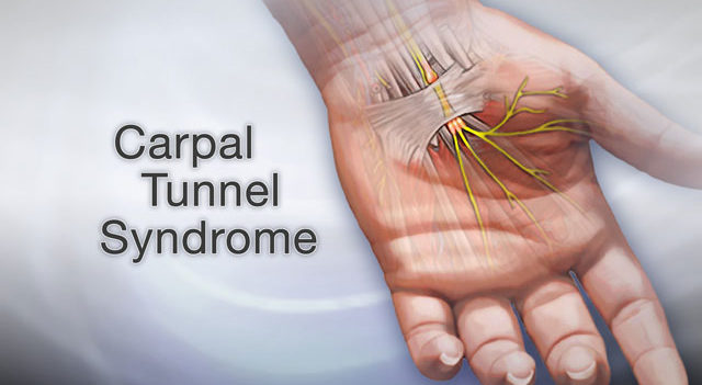
Compression of the median nerve in the carpal tunnel can cause carpal tunnel syndrome (CTS).
It is the most common mononeuropathy. risk factors for CTS are many more such as diabetes, pregnancy, and obesity, loss of protein but exact the underlying etiology is not well understood.
Clinical features include numbness, tingling, and pain in the whole area which is supplied by the median nerve. Symptoms present when the patient wake from sleep and are usually worse in the morning. If it is untreated, chronic CTS can cause weakness and atrophy of the thenar muscles.
Tests for carpal tunnel syndrome to confirm the diagnosis:
- Tinel’s sign – tapping the nerve in the carpal tunnel so that pain arises in the median nerve distribution.
- Phalen’s test – holding the wrist in flexion for 60 seconds to produce numbness/pain in the median nerve distribution.
Treatment is the use of a splint, holding the wrist in extension overnight to relieve symptoms. If symptoms are not relieved, and the condition becomes severe then surgery for carpal tunnel is required.

Hand of Benediction:
The median nerve is sometimes damaged in a supracondylar fracture at the elbow. at the elbow. Because of that results the radial head of flexor digitorum profundus muscle is denervated. The forearm is in a supinated position and the lateral two lumbricals have also been affected, the flexion at the metacarpophalangeal and interphalangeal joints of 2 & 3 digits is affected. It is unable to make a fist as both of these fingers are extended and the hand is in a classic or extended position known as the ‘hand of benediction’ (when the person make tries to flex their fingers).
Simian/ape hand deformity:
When the recurrent motor branch of the median nerve is damaged, the thenar eminence muscles become denervated. Thenar muscles are including abductor pollicis brevis, flexor pollicis brevis, opponens pollicis, and the two radial lumbricals. Due to that, the person becomes can’t oppose the thumb i.e. can’t do the tip of the thumb to the tip of the other fingers. It’s called an ape’s hand deformity.
Pronator syndrome:
The median nerve can become entrapped in between the two heads of pronator teres muscle, symptoms arise like tingling, numbness, etc. Because of that entrapment, the wrist flexor muscles are wasting and the pronation movement of the forearm is also affected.

21 Comments