
Thenar muscles
Table of Contents
Description
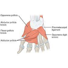
The thenar musculature is made up of 4 short muscles situated on the lateral aspect of the hand. These muscles are the adductor pollicis, abductor pollicis brevis, flexor pollicis brevis, and opponens pollicis.
The thenar muscles work together to form the thenar eminence, a fleshy prominence on the radial side of the palm. These muscles arise from various carpal bones and distally join the thumb. Most of the thenar muscles are innervated by the median nerve (T1). The exceptions are the deep head of the flexor pollicis brevis and adductor pollicis muscle that receive their innervation through the ulnar nerve (C8, T1).
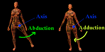
The primary function of the thenar muscles is to generate the movements of the thumb. More specifically, these muscles are accountable for the adduction, abduction, and flexion of the thumb at the MCP and CMC joints. Their combined movement may generate the opposition of the thumb, which is the combination of flexion, adduction, and medial rotation.
Flexor pollicis brevis muscle
Description
This muscle is the short tiny and broad intrinsic muscle. It is a part of the thenar muscles group together with the opponent pollicis, adductor pollicis, and abductor pollicis brevis. The heads of the FPB are superficial and deep. The deep head, on the other hand, may be undersized or even absent at times.
Origin of Flexor pollicis brevis muscle
- The trapezium and flexor retinaculum’s range is the source of the superficial Head.
- The trapezoid and capitate bones are the roots of the deep head.
Insertion
- The profound head further separates from the superficial head after parting profoundly from the Flexor Pollicis Longus ligament on the spiral sesamoid bone and the foundation of the proximal phalanx of the thumb.
Relations
- The thenar muscle with the vastest is the flexor pollicis brevis.
- It is lateral to the AP muscle and is medial to the abductor pollicis brevis and opponens pollicis muscles.
- Also, the OP muscle and the superficial head are generally interconnected.
- The motor branch of the median nerve goes across the muscle’s shallow surface.
Innervation
- The innervation of the fFPB’s two heads generally opposes.
- The recurrent branch of the median nerve provides supply to the flexor pollicis muscle’s superficial head, whereas the deep head provides the innervation from the deep branch of the ulnar nerve which emerges from the spinal roots C8 and T1.
Blood supply
Flexor pollicis brevis take a blood supply from the various regions of the radial artery; branches of the princeps pollicis artery, the superficial palmar artery, and the radialis indicis artery.
The function of Flexor pollicis brevis muscle
- The thumb is flexed at the MCP and CMC joints by the FPB, a member of the thenar muscles.
- This helps the thumb move in the opposite direction, and if it continues, it will cause the thumb to rotate in the internal direction.
Abductor pollicis brevis muscle
Description
The APB muscle is a tiny, triangular muscle located in the hand. APb is one of the intrinsic muscles of the hand, meaning that it forms and inserts within the hand itself. The primary role of the APB muscle is to bring the thumb laterally, meaning it moves the thumb out from the palm of the hand. It also supports in opposition & extension of the thumb.
This muscle is innervated by the median nerve, which is a nerve that arises from the brachial plexus and runs down the arm and into the hand. This muscle is crucial for fine motor control of the hand, and its proper function is necessary for doing many routine activities, such as grasping and manipulating objects.
Under the skin is the most lateral and external to the thenar muscles, the abductor pollicis brevis. It connects the thumb’s proximal phalanx to the scaphoid and trapezium carpal bones through the flexor retinaculum. The abduction of the thumb at the CMC joint is the preliminary function of the abductor pollicis brevis.
Origin of Abductor pollicis brevis muscle
It takes its origin from the front of the transverse carpal ligament, extending into the tubercles of the scaphoid and trapezium with an occasional contribution from the tendon of the abductor pollicis longus.
Insertion
A short tendon connects the muscle to the base of the thumb’s proximal phalanx on the little finger side.
Relations
- The fusiform APB muscle is directly lateral to the OP and FPB muscles at the grade of the thenar eminence.
- The thenar branch of the median nerve goes through a gap generated by these three muscles.
- The abductor pollicis brevis’s external component is traversed by the external palmar branch of the radial artery.
Innervation
The median nerve’s thenar branch arises the abductor pollicis brevis (root values C8 and T1).
Blood supply
The APB is taken its supply from the superficial palmar branch that comes from the radial artery.
The function of the APB muscle
- The abduction of the thumb at the CMC and MCP joints is the foremost role of the abductor pollicis brevis muscle.
- The abductor pollicis longus muscle and this movement work merged.
- Moreover, the abductor pollicis brevis creates it more leisurely to drive the thumb toward the fingertips in opposition to the CMC joint and to bend the MCP joint.
Adductor pollicis muscle
Description
A hand muscle called the AP is found in the palm of the hand, especially near the base of the thumb. The oblique head and the transverse head are its two components. The transverse head originates from the lower third of the ulna and the interosseous membrane, whereas the oblique head derives from the capitate bone and the bases of the second and third metacarpal bones.
- The base of the thumb’s proximal phalanx is where the adductor pollicis muscle’s 2 heads attach.
- The APB muscle helps in both thumb adduction (taking the thumb toward the palm) and thumb opposition (bringing the thumb toward the other digits).
- It also plays an important role in movement such as gripping and grasping objects, particularly those that need a strong grip.
- The deep branch of the ulnar nerve, which comes from the medial chord of the brachial plexus, innervates the adductor pollicis muscle.
- The natural adductor pollicis of the hand is a triangle muscle.
- It is a component of the thenar muscles group, which also includes the abductor pollicis, flexor pollicis brevis, and opponens pollicis.
- The thenar muscles on the outer side of the hand make an elevation known as the thenar eminence.
- Tangential and transverse heads are noticed on the adductor pollicis. This muscle extends from the base of the 1st proximal phalanx to the capitate and 3rd metacarpal bones.
- The main motion of this muscle is the adduction of the thumb at the carpometacarpal joint.
Origin of adductor pollicis muscle
The oblique head forms from the bases of the 2nd and 3rd metacarpals, while the transverse head originates from the shaft of the 3rd metacarpal.
Insertion of adductor pollicis muscle
The inner side of the proximal phalanx of the thumb.
Relations
- The biggest and deepest muscle in the thenar muscle group is the AP.
- The adductor pollicis’ ventral part is crossed by the flexor tendons of the FPB muscle, first lumbrical, and index finger.
- On its upper side, the muscle is interconnected to, or typically attached to the 1st dorsal interosseous muscle.
- Furthermore, the pathway formed by the two heads of the adductor pollicis is used by the deep branch of the ulnar nerve, the deep palmar arch, and the radial artery.
Innervation
The profound ulnar nerve branch that innervates the adductor pollicis muscle (root value C8, T1)
Blood supply
The anastomosis of the radial artery and the deep palmar branch of the ulnar artery creates the deep palmar arterial arch, which gives supply to the adductor pollicis.
The function of adductor pollicis muscle
- The AP is the intrinsic hand muscle with the most significant influence.
- The thumb’s important function is adduction or the movement of the thumb from an abducted posture towards the 1st finger.
- This movement is crucial whrease completing activities that demand pinching and grabbing.
- Further, the adductor pollicis supports the thumb’s resistance in its last stages.
- This development involves a mixture of actions, including the thumb’s adduction, average rotation, flexion, and adduction in order for it to feel the tips of each finger on the same hand.
- As a result, the therapist can attempt to draw them apart whrease pressing the thumb on the index finger to gauge the strength of the adductor pollicis.
Opponens pollicis muscle
The opponens pollicis is a short muscle situated deep to the abductor pollicis brevis muscle. It comes from the tubercle of the trapezium bone and flexor retinaculum. The muscle fibers then run distally to insert onto the outer part of the 1st metacarpal bone.
Origin of OP
Tubercle of trapezium bone and flexor retinaculum
Insertion of OP
The opponens pollicis muscle obtains its innervation via the recurrent branch of the median nerve (T1) and its blood supply via the superficial palmar branch of the radial artery.
Function Of OP
- The main function of opponens pollicis is to create the opposite of the thumb in the 1st CMC joint.
- The opposition is a difficult movement in which the flexion, adduction, and medial rotation happen together.
- This motion is of key importance for fine motor skills and precise movements of the hand (e.g. writing or pinching).
Innervation of OP
Recurrent branch of median nerve(C8, T1)
Blood supply of OP
Superficial palmar branch of radial artery
Thenar muscle exercises
Thumb flex and extension
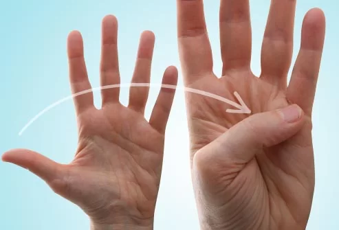
- Raise the hand, be sure that the thumb is positioned away from the fingers.
- Move the thumb across the palm so that it is touching just below the little finger.
- Hold each position for 5 to 10 seconds, performing 15 reps with each hand.
Thumb extension with a rubber band
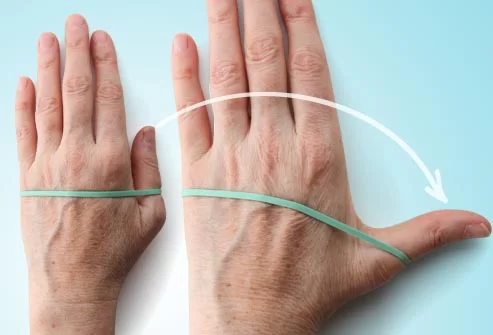
- Put the hand flat on a table or other hard surface.
- Put a rubber band around the hand so it sits at the base of the finger joints.
- Slowly move the thumb away from the other fingers as far as it may go.
- Maintain this motion for 10 to 20 seconds and then release. Do it again 5 to 10 times with each hand.
Hand grip exercise

- Take a tennis or similar-sized ball in your hand.
- Squeeze the ball as hard as a person may for between 5 and 10 seconds prior to slowly relaxing the grip.
- Do it again 5 to 10 times with the same hand and then with the other hand.
Pinch strength exercise

- Take up a soft foam ball in the middle of your thumb and index finger.
- Pinch the ball, holding the position between 20 and 40 seconds. Gently release the pinch.
- Repeat 5 to 10 times with the same hand and again with the other hand.
FAQ
The thenar muscle, or thenar eminence, is a combination of 3 muscles at the fleshy base of the thumb (first digit) on the palmar aspect that performs as a exert movement about the thumb. The thenar muscles involve abductor pollicis brevis (APB), flexor pollicis brevis (FBB), and opponens pollicis (OPP).
The primary function of the thenar muscles is to generate the movements of the thumb. More particularly, these muscles are liable for the adduction, abduction, and flexion of the thumb at the MCP and carpometacarpal CMC joints.
Put a rubber band around the hand so it sits at the base of the finger joints. Slowly move the thumb away from the other fingers as far as it, may go. Hold this position for 20 to 40 seconds and then release. Do it again 5 to 10 times with each hand.
Thenar eminence pain is generally because of overuse syndrome brought on by repetitive thumb movements. It generally improves with a combination of medical treatments and home remedies. A person may sometimes stop thenar eminence pain by bypassing activities that required repetitive thumb motion.

5 Comments