
Brachial Artery
The brachial artery is a major blood vessel situated in the upper arm. It is one of the main arteries responsible for supplying oxygenated blood to the arm and forearm regions.
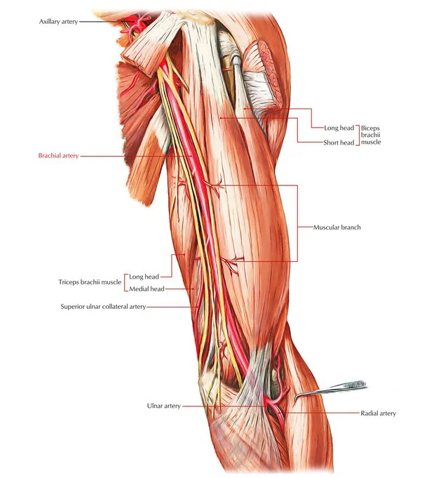
Arising from the axillary artery at the lower border of the teres major muscle, the brachial artery descends along the medial aspect of the arm, coursing down towards the elbow joint.
Table of Contents
Introduction
The arm contains two muscular components: The coracobrachialis, biceps brachii, and brachialis muscles are located in the anterior portion of the arm, whereas the triceps brachii muscle is located in the posterior portion.
The brachial artery, which is the primary artery supplying the arm, is situated inside the anterior compartment. Because of its close relationship to the humerus, it can be used for a variety of clinical exams, including blood pressure and pulse checks, but it is also vulnerable to injuries that predominantly affect the bone, including fractures.
The main blood vessel that supplies blood to your upper arm, elbow, forearm, and hand is the brachial artery. It begins at the area right behind your shoulder in your upper arm and travels through the crease directly in front of your elbow. It splits into multiple branches as it travels.
If you’ve ever had your blood pressure checked, your doctor will wrap an arm cuff over you. Your artery pressure is measured by that cuff by means of your brachial artery. To take your brachial pulse, a medical professional may occasionally apply pressure to this artery.
What is a Brachial Artery?
The aortic and brachiocephalic trunks are the respective sources of the left and right subclavian arteries. These arteries known as the axillary arteries, go through the shoulder blade. The same blood vessels are known as the brachial arteries once they reach the arm. Each brachial artery in your body is separate from the other.
All areas of the body receive oxygenated blood via aortic branches. This action is carried out for the upper limbs by the brachial arteries.
The structure of the brachial artery makes it highly helpful for many medical treatments. It is most often used by your doctor to take your blood pressure. Your arm is bound with a cuff, and they monitor the blood flow in the brachial artery located in front of your elbow. Occasionally, medical professionals will locate the brachial artery on the inside of the arm to feel the pulse. It’s also helpful to sense a baby’s pulse here.
Your doctor may occasionally take your arm and lower limb blood pressure readings. The ratio of the pressures recorded at these two locations is known as the ankle-brachial index. It facilitates peripheral artery disease diagnosis. Blood pressure testing can be used to detect coarctation of the aorta, a congenital cardiac disease that can produce a difference in blood pressure in the upper and lower limbs in children.
A further use for the brachial artery is in interventional radiology techniques. A thin, flexible catheter is pushed into the major vessels close to the heart by doctors after being passed through the elbow artery. This occasionally prevents the need for extensive heart surgery by enabling them to identify and treat disorders including blood clots, blockages, and aneurysms in the main blood channels.
What is the composition of the brachial artery?
Every artery in your body is composed of three layers:
- Tunica intima: Your blood flows freely because of this inner layer. It keeps toxins out of your circulation, controls blood pressure, and avoids blood clots.
- Media: The layer in the centre helps blood vessels dilate and constrict so blood flows in a single direction.
- Adventitia: Blood vessels are supported and given structure by their outer layer. It has tiny capillaries that carry nutrition and oxygen to your cells while also eliminating trash.
Anatomy
Where is the brachial artery situated?
The front portion of your bicep is where the brachial artery travels. It is the axillary artery’s continuation in your shoulder and armpit. The cubital fossa, which is the indentation at the front of the elbow that separates your upper and lower arms, is where it stops.
It then splits into your forearm’s radial and ulnar arteries. The major nerve that supplies your forearm, the median nerve, is parallel to the brachial artery.
Course
It starts at the inferior border of the teres major tendon and finishes at the level of the radius neck, approximately 1 cm distal to the elbow joint, continuing the axillary artery
The main blood vessel in the upper arm is the brachial artery. It is the axillary artery’s continuation beyond the teres major muscle’s lower border. It proceeds to the cubital fossa at the elbow via the ventral surface of the arm. The radial and ulnar arteries, which travel down the forearm, are where it splits after that.
In certain people, the radial and ulnar arteries run via the upper arm, and the bifurcation happens considerably sooner. The brachial artery pulse can be felt on the anterior portion of the elbow, medial to the biceps tendon. It is also frequently used to take blood pressure readings with a stethoscope and sphygmomanometer (blood pressure cuff).
The median nerve and the brachial artery are intimately connected; in proximal locations, the median nerve is located directly lateral to the brachial artery. The median nerve is located anterior to the elbow joint and extends distally across the medial side of the brachial artery.
Together with the axillary and subclavian arteries, the brachial artery is one of the main blood vessels supplying the arm and hand. It is also a vital part of the cardiovascular system. It is located in the arm between the teres major muscle and the elbow and is the continuation of the axillary and subclavian arteries.
Anatomical Relationships
The brachial artery’s anatomical path varies from person to person, just like many other structures in the human body do.
The brachial artery may travel more medially toward the medial epicondyle of the humerus rather than following its typical path along the medial portion of the biceps. In this instance, the brachial artery runs through or posterior to the pronator teres muscle after passing posterior to the supracondylar process of the humerus.
Additionally, the brachial artery may branch more proximally than typical or generate anastomoses. In this instance, the artery splits into three branches, known as the radial, ulnar, and common interosseous arteries. The common interosseus and ulnar arteries share a division with the radial artery, which normally grows more proximally from the brachial artery. On rare occasions, the ulnar artery may diverge more proximally, leaving the radial and common interosseous arteries with a shared division.
Vasa aberrantia are microscopic, thin arteries that can occasionally be seen connecting the axillary and brachial arteries.
In clinical practice, the brachial artery’s relationships with other arm structures might be significant. With a few notable exceptions, the brachial artery is a superficial vessel that is only covered by the skin’s layers as well as the superficial and deep fasciae.
- The first instance of this not being the case is at the cubital fossa, where the artery is covered and the median cubital vein is divided by the bicipital aponeurosis or the aponeurosis of the biceps brachii muscle.
- The second exception occurs when the median nerve passes via the brachial artery close to the coracobrachialis’ distal connection.
Posteriorly, the profunda brachii artery, and the radial nerve divide the brachial artery from the long head of the triceps brachii muscle. The brachial artery is situated posteriorly to the attachments of the coracobrachialis and brachialis muscles, as well as the medial head of the triceps brachii muscle.
The brachial artery lies laterally to the coracobrachialis muscle and median nerve at its proximal side, whereas the ulnar nerve and the medial cutaneous nerve of the forearm are located medially to the artery proximally.
The basilic vein and median nerve are located medially at the distal portion of the brachial artery. The brachial artery runs parallel to two venae comitantes, or accompanying veins, which are joined by oblique and transverse branches.
Function:
The brachial artery is mainly responsible for supplying the arm and hand with oxygen-rich blood. As a result, it’s critical for almost all aspects of upper limb movement, guaranteeing that tendons and muscle groups have the nourishment they require to work properly.
The brachial artery is used by doctors to take blood pressure since it is somewhat below skin level, particularly around the elbow. This explains why the typical blood pressure gauge’s inflated cuff is positioned on the elbow.
In trauma patients, brachial artery compression may also be necessary for surgeons to reduce blood loss. This is done above the injury site, proximally, to halt blood flow until the patient reaches the operation room. The more time a tourniquet is blown up, the higher the risk of loss of tissue.
The brachial artery and its branches are responsible for supplying blood to your upper extremities, such as your:
- Biceps brachii muscles, or just biceps.
- Brachialis muscles (behind your biceps).
- Elbow joint
- Triceps brachii muscles, or just triceps.
- Your arm’s soft tissues, bones, and nerves depend on the oxygen and nutrients in your blood to function and heal. Like all other arteries in your body, the brachial artery removes blood that is rich in oxygen from your heart.
Branches:
The brachial artery divides into eight branches, which are the:
- Deep brachial artery (Arteria profunda brachii)
- Radial collateral artery (Arteria collateralis radialis)
- Deep brachial artery
- Arteria profunda brachii
- Profunda brachii artery
- Nutrient artery of the humerus
- Superior ulnar collateral artery
- Middle ulnar collateral artery
- Inferior ulnar collateral (supratrochlear) artery
- Deltoid (ascending) artery
- Radial artery
- Ulnar artery
Deep Brachial Artery: Distal to the teres major muscle, the deep brachial artery is a large posteromedial branch of the brachial artery. It travels beside the radial nerve. The profunda brachii artery enters the humerus through the spiral groove after first passing posteriorly between the long and medial heads of the triceps brachii muscle. After that, it splits into two branches: the radial collateral, which descends anteriorly, and the middle collateral, which descends posteriorly.
Collateral artery: The bigger of the two collateral branches, the middle one, emerges behind the humerus and descends behind the lateral intermuscular septum to the elbow. This branch is located near the lateral heads of the triceps brachii and the brachialis. It extends anteriorly to the lateral head of the triceps brachii and posteriorly to the brachioradialis. The middle collateral branch, which either stays deep to the fascia or crosses it to become cutaneous before the interosseus recurrent artery is reached posterior to the lateral epicondyle. It releases five fasciocutaneous perforators, which are tiny arteries that nourish the skin by piercing the fascia separating muscles.
Between the brachialis and brachioradialis, the radial collateral branch descends anteriorly to the lateral epicondyle after running parallel to the radial nerve and crossing the lateral intermuscular septum. After that, it joins the radial recurrent artery and supplies several fasciocutaneous perforators, the brachioradialis and brachialis muscles, and the radial nerve.
Nutrient artery: The nutrient artery emerges from the brachial artery in the midpoint of the arm and proceeds into the nutrient canal, a large opening or foramen in the humerus. It enters this canal close to the coracobrachialis attachment, posterior to the deltoid tuberosity.
Superior ulnar collateral artery: Though it can also occasionally emerge as a branch of the profunda brachii artery, the superior ulnar collateral artery originates from the brachial artery slightly distal to the arm’s mid-level. It travels along the ulnar nerve and enters the posterior compartment of the arm through the medial intermuscular septum, supplying the medial head of the triceps brachii muscle. It then passes between the ulnar epicondyle and the medial epicondyle of the humerus. The inferior collateral artery and the posterior branch of the posterior ulnar recurrent artery are joined deep to the flexor carpi ulnaris muscle. A branch may occasionally proceed anteriorly to the medial epicondyle before anastomosing with the anterior ulnar recurrent artery.
Middle ulnar collateral artery: The brachial artery, which is situated between the superior and inferior ulnar collateral arteries, gives rise to the middle ulnar collateral artery, which in certain individuals supplies the triceps brachii muscle. It then proceeds anteriorly to the medial epicondyle and anastomoses with the anterior branch of the anterior ulnar recurrent artery. It releases a few tiny fasciocutaneous perforators, just like the profunda brachii artery’s middle collateral branch.
Inferior ulnar collateral artery: The brachial artery gives rise to the inferior ulnar collateral (supratrochlear) artery approximately 5 centimetres forward to the elbow joint. Before crossing the medial intermuscular septum, it passes medially between the median nerve and the brachialis muscle. It then forms an arch proximally to the olecranon fossa, a depression on the posterior face of the humerus, by spiralling around the bone and the triceps brachii muscle of the humerus until it anastomoses with the middle collateral branch of the profunda brachii artery. The inferior ulnar collateral artery splits anteriorly to the brachialis muscle, forming a few branches that either flow posteriorly to anastomose with the posterior ulnar recurrent artery and superior ulnar collateral artery or anteriorly to the medial epicondyle, where they anastomose with the anterior ulnar recurrent artery.
Deltoid artery: Between the lateral and long heads of the triceps brachii, the deltoid artery is a muscular branch of the brachial artery that runs until it meets the descending branch of the posterior humeral circumflex artery.
Radial artery: To supply the radial recurrent artery distal to the elbow joint, the radial artery descends deeply to the brachioradialis. Before passing superiorly posteriorly to the brachioradialis muscle and anteriorly to the supinator and brachialis muscles, it crosses between the superficial and deep branches of the radial nerve. It serves the elbow joint, the brachioradialis, the supinator, and the brachialis muscles before anastomosing with the profunda brachii artery’s radial collateral branch.
Ulnar artery: The biggest of the brachial artery’s terminal branches is the ulnar artery. The ulnar recurrent artery is its first branch, and it splits into anterior and posterior branches further on. The common interosseous artery, dorsal, palmar, and deep carpal branches, as well as the superficial palmar arch, are all produced by the ulnar artery as it descends the forearm and contributes to the blood supply to the hand and forearm along with the radial artery’s branches.
Clinical Importance:
The brachial artery can be involved in several illnesses or conditions due to its vital role in supplying blood to the upper limbs. It is very susceptible to injury; because of its susceptibility, it is the artery in the upper body that sustains injuries the most frequently.
In addition to being used for dialysis access operations, the brachial artery can occasionally be used as a point of access for endovascular procedures in patients who have peripheral arterial disease or aneurysms. Patients with diabetes or chronic renal illness may have brachial artery atherosclerosis.
In what ways are examinations and procedures performed on the brachial artery?
An essential entry point for interventional radiology techniques is the brachial artery. A catheter—a thin, flexible tube—may be inserted into your brachial artery by your healthcare professional. They thread it up to blood vessels close to your heart using imaging guidance. Your provider can treat conditions including blood clots, aneurysms, or constricted arteries with this minimally invasive method without the need for extensive open heart surgery.
Additionally, your physician might employ an ankle-brachial index test (ABI). This examination compares the blood pressure in your ankle and brachial arteries. Peripheral artery disease (PAD) may be the cause of inadequate blood flow to your legs.
What conditions and disorders affect the brachial artery?
The following medical issues may have an impact on the brachial artery:
Injury: Because it passes near the skin, injuries to the brachial artery frequently cause harm. So, it can be harmed by acute injuries like cuts or fractures to the arms. Workplace accidents, window glass injuries, and stabbing wounds are common causes of injury to this artery. Gunshots and auto accidents are two other causes. If necessary, your doctor might recommend immediate surgery to save your hand and arm.
Acute compartment syndrome in the arm can also be brought on by trauma. This is extreme oedema that stops blood flow to the muscles. This could be the consequence of trauma, internal bleeding in the arm, or arm surgery. Volkmann ischemia contracture may result from compartment syndrome. This disorder causes a malformation in your hand and lower arm that resembles claws. Muscle injury from reduced blood supply is the cause of it.
Ischaemic compartment syndrome: Compartment syndrome is another illness. Arm injuries result in tissue oedema, which compresses the brachial artery and the surrounding muscles and nerves and stops blood flow. When an individual has ischemic compartment syndrome, scarring can begin anywhere between 30 minutes and 12 hours after the injury, resulting in a permanent shortening of muscle. This may result in localized discomfort, paralysis, and paraesthesia (sometimes known as “pins and needles”).
Volkmann’s Ischaemic Contracture: Injury-related ischemia muscle damage can develop in Volkmann’s ischemic contracture, which is a claw-like hand if treatment is delayed. Inappropriate use of a tourniquet or plaster cast, or direct damage to the artery, might potentially result in this kind of problem. This is a permanent wrist contracture in the hand. The illness can have a variety of causes, but injury or brachial artery blockage are two common ones. Moreover, ischemic compartment syndrome may be the cause.
Peripheral arterial disease: This disorder usually affects the arteries in the lower limbs, although it can also cause blood flow reduction in the brachial artery in the arms. Blood clots that originated elsewhere in your body—often the aorta or heart—and then made their way to your arms are typically the cause of blockages in the arteries in your arms. PAD is the result of in one or more limb-supplying arteries, usually brought on by atherosclerosis or the accumulation of plaque in blood vessels. Dialysis access procedures or atherosclerosis can potentially result in upper extremity artery disease.
Ankle-brachial index: a non-invasive way to screen for peripheral artery disease (PAD) by measuring blood pressure in the brachial artery as well as the ankle.
Blood clots: In the brachial artery, they hardly ever form. The majority of clots are emboli, which are clots that originated elsewhere but pass via the bloodstream. Emboli obstruct blood flow at the point where the artery narrows. The aorta, or heart, is the most common place where blood clots in the brachial artery begin.
Aneurysm: This is the consequence of a brachial artery wall weakening. Bulges known as aneurysms, can form in weaker or damaged arteries. An aneurysm is a bulging balloon that forms in the vessel. Although they are uncommon, brachial artery aneurysms can occur. The majority of brachial aneurysms are caused by trauma. However, occasionally they are brought on by hereditary conditions, infectious endocarditis, atherosclerosis (an arterial disease), Kawasaki disease (an illness that causes inflammation of the blood vessels), dialysis access-related brachial artery aneurysms, infectious aneurysms from septic emboli, and pseudoaneurysms resulting from brachial artery damage during an IV attempt.
An aneurysm may initially appear as a bulge that pulses. Usually painless, your doctor may perform an ultrasound check to rule out an arteriovenous malformation, which also pulses, or a bone tumour as the cause of the protrusion.
Aneurysms can rupture and result in severe bleeding and shock if treatment is not received.
Fracture of the Humerus Shaft Supracondylar: Supracondylar fractures of the humerus shaft are particularly common in children and are brought on by falls onto the elbow or extended hand. This may result in displacement of the humerus’s distal component, or the portion of the bone farthest from the body, endangering the brachial artery.
In general, brachial artery function might be impacted by upper arm fractures or nerve issues.
Complications from a medical procedure: Access to the vascular system is preferred through the brachial artery. Catheters can be inserted into this artery to access the heart and the large vessels around it, enabling the performance of numerous intricate procedures. However, these procedures can occasionally cause blockage of the brachial artery. This is a dangerous condition that your doctor will treat to restore blood flow and remove blood clots via surgery or balloon angioplasty.
Indications of Disorders of the Brachial Artery:
Disorders of the brachial artery will lessen the blood flow to the upper limb, which will lessen the limb’s supply of nutrients and oxygen. This may result in enduring symptoms such as:
- Spasms in your forearm or arm muscles
- Arm edema
- Fingers or hands that are red or blue
- Skin that is pale, crimson, or blue on your forearm or arm
- Consult your physician if you observe any of these symptoms. Such symptoms can be caused by brachial artery diseases such as aneurysms, peripheral artery disease, or obstruction by clots or atherosclerosis. All of these conditions require immediate medical attention.
Summary
The main blood vessel that supplies blood to your arms is the brachial artery. It originates somewhat behind your shoulder, passes through your elbow, and ends where your forearm starts.
Because the brachial artery is so near the skin’s surface, trauma is the most frequent cause of injury to this blood vessel. This artery in your arm may potentially be affected by vascular conditions such as peripheral artery disease (PAD), aneurysms, or blood clots, however, these are not common.
FAQs
Where is the brachial artery pulse located?
Feeling the bicep tendon around the antecubital fossa will allow you to detect the brachial pulse. Place your three finger pads 2 cm medially from the tendon and 2-3 cm above the antecubital fossa to locate the pulse.
What happens if the brachial artery is cut off?
Whether or not a brachial artery injury occurs close to or far from the profunda brachii determines the extent of ischemia that results. All brachial artery injuries are successfully treatable unless they are accompanied by significant concurrent nerve injury. Along its entire length, the brachial artery and the median nerve run parallel.
What is the brachial artery used for?
The main artery giving blood to the arm, hand, and forearm is the brachial artery. It provides nutrition and oxygen to the upper limb’s tissues. The forearm and hand are supplied by the ulnar and radial arteries, two terminal branches of the brachial artery.
Can you feel the brachial artery?
Brachial artery pulse: Using the middle and index fingers, gently press the brachial artery against the underlying bone on the anterior part of the elbow to feel its pulse. A stethoscope and sphygmomanometer are frequently used to assess blood pressure using the brachial artery pulse.
References:
Brachial Artery. (2019, March 6). Healthline. https://www.healthline.com/human-body-maps/brachial-artery#1
Mehta, P. (2022, September 5). Brachial Artery: What to Know. WebMD. https://www.webmd.com/heart/brachial-artery-what-to-know
Gurarie, M. (2022, October 25). The Anatomy of the Brachial Artery. Verywell Health. https://www.verywellhealth.com/brachial-artery-anatomy-function-and-significance-4686973
Brachial artery. (2023, July 7). Wikipedia. https://en.wikipedia.org/wiki/Brachial_artery
Professional, C. C. M. (n.d.). Brachial Artery. Cleveland Clinic. https://my.clevelandclinic.org/health/body/22193-brachial-artery
Brachial artery. (2023, October 10). Kenhub. https://www.kenhub.com/en/library/anatomy/brachial-artery

3 Comments