List of Arteries of the Human Body
Table of Contents
Introduction
Three different types of blood vessels make up our circulatory system: capillaries, veins, and arteries. Unlike veins, which bring blood to the heart, arteries move blood away from the heart.
The walls of arteries are more elastic and thicker than those of veins because they carry the blood that the heart pumps out.
This is because the pressure at which blood flows through arteries is higher than that of veins. Arteries thick, elastic walls can withstand that pressure.
What is an Artery?
The blood channels that carry oxygen-rich blood from your heart to every cell in your body are called arteries, and they are a component of your circulatory (cardiovascular) system. They are necessary for your body to properly distribute nutrition, oxygen, and hormones throughout it. Arteries keep your body alive and healthy by delivering nutrients to the cells and tissues.
Large vessels called arteries are responsible for removing oxygenated blood from the heart (except the pulmonary circuit, where arterial blood is deoxygenated). The distribution of the systemic arteries resembles a tree with ramified branches; the aorta forms the common trunk, which starts at the left ventricle, and the smaller branches reach the organs and the body’s outside regions
As the arteries spread, they connect with one another (forming anastomoses) and end in tiny vessels called arterioles. These arterioles then open up into a tightly knit network of microscopic vessels known as capillaries, which are the actual conduits for nutrients and oxygen to the cells.
Every component of the body has arteries, with the exception of the eye, hairs, nails, epidermis, and cartilages. The larger trunks are typically found in the most protected positions; in the limbs, they are less likely to be injured because they run along the flexor surface.
Microscopic Anatomy
The walls of arteries are composed of three distinct layers:
- Tunica Intima: The deepest layer, which is composed of elastic fibres and endothelial cells.
- Tunica Media: The middle layer, which is frequently the thickest, and is composed of elastic fibres and smooth muscle cells that can assist regulate the blood vessel’s diameter.
- Tunica External: The outermost layer, composed of collagen and elastic fibres. The main functions of this layer are support and structure.
It is quite easy to remove the outer coat from the two interior coats combined.
Thin fibroareolar investments, which serve as the arteries’ sheaths, are present throughout the arteries’ distribution throughout the body. Delicate areolar tissue binds the vessel loosely to its sheath, which often encloses the adjacent veins and occasionally a nerve.
Like the body’s other organs, all of the bigger arteries are supplied with blood. These nutrient channels, known as the vasa vasorum, emerge from a nearby vessel or an artery branch and are delivered to the external coat. Nerves from the sympathetic nervous system are also responsible for supplying arteries with blood.
These nerves run as single filaments along the smaller arteries and form intricate plexuses onto the surfaces of the bigger trunks. The branches that emerge from these plexuses pierce the outer coat and are mostly found in the middle coat’s muscle tissue. By inducing contraction and relaxation in this tissue, these branches control the quantity of blood delivered to any particular area.
Arteries and Circulatory System:
There are two different ways that arteries remove blood from the heart:
- The systemic circuit: Blood that is rich in oxygen is transported by this channel from the heart and into the body’s tissues.
- The pulmonary circuit: In the pulmonary circuit, Blood that has lost oxygen is sent from the heart to the lungs, where it may acquire more oxygen and remove carbon dioxide.
Your arteries are divided into two types, each with a slightly different function. Both play a part in delivering blood that is rich in oxygen from your heart to the tissues in your body.
The first kind, elastic, absorbs the force of the toss, much like when a football player catches the ball. This kind is the one that collects blood from the heart and transfers it to other arteries.
Muscular in nature, the second type is similar to when players run down the field to deliver the ball to its targeted spot. This is blood flowing to the tissues in your body.
Based on the composition of their tunica media, or middle layer, arteries can also be separated into elastic and muscular arteries.
There are two varieties of arteries:
- Elastic: Elastic: Compared to muscular arteries, they are located close to your heart and have more elastic tissue. The aorta and pulmonary artery are two examples. More elastic fibres are found in the elastic arteries around the heart, where blood pressure is highest. This permits the arteries to expand and contract in response to blood surges that happen when the heart beats.
- Muscular: Smooth muscle is more prevalent than elastic arteries in the muscle. Brachial, radial, and femoral arteries are a few examples. More smooth muscle tissue and less elastic fibres can be found in the arteries that are far from the heart, where blood pressure is lower.
Arterial sizes:
There are many sizes of arteries. The aorta, which starts at the heart, is the biggest artery in the body.
Arteries split off and get progressively smaller as they get farther away from the heart. Arterioles are the tiniest arteries.
The smallest blood vessels, called capillaries, are where arterial connections to the body’s cells take place for the exchange of waste products, nutrients, and oxygen.
The blood then moves back toward the heart through the venous system following this exchange.
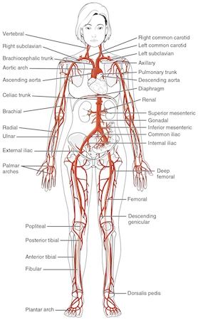
Major blood vessels in the body:
The body’s main arteries are listed here, along with the tissues and organs they support.
- The aorta
- The arteries of the head and neck
- The common carotid artery
- The external carotid artery
- The triangles of the neck
- The internal carotid artery
- The arteries of the brain
- The arteries of the upper extremity
- The subclavian artery
- The axilla
- The axillary artery
- The brachial artery
- The radial artery
- The ulnar artery
- The arteries of the trunk
- The descending aorta
- The thoracic aorta
- The abdominal aorta.
- The common iliac arteries
- The hypogastric artery.
- The external iliac artery
- The arteries of the lower extremity
- The femoral artery.
- The popliteal artery
- The anterior tibial artery.
- The arteria dorsalis pedis
- The posterior tibial artery
Aorta
The aorta is the biggest and most significant artery in the circulatory system. Because it acts as the first route for blood to leave the heart and travel through smaller, branching arteries to the rest of the body, it is extremely significant.
The body’s tissues wouldn’t receive the oxygen and nourishment they require without the aorta.
The aortic valve connects your heart to the aorta. It consists of the following components:
- Aorta ascending: Through the coronary arteries, the ascending aorta supplies the heart with nutrition and oxygen.
- Aortic arch: Its three principal branches are the left subclavian artery, the brachiocephalic trunk, and the left common carotid artery. Blood is sent to the arms, neck, and head as well as the upper body.
- Aorta descending: Blood is sent to your lower body, belly, and torso by the descending aorta. The aorta above the diaphragm is called the thoracic aorta, but it changes its name to the abdominal aorta once it passes it.
Arteries in the head and neck
The head and neck contain many arteries:
- Left and right common carotid vessels: The brachiocephalic trunk is the origin of the right common carotid, whereas the left common carotid comes straight from the aortic arch.
- External carotid: The common carotid arteries give rise to these paired arteries. The face, lower jaw, and neck receive blood supply via the external carotid.
- Internal carotid: These paired arteries originate from the common carotid arteries, just as the external carotid. These are the main blood vessels that supply the brain with blood.
- Vertebral: These paired arteries originate from the subclavian arteries and ascend the neck, providing blood flow to the brain.
- Thyrocervical trunk: The thyrocervical trunk, which is also derived from the subclavian arteries, divides into many vessels that supply blood to the thyroid, neck, and upper back.
Arteries of the torso
The abdominal aorta, heart, lung, and pelvic arteries
Among the torso arteries are:
- Bronchial: Bronchial arteries usually have two branches, one on the left and one on the right. They provide the lungs with blood.
- Esophageal: The oesophagus receives blood flow from the oesophageal artery.
- Pericardium: This artery provides blood to the membrane that envelops the heart, known as the pericardium.
- Intercostal: The intercostal arteries are two arteries that run parallel to each other on either side of the torso. They supply blood to the skin, back muscles, spinal cord, and vertebrae.
- Superior Phrenic: The superior phrenic arteries are paired, just like the intercostal arteries, and they supply blood to the skin, diaphragm, spinal cord, and vertebrae.
Abdomen arteries
Among the arteries in the abdomen are:
- Celiac trunk: The celiac trunk splits into smaller arteries that nourish organs like the stomach, liver, and spleen after branching off from the abdominal aorta.
- Superior mesenteric: It also branches off of the abdominal aorta and supplies blood to the pancreas, the majority of the large intestine, and the small intestine.
- Inferior mesenteric: This artery, like the superior mesenteric artery, emerges from the abdominal aorta and provides blood to the rectum and the remaining segment of the large intestine.
- Inferior phrenic. The diaphragm receives blood flow from these paired arteries.
- Adrenal: Blood is sent to the adrenal glands via the paired adrenal arteries.
- Renal: The kidneys receive their blood supply from these two arteries.
- Lumbar: The spinal cord and vertebrae receive blood supply from these two arteries.
- Gonadal: The paired gonadal arteries supply blood to the female ovaries and the testes in men.
- Common iliac: The internal and external iliac arteries split off from this branch of the abdominal aorta.
- The internal iliac: This artery, which receives its blood supply from the common iliac artery, serves the bladder, pelvis, and external part of the genitalia. In females, it also supplies the vagina and uterus.
- The external iliac: This artery, which also originates from the common iliac artery, eventually transforms into the femoral artery.
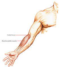
Arteries of the arms
The arm’s arteries are the following:
- Axillary. The subclavian artery is known by this term as it leaves the body and enters the arm.
- Brachial. Blood is then sent to the arm’s upper area.
- Ulnar and radial. These eventually split to supply blood to the wrist and hand, running parallel to the forearm’s two bones.
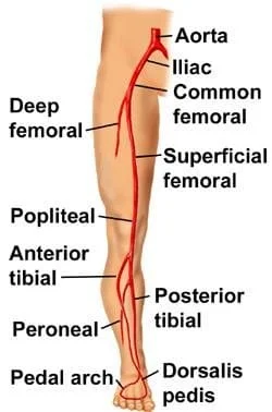
Leg arteries
Among the leg arteries are:
- Femoral. This artery, which receives its blood supply from the external iliac artery, branches off into smaller arteries that serve the legs and the thigh.
- Genicular. This provides blood to the area around the knee.
- Popliteal. The femoral artery is referred to by this name as it travels below the knee.
- Anterior and posterior tibial: These arteries, which give blood to the lower leg, are derived from the popliteal artery. They split off once they get to the ankle to supply the ankle and foot area.
Which artery diseases are more common?
Systemic problems can result from a variety of conditions that impact the cross-section and structure of the arteries, including:
- Peripheral vascular disease: The arteries constrict or clog for a variety of reasons (such as smoking, ageing, scarring, or autoimmune illnesses), which compromises the blood supply to the organs they supply.
- Atherosclerosis: This illness is characterized by the buildup of cholesterol plaques on the inside walls of the arteries, which results in a narrowing of the cross-sections or the production of clots that damage the organs they serve. Atherosclerosis can be caused by ageing, an aberrant lipid profile, hereditary predisposition, etc.
- Arterial aneurysm: An arterial aneurysm is a localized enlargement of the artery brought on by the weakening of the arterial wall.
- The reasons could include birth defects, trauma, etc.
- Sometimes the aneurysm bursts, causing a risk to life-threatening condition or sudden death.
Summary
In the circulatory system, the arteries are blood channels that transport blood away from the heart. Two distinct circuits are used to achieve this.
The body’s organs and tissues receive oxygen and other nutrients from the systemic circuit. The pulmonary circuit eliminates carbon dioxide from the circulation and permits new oxygen to enter it.
Maintaining the health of arteries is crucial due to their essential function. A heart attack or stroke may occur as a result of damaged or restricted arteries, which can prevent the body from receiving enough blood.
FAQs
Which artery is the smallest?
The smallest arteries are called arterioles, and their diameter ranges from 0.3 mm to 10 microns (µm).
Which artery is nearest to the skin surface?
One of the two main blood vessels that supplies blood to the hand and forearm is the radial artery. The radial artery passes through the elbow’s front and passes through a substantial amount of muscle before ending at the wrist. This artery approaches the epidermis.
Which four cardiac arteries are the primary ones?
The four major coronary arteries are the left anterior descending, left circumflex, right coronary, and left main coronary. Heart failure, angina, heart attacks, and heart disease are frequently brought on by blockage of these arteries.
How many arteries does a human body contain?
The human body contains about 20 major arteries. There are three layers to the muscle tissue that makes up each artery: the larger, more muscular layer, the stiff layer, and the smooth layer.
What’s the biggest artery in the body?
Blood is transported from the heart to the circulatory system via the aorta, which is the biggest artery in the body. It is divided into multiple sections: The body’s principal water source is the Aortic Root, the point at which blood leaves the heart for the first time.
References:
- Mbbs, K. K. (2021, August 5). Major Arteries of the Body: The Aorta, Head, Neck & Torso. MedicineNet. https://www.medicinenet.com/what_are_the_major_arteries/article.htm
- Professional, C. C. M. (n.d.). Arteries. Cleveland Clinic. https://my.clevelandclinic.org/health/body/22896-arteries
- Seladi-Schulman, J. (2019, February 26). Arteries of the Body. Healthline. https://www.healthline.com/health/arteries-of-the-body#arm-arteries
- List of arteries of the human body. (2024, February 15). Wikipedia. https://en.wikipedia.org/wiki/List_of_arteries_of_the_human_body