
Popliteus muscle
Table of Contents
Origin of Popliteus muscle
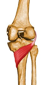
The popliteus muscle fibers originate from the lateral condyle of the femur or the posterior horn of the lateral meniscus, a tendon called the popliteus tendon. In the dissections have also shown fibers originating from the styloid portion of the fibular head, which runs obliquely blending with the main muscular structure.
From there it goes inferiorly and mediolaterally towards the Tibia. It courses diagonally across the posterior upper tibia and a portion of the joint capsule, to lie as the deepest muscle of the posterior knee part. The popliteal tendon pierces the joint capsule but didn’t enter the synovium. The popliteus tendon goes through the lateral collateral ligament (LCL) and the biceps femoris tendon.
The popliteal bursa, which is usually an extension of the synovial membrane, is divided from the lateral femoral condyle. Although the popliteus muscle has extra-articular regions, it is a capsular structure separating the lateral meniscus of the knee joint from the lateral collateral ligament. An additional head of popliteus might arise from the sesamoid bone in the lateral head of the gastrocnemius muscle. an additional inconstant muscle known as the popliteus minor is seen which arises from the femur on the inner side of the plantaris muscle and inserts into the posterior ligament of the knee.
Insertion of Popliteus muscle
It inserts on the tibia just proximal to the sole line but below the tibial condyles.
Innervation
The popliteal muscle receives nervous supply from the L4-S1(tibial nerve), a branch of the sciatic nerve.
Nerve supply
The popliteus receives arterial blood supply from branches of the popliteal artery, namely the inferior medial and lateral genicular arteries. Moreover, it receives smaller contributions from the posterior tibial artery, the proximal part of the posterior tibial artery, and the nutrient artery of the tibia.
Blood supply
Arterial supply to the popliteus is provided by the medial inferior genicular branch of the popliteal artery and the muscular branch of the posterior tibial artery.
Function of Popliteus muscle
Despite its anatomical classification, the popliteus muscle’s main role as a knee flexor is insignificant. Whereas, the popliteus muscle plays a vital role in initiating flexion of the fully extended locked knee. Thus, the popliteus is preferred as the key to unlocking the knee, making it necessary for the processes of standing up, walking, and sitting down.
The popliteus muscle can produce 2 types of actions; lateral rotation of the femur or medial rotation of the tibia. The phase of the gait cycle observes these actions:
During the closed chain of the gait cycle, the foot is in contact with the ground. The tibia is laterally rotated about 5 degrees on the femur, which locks the knee joint. The tibia is fixed in position, the popliteus muscle acts on its origin and laterally rotates the femur on the tibia. Afterward, Movement unlocks the knee and allows flexion to occur.
During the open chain swing phase of the gait cycle, the foot is above the floor and the tibia is free to move. Therefore, the popliteus can act on its insertion to medially rotate the tibia on the femur. This action gives stability to the tibia during knee flexion.
The popliteus muscle is also the primary stabilizer of the dorsal knee area via its connections to the posterior joint capsule and popliteofibular ligament. moreover, the popliteus muscle pulls the lateral meniscus dorsally during knee flexion and femoral lateral rotation, thus preventing its entrapment.
Assessment
Diagnostic Test
This test is where the pain is felt when trying to remove the shoe on another side of the affected side of the knee. This action requires internal rotation of the affected leg to reach the heel of the other leg and can cause pain during the maneuver when there is an injury to the popliteal muscle.
Clinical relevance
It is commonly involved in the posterolateral corner injuries of the knee, which occur secondary to –
- A direct blow to the knee.
- Varus force applies to a flexed knee.
- Dislocation of the knee.
- Varus/hyperextension.
Whatever the mechanism of injury to the postero-lateral corner, urgent asses of the suspect’s neurovascular status of the limb are performed. In the case of knee dislocations, vascular status is assessed followed by closed reduction of the knee which is then again followed by assessing the vascular status.
In further trauma, incorrect movement patterns and posture can often weigh heavily on the popliteus muscle leaving it prone to weakness and injury. Iatrogenic injury to this muscle is common, which can lead to unbalance functional prognosis and becomes vital to be addressed – especially following knee reconstruction surgery. Smaller knees also need extra attention as the risk of popliteal injury is enhanced.
Popliteal tendinitis could also occur as posterolateral knee pain. However, it cannot be easy to single it out due to other more common knee pain etiologies in the vicinity. As this muscle inhibits over tibial rotation along with preventing significant anterior translation of the knee, it can be pathologically overcome secondary to over sprinting or running downhill, and such activities should be avoided or modified to run on flat surfaces like a treadmill. If the lateral hamstrings are stronger than the medial(inner) hamstrings, the popliteal muscle will be weaker. over pronation or collapse of the inner foot when walking or running will stress the popliteus in the opposite direction.
Many different EMG studies have shown that popliteus muscle activity increases with knee extension and downhill walking, thereby consolidating its role in the control of hyperextension of the knee. The referred pain pattern in case of trigger point of popliteal muscle is the back of the knee. Due to its deep location, isolated injuries to the popliteus muscle are rare but can be associated with other knee joint injuries such as ACL injuries, and meniscus injuries. There are some general symptoms of muscle injury which include swelling, tenderness, edema, bleeding, and suspect keeping the leg (tibia) in lateral rotation during knee flexion. The following should be checked to rule out popliteus muscle injury.
Tenderness – As many neurovascular structures lie over it, only terminal portions of the popliteal muscle can be palpated. Proximal tendon tenderness is checked in a prone lying position, whilst tenderness over the posterolateral knee may be a sign of biceps femoris tendon strain and/or lateral meniscus injury.
Garrick test- Pt is in a high sitting position with hip and knee flexed at 90 degrees. Resistance to external rotation of the lower leg is applied. Pain during this maneuver is considered to be a positive test
Shoe removal maneuver Patient tries to remove the contralateral shoe by internally rotating the affected leg to reach the heel of the opposite leg. Pain during this maneuver indicates injury to the popliteus muscle. As said earlier, isolated injuries to the popliteal muscle are rare, and only 2 out of 2412 knee MRI studies showed isolated acute rupture of the popliteus tendon.
The popliteus muscle, along with PCL (posterior cruciate ligament ), stabilizes the femur over the fixed tibia in the stance phase especially when extra stability is needed for activities like running downhill. Hence running downhill especially on banked surfaces with hyperpronation can lead to popliteus muscle injuries like tenosynovitis, tendinopathy, rupture, and strain.
Exercise for Popliteal muscle
Popliteus muscle exercises are not focused on this small muscle. They strengthen the entire leg, including your thighs and calves.
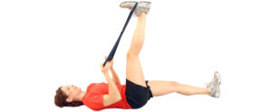
1: Supine Hamstring Stretch
Lie on your back on the floor or on an exercise mat with both knees bent and your feet planted.
Bring your right knee toward your chest and hold on to the back of your thigh with both hands.
Lengthen the leg up to the ceiling as you gently pull the leg toward your head.
Hold for 30 to 50 seconds and repeat on the other side.
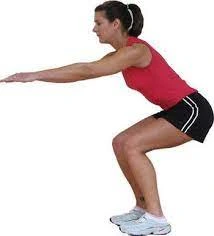
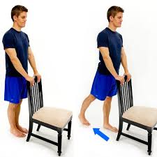
2: Half Squats
Stand with your feet about hip distance apart. If you feel unstable, hold on to the wall or the back of a chair for balance.
Maintain a long spine and lifted chest as you bend gently through your hips and knee. Let the hips bend just about 10 to 12 inches as if you’re sitting toward a chair. Keep your feet planted, and keep weight in your heels. After that, Pause for three to five counts and straighten again to a stand. Repeat 10 to 12 times.
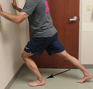
3: Heel Cord Stretch
the suspect has to Stand facing a wall.
Place your good leg forward and bend the knee slightly.
Place the injurious leg straight behind you with the heel flat and the toes pointed in a little bit.
Keep your heels flat on the base and press your hip complex forward, toward the wall.
Hold for 20 to 30 seconds.
4: Standing Quadriceps Stretch
Standing behind the back of a chair and place your hands on it for stability.
Bend your right knee and pull the heel up toward your right hip.
Grab your ankle with the right side of the hand and pull it closer to your body.
Hold 30 to 60 seconds. Repeat on the other side.

5: Leg Extensions
Sit at the corner of a firm chair with a straight spine.
Straighten the right side of the leg and contract the muscles of your thigh as you raise the leg as high as you can.
Hold the lifted leg up for about 5 to 10 seconds.
Told the to patient relax the leg and lower back toward the floor.
Repeat ten to fifteen times; perform on the other side.
6: Hamstring Curls
Standing facing the back of a chair. Through the hands Hold on for support.
Bend your right side of the knee behind you and raise the heel toward the ceiling as far as possible. Stop if you feel pain.
Hold for five to ten seconds. Repeat ten to fifteen times. Perform on the other leg.

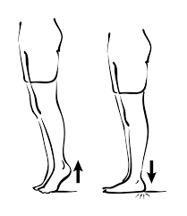
7: Calf Raises
Standing and facing the back of a chair; hold on for balance.
Distribute your weight evenly over both feet.
Bend your left knee behind you and place your all weight on your right foot.
Raise the right side of the heel as high as you can and then lower it slowly.
Repeat ten times
FAQ
How important is the popliteus muscle?
The popliteus muscle plays a vital role in the gait cycle by initiating the flexion of the fully extended locked knee. Thus, the popliteus is referred to as the key to unlocking the knee. Additionally, the popliteus muscle is the primary stabilizer of the dorsal knee region.
What actions does the popliteus muscle perform?
Despite it is small size, the popliteus is a major stabilizing muscle of the knee. The popliteus is involved in both the closed chain and open chain of the gait cycle. During the closed chain, which is when the foot is in contact with the base, the muscle externally rotates the femur on the tibia.
How do I know if I tore my popliteus?
Popliteus injury symptom
Your knee will feel tender point when pressing in at the back. It is likely to be painful when trying to bend your knee against resistance, while your shin bone is rotated outward. If you have a severe injury then simply straighten your knee function.What causes a tight popliteus muscle?
Popliteus muscle strains tendinopathies mostly occur in downhill skiers, and in runners and triathletes who compete on hills or uneven ground. The typical cause of injury is a direct blow to the inside of the knee or a sudden forceful overextension or over-straightening of the knee.
Which muscle is the deepest in the popliteal space?
popliteus courses diagonally across the posterior upper tibia and an area of the joint capsule to lie as the deepest muscle of the posterior knee part

One Comment