

Skeletal Muscle
Table of Contents
What is a Skeletal Muscle?
Skeletal muscles (often referred to as muscles) are the organs of the vertebrate muscular system and are usually connected to the bones of the skeleton by tendons. Muscle cells in skeletal muscle are much longer than in other types of muscle tissue and are often called muscle fibers. Skeletal muscle tissue is striated – it has a striated appearance due to the arrangement of sarcomeres.
Skeletal muscles are classified as voluntary muscles under the control of the somatic nervous system. Other muscle types include cardiac muscle, which is also striated, and smooth muscle, which is striated; both of these types of muscle tissue are classified as involuntary or under the control of the autonomic nervous system.
Skeletal muscle contains several connective tissues – bundles of muscle fibers. Each individual fiber and each muscle is surrounded by a type of connective tissue called fascia. Muscle fibers are formed by the fusion of developing myoblasts in a process called myogenesis, resulting in long, multinucleated cells. These cells have nuclei called myonuclei on the inside of the cell membrane. Skeletal muscle fibers also have multiple mitochondria to meet their energy needs.
But muscle fibers are made up of myofibrils. Myofibrils are composed of actin and myosin filaments called myofilaments, which repeat in units called sarcomeres, the basic functional contractile units of a muscle fiber, essential for muscle contraction. Muscles derive their power primarily from the oxidation of fats and carbohydrates, but anaerobic chemical reactions are also used, especially by fast-twitch fibers. These chemical reactions generate molecules of adenosine triphosphate (ATP), which are used to move the myosin heads.
Skeletal muscles make up approximately 35 percent of the human body mass. Skeletal muscle functions include producing movement, maintaining body position, regulating body temperature, and stabilizing joints. Skeletal muscle is also an endocrine organ. 654 different protein subsets, as well as lipids, amino acids, metabolites, and small RNAs, can be found in skeletal muscle secretion under different physiological conditions.
Skeletal muscles consist mainly of multinucleated contractile muscle fibers (myocytes). However, skeletal muscle also contains significant numbers of quiescent and infiltrating mononuclear cells. By volume, myocytes make up the majority of skeletal muscle. Skeletal muscle myocytes are usually very large, approximately 2–3 cm long and 100 μm in diameter.
In comparison, skeletal muscle mononuclear cells are much smaller. Some of the mononuclear cells in muscle are endothelial cells (which are approximately 50–70 μm long, 10–30 μm wide, and 0.1–10 μm thick), macrophages (21 μm in diameter), and neutrophils. (12 -15 μm in diameter).
However, in skeletal muscle nuclei, myocyte nuclei may account for only half of the nuclei present, while persistent nuclei and infiltrating mononuclear cells account for the remaining half.
Structure Of Skeletal Muscle
Gross Anatomy
There are more than 600 skeletal muscles in the human body, which make up about 40% of the body weight of healthy young adults. In Western countries, men have on average about 61% more skeletal muscle than women. Most muscles are arranged in bilateral pairs to serve both sides of the body. Muscles are often classified into groups of muscles that work together to perform a specific function.
There are several major muscle groups in the body, including the pectoral and abdominal muscles; Intrinsic and extrinsic muscles are subdivisions of the muscle groups of the arms, legs, tongue, and extraocular muscles. Muscles are also grouped into sections, including four groups in the arms and four groups in the legs.
In addition to the contractile part of the muscle consisting of fibers, the muscle has a non-contractile part of dense connective tissue that forms a tendon at each end. Tendons attach muscles to bones to allow skeletal movement. The length of the muscle includes the tendons. Connective tissue is present in all muscles as deep connective tissue.
The deep fascia is specialized for the muscle to surround each muscle fiber like an endomysium; each muscle bundle is a perimysium and each individual muscle is an epimysium. These layers together are called mysia. Deep fascia also separates muscle groups into muscle compartments.
There are two types of sensory receptors in muscles: muscle spindles and Golgi tendon organs. Muscle spindles are stress receptors located in the muscle belly. Golgi tendon organs are proprioceptors located at the myotendinous junction that signal muscle contraction and tension.
Skeletal Muscle cells
Skeletal muscle cells are the single contractile cells of a muscle and are often called muscle fibers. A single muscle, such as the biceps of a young adult male, contains approximately 253,000 muscle fibers.
Skeletal muscle fibers are the only muscle cells that are multinucleated with nuclei often called myonucleotides. This occurs during myogenesis when myoblasts fuse to form each nucleus. Fusion depends on muscle-specific proteins called fusogens, called myomaker and myomerger.
Skeletal muscle cells require many nuclei to produce large amounts of proteins and enzymes necessary for normal cell function. A single muscle fiber can contain hundreds of thousands of nuclei. For example, a 10 cm long human bicep muscle tissue can contain up to 3000 nuclei. Unlike non-muscle cells, where the nucleus is in the center, the myonucleus is elongated and located close to the sarcolemma.
The myonuclei are fairly evenly spaced along the filament, and each nucleus has its own myonuclear domain, which is responsible for supporting the cytoplasmic volume in that part of the myofiber.
A group of muscle stem cells known as myosatellite cells, also known as satellite cells, are found between the basement membrane and the sarcolemma of muscle fibers. These cells are normally inactive but can be activated by exercise or pathology to provide additional myonuclei for muscle growth or repair.
Attachment to Tendons
Muscles attach to tendons at a complex interface region, also known as the muscle-tendon junction, which is specialized for the primary transmission of force. At the muscle-tendon interface, force is transmitted from the sarcomeres of the muscle cells to the tendon. Muscles and tendons form in close association, and after joining at the myotendinous junction, they form a dynamic unit to transmit the force of muscle contraction to the skeletal system.
Arrangement of muscle fibers
Muscle architecture refers to the arrangement of muscle fibers relative to the axis of force generation that runs from the origin of the muscle to its insertion. Common arrangements are the parallel and extensor muscle types. In parallel muscles, the ligaments run parallel to the axis of force generation, but the ligaments can differ in their relationship to each other and to the tendons.
These variations are seen in the fusiform, strap, and adductor muscles. A converging muscle is triangular or fan-shaped because the fibers converge at their insertion and point widely outward toward the point of origin.
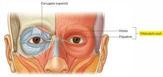
A less common example of a parallel muscle is a circular muscle such as orbicularis oculi, where the fibers are arranged lengthwise but form a circle from origin to insertion. These different architectures can cause differences in the tension a muscle can generate between its tendons.
Penned muscle fibers run at an angle to the axis of force generation. This pin angle reduces the effective strength of the individual fiber as it effectively pulls away from the stem. On the other hand, this angle allows for the packing of more fibers into the same muscle volume, resulting in an increase in the physiological cross-sectional area (PCSA). This effect is known as fiber packing, and in terms of force generation, it exceeds the reduction in efficiency in the off-axis orientation.
The trade-off comes in the overall speed and full excursion of muscle shortening. The total velocity of muscle shortening is less than the velocity of fiber shortening, as is the total distance of shortening. All these effects vary with pen angle; Larger angles result in greater strength due to increased fiber packing and PCSA but at the expense of increased speed and shortening of the range of motion.
The types of adductor muscles are single-, double-, and multipenny. A unilateral muscle has fibers at a similar angle to those on the other side of the tendon. A bilateral muscle has fibers on both sides of the tendon. Striated muscles have fibers oriented at multiple angles along the force-producing axis and are the most common and common architecture.
Muscle fiber growth
Muscle fibers grow during exercise and shrink when not in use. This is because exercise stimulates the proliferation of myofibrils, which increases the overall size of muscle cells. Well-trained muscles can not only grow, but also more mitochondria, myoglobin, glycogen, and denser capillaries. However, muscle cells are unable to divide to produce new cells, and as a result, an adult has fewer muscle cells than a newborn.
Naming Muscles
Many terms are used to name muscles, including terms related to size, shape, function, location, and orientation. and the number of their heads.
In size
brevis means short; fall means long; longissimus means longest; Magnus means excellent; big means bigger; maximus means greatest; minor means smaller and minimum the smallest; latissimus means widest and resistus means huge. These terms are often used after specific skeletal muscles, such as gluteus maximus and gluteus minimus.
In relative form
means a triangle in the deltoid muscle; quadratus means four sides; rhomboideus means diamond-shaped; teres means round or cylindrical and trapezoid; serratus means serrated; orbicularis means round; pectinate means comb-like; piriformis means pear-shaped of muscle; Platys means flat shaped and gracilis means slender. Examples include pronator teres and pronator quadratus.
Action
moves the trap away from the center line; the adductor moves to the midline; the wrestler moves down; the elevator moves up; flexion movements that reduce the angle; extension movements that increase the angle or extend; pronator moves face down; the supinator moves to face up; the internal rotator turns towards the body; external rotator, which rotates away from the body; sphincter reduces size and tensor provides tension; Anchor’s muscles fix a joint in a certain position, stabilizing the main movement while other joints move.
by the number of heads
biceps two; triceps three and quadriceps four.
Location
named after a nearby head structure, such as the temporal muscle (temporalis) near the temporal bone. Also above; infra-low and low-low.
By fascicle orientation
Straight to the central line means parallel to the central line; transverse means perpendicular to the center line and oblique means diagonal to the center line. In relation to the axis of force production – parallel types and pennate muscle types.
Pathophysiology Of Skeletal Muscle
Diseases affecting the neuromuscular junction
- Myasthenia Gravis is an autoimmune disease caused by antibodies against ACh receptors in the neuromuscular junction. These antibodies inhibit ACh binding and reduce the depolarization transmitted to the muscle cell. Because repeated use depletes ACh stores, the lower concentrations of ACh released at the NMJ cannot saturate the binding sites and generate an action potential in the muscle. This is manifested in the variable weakness of the patient, which is worse with exercise and better at rest. Extraocular muscles are often the first muscles affected during the disease. Edrophonium, a short-acting acetylcholinesterase inhibitor, can be used to diagnose myasthenia gravis disease. When administered, edrophonium prolongs the effect of ACh at the NMJ and prevents short-term muscle fatigue.
- Lambert-Eaton myasthenic syndrome (LEMS) is a disease of the NMJ that can present as a paraneoplastic phenomenon, with more than half of cases associated with small cell lung cancer (SCLC). The main clinical manifestation is muscle weakness. The pathology is due to the formation of antibodies against voltage-gated ka channels in presynaptic nerve endings, which leads to a decrease in the neurotransmitter ACh. In addition to muscle weakness, LEMS patients may present with eyeball weakness, dysphagia and dysarthria, and autonomic dysfunction. Rarely, respiratory failure can occur in the late stages of LEMS. Post-exercise or post-activation facilitation is a phenomenon observed in LEMS associated with improvement in muscle weakness after exercise. Repetitive muscle contractions increase Ca influx into the presynaptic membrane, which facilitates the release of ACh by binding to multiple vesicles. This effect is temporary because mitochondria eventually remove excess calcium.
- Toxins can also affect excitation-contraction connections at the NMJ. Botulinum toxin produced by C. botulinum inhibits the release of ACh from the presynaptic neuron at the NMJ, preventing skeletal muscle excitation and causing flaccid paralysis. Tetanospasmin, a neurotoxin released by C. tetanus, prevents relaxation by preventing the release of inhibitory neurotransmitters from synapsing interneurons at the NMJ, resulting in spastic paralysis.
Muscular dystrophies
Muscular dystrophies include more than 30 inherited diseases that cause permanent muscle weakness. The two most common and well-known forms of muscular dystrophy are Duchenne muscular dystrophy (DMD) and Becker muscular dystrophy (BMD).
- Duchenne muscular dystrophy (DMD) is an X-linked inherited neuromuscular disease caused by mutations in the dystrophin gene. The disease is characterized by progressive muscle wasting and weakness caused by a lack of dystrophin, a protein that causes skeletal and heart muscle degeneration. DMD affects approximately 1 in 3,600 male births worldwide and has no clinical symptoms at birth. The average age of diagnosis and first symptoms is around 4 years. DMD is a rapidly progressive disease that makes patients wheelchair dependent and develop severe cardiomyopathy around the age of ten. Death is usually caused by cardiovascular and respiratory complications.
- Becker muscular dystrophy (BMD) is described as a milder form of DMD with an incidence of 1,18,518 male births. BMD usually appears later than DMD, between the ages of 5 and 15 years, and clinical progression is slower. People with BMD usually begin to show symptoms after age 30 and may remain active until age 60. Despite mild skeletal muscle involvement, heart failure due to dilated cardiomyopathy is a common cause of morbidity and mortality in patients with CML.
Idiopathic inflammatory myopathies (myositis)
- Dermatomyositis (DM) causes symmetrical proximal muscle weakness that develops over weeks or months with erythematous changes that may precede or follow the myopathy. Erythematous changes include a heliotrope rash, eyelid edema, Gottron’s papules on the extensor surfaces, and subcutaneous calcifications. Myalgia is not usually seen, but patients may develop dysphagia, dysarthria, and interstitial lung disease. DM shows an increased prevalence in women and elderly patients.
- Polymyositis (PM) causes a subacute onset of proximal muscle weakness, most prominent in the pelvic girdle and shoulders, and marked elevation of creatine kinase (CK). The neck flexors and in some cases, the coccyx are also usually affected. A biopsy showing cellular infiltrates composed of macrophages and cytotoxic CD8+ T cells distinguishes PM from DM.
- Necrotizing myopathy (NM) is clinically indistinguishable from PM and presents with progressive symmetrical weakness of the proximal muscles of the arms and legs. Myalgia occurs in up to 80% of patients, and dysphagia and dysarthria may occur in severe cases. NM shows a male predominance, with a mean age starting in the 5th decade. Diagnosis of NM requires a biopsy showing scattered necrotic muscle fibers, sparse inflammatory cells surrounding the necrosis, predominant macrophages, and some lymphocytes (CD4+ and CD8+ T cells).
- Inclusion body myositis (IBM) is the most common acquired myopathy after age 50. IBM is a slowly progressive disease that usually affects the flexion of the hand and fingers and extension of the knee. Dysphagia is typical, and at least mild swallowing difficulties have been observed in up to 80% of affected individuals. Dysphagia may precede arm and leg weakness. Finally, patients are usually wheelchair-dependent, but life expectancy is normal.
Rhabdomyolysis is a direct injury to skeletal muscle structures that results in the release of intracellular contents such as electrolytes and myoglobin into the extracellular space. Injuries that lead to musculoskeletal injuries are numerous and beyond the scope of this article, but some examples include overuse (such as marathon running), compartment syndrome, and the use of prescription, over-the-counter, and illegal drugs. Cell damage is directly caused by the release of large amounts of ionized calcium from terminal reservoirs, which activates degradation processes. Later consequences of rhabdomyolysis can be systemic and life-threatening, and skeletal muscle damage leads to the release of breakdown products that impair kidney function and, in more severe cases, acute renal failure requiring dialysis.
Skeletal muscle atrophy can be caused by many things, including abuse, denervation, systemic disease, chronic use of glucocorticoids, and malnutrition. Although the pathways may differ, in all these cases atrophy is caused by increased proteolysis and decreased protein synthesis, mediated by the ubiquitin system, which reduces muscle mass by reducing the diameter of individual muscle fibers.
Skeletal Muscle Fibers
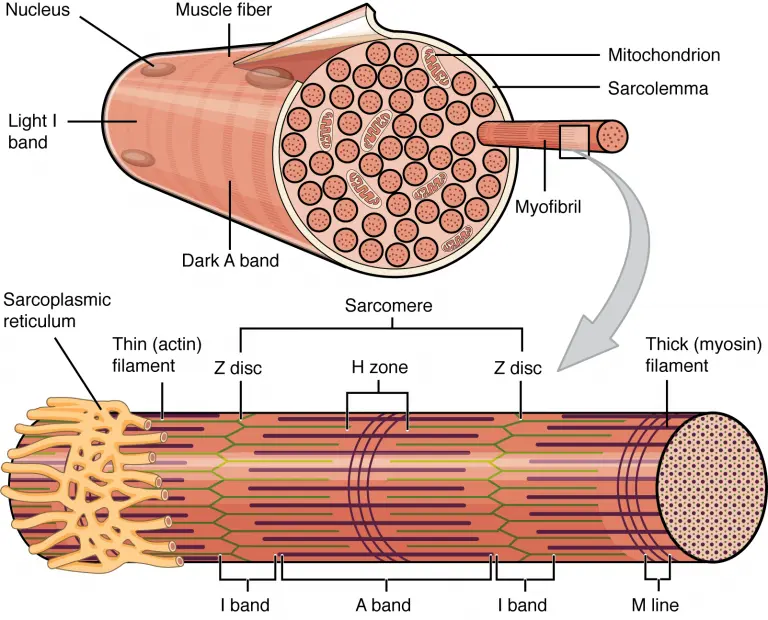
Because skeletal muscle cells are lengthy and cylindrical, they’re typically known as muscle fibers (or myofibers). Skeletal muscle fibers may be pretty big as compared to different cells, with diameters as much as a hundred μm and lengths as much as 30 cm (11. eight in) inside the Sartorius of the higher leg. Having many nuclei permits the manufacturing of big quantities of proteins and enzymes wished to keep the regular features of those big protein-dense cells. In addition to nuclei, skeletal muscle fibers additionally include mobile organelles determined in different cells, which include mitochondria and endoplasmic reticulum. However, a number of those systems are specialized in muscle fibers. The specialized easy endoplasmic reticulum, known as the sarcoplasmic reticulum (SR), stores, releases, and retrieves calcium ions (Ca++).
The plasma membrane of muscle fibers is known as the sarcolemma (from the Greek sarco, which means “flesh”) and the cytoplasm is known as sarcoplasm. Within a muscle fiber, proteins are prepared into organelles known as myofibrils that run the duration of the cellular and include sarcomeres linked in series. Because myofibrils are handiest about 1.2 μm in diameter, loads to heaps (every with heaps of sarcomeres) may be determined interior one muscle fiber. The sarcomere is the smallest useful unit of skeletal muscle fiber and is a pretty prepared association of contractile, regulatory, and structural proteins. It is the shortening of those character sarcomeres that results in the contraction of character skeletal muscle fibers (and in the long run the complete muscle).
Sarcomere
A sarcomere is defined as a region of myofibrils located between two cellular skeletal structures called Z-sheets (also called Z-lines or Z-bands), and the striated appearance of skeletal muscle fibers is due to the arrangement of thick muscles. fiber and thin myofilaments in each sarcomere. The dark-striped A-zone consists of myosin-containing thick filaments that extend toward the center of the sarcomere, extending toward the Z-discs. Thick filaments are anchored to the center of the sarcomere (M line) by a protein called myomesin.
The lighter I-zone regions contain thin actin filaments anchored to Z discs by a protein called α-actinin. The thin filaments extend towards the M line of the A zone and overlap the regions of the thick filament. The A zone is dark due to thicker myosin filaments and overlaps with actin filaments. The H zone in the middle of the A band is slightly lighter in color because it contains only the part of the thick filament that does not overlap with the thin filaments.
Because a sarcomere is defined by Z discs, a single sarcomere contains a single dark A band flanked by half of a lighter I band at each end. During contraction, the myofilaments themselves do not change length, but actually slide over each other, shortening the distance between the Z plates, resulting in sarcomere shortening. The length of the A-band does not change (the thick myosin filament remains a constant length), but the H-band and I-band regions shrink. These regions represent regions of non-overlapping filaments, and as filament overlap increases during contraction, these non-overlapping regions decrease.
Myofilament Components
Thin filaments consist of two filamentous actin chains (F-actin) composed of individual actin proteins. These thin filaments are anchored to the Z plate and extend toward the center of the sarcomere. Within the filament, each globular actin monomer (G-actin) contains a myosin binding site and is also associated with the regulatory proteins troponin and tropomyosin protein. The troponin protein complex consists of 3 polypeptides. Troponin I (TnI) binds to actin, troponin T (TnT) binds to tropomyosin, and troponin C (TnC) to calcium ions. Troponin and tropomyosin move along actin filaments and regulate when actin-binding sites come into contact to bind to myosin.
Thick myofilaments are composed of myosin protein complexes consisting of six proteins: two myosin heavy chains and four light chain molecules. Heavy chains consist of a tail region, a flexible hinge region, and a globular head that contains an actin-binding site and a binding site for the high-energy ATP molecule. The light chains play a regulatory role in the hinge region, but the head region of the heavy chain interacts with actin and is the most important factor in force generation. Hundreds of myosin proteins are arranged in each thick filament, with their tails toward the M line and their ends extending toward the Z discs.
Other structural proteins are associated with the sarcomere but don’t play a direct role in active force production. Titin, the largest protein known, helps align the thick filament and adds an elastic element to the sarcomere. Titin is anchored to the M-line, runs the length of myosin, and extends to the Z-sheet. The thin filaments also have a stabilizing protein called nebulin that runs the length of the thick filaments.
Sliding Filament Model of Contraction
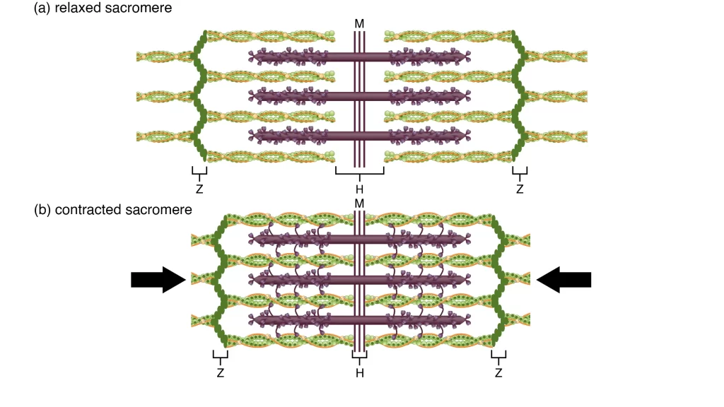
The arrangement and interaction of thin and thick filaments allow sarcomeres to generate force. When a motor neuron sends a signal, a skeletal muscle fiber is activated. Bridges form between the thick and thin filaments, and thin filaments are pulled, sliding along the thick filaments in the sarcomeres of the fibers. It is important to note that when the sarcomere shortens, the individual proteins and filaments do not change length, but simply slide next to each other. This filament process is called the sliding filament model of muscle contraction.
The process of filament sliding contraction can only occur when the myosin binding sites on actin filaments are exposed through steps that begin with the entry of Ca++ into the sarcoplasm. Tropomyosin wraps around actin filament chains and covers myosin binding sites to prevent actin binding to myosin. The troponin-tropomyosin complex uses the binding of calcium ions to TnC to regulate when myosin moves across actin filaments. Cross-bridge formation and filament sliding occur in the presence of calcium, and the signaling process leading to calcium release and muscle contraction is called excitation-contraction coupling.
Properties Of Skeletal Muscle
The skeletal muscular tissues have the following properties:
- Extensibility: The muscular tissues can increase while it’s miles stretched.
- Elasticity: The muscular tissues can go back to their authentic shape while launched.
- Excitability: The muscle can reply to a stimulus.
- Contractility: A muscle can settle while in touch with a stimulus.
Types Of Skeletal Muscle
There are styles of skeletal muscular tissues named pink and white muscular tissues-
Red Muscles
Red muscular tissues are because of the pink pigment referred to as myoglobin, which is in excessive quantities inside the human frame. These muscular tissues are smaller in diameter and feature a massive quantity of mitochondria. The myoglobin shops the oxygen, that is utilized by the mitochondria for the synthesis of ATP. Red muscular tissues have a massive quantity of blood capillaries in them.
White Muscles
Unlike the pink muscular tissues, the white muscular tissues are larger in diameter and feature a small quantity of myoglobin in them. They additionally have much less quantity of mitochondria in them.
Functions Of Skeletal Muscle
The following are the essential skeletal muscle features:
- The skeletal muscular tissues are liable for frame actions, including typing, breathing, extending the arm, writing, etc. The muscular tissue settlement draws the tendons at the bones and reasons movement.
- The frame posture is maintained through the skeletal muscular tissues. The gluteal muscle is liable for the erect posture of the frame. The Sartorius muscular tissues in the thighs are liable for frame movement.
- The skeletal muscular tissues shield the inner organs and tissues from any damage and additionally offer aid to those sensitive organs and tissues.
- These additionally aid the access and go-out factors of the frame. The sphincter muscular tissues are gifted across the anus, mouth, and urinary tract. These muscular tissues settle which reduces the dimensions of the openings and helps the swallowing of food, defecation, and urination.
- The skeletal muscular tissues additionally adjust frame temperature. After a strenuous exercise, the frame feels hot. This is because of the contraction of skeletal muscular tissues which converts power into heat.
Smooth Muscle
Smooth muscle is a kind of muscle mass that is used by special structures for the utility of stress to organs and vessels. It is an involuntary muscle, which suggests no-go stripes even if tested beneath a microscope.
A clean muscle consists of cells that can be slender and spindle-formed with an unmarried nucleus that is positioned centrally.
The cells of clean muscular tissues are made from fibers of myosin and actin that run via the cells and are sponsored through a framework of diverse proteins, in which the filaments are organized in a stacked sample throughout the molecular.
It reveals a ‘staircase’ association of the fibers-actin and myosin, which is commonly special in the shape located in cardiac and skeletal muscular tissues.
Unlike the striated muscular tissues, the clean muscle groups settle at a slower tempo automatically. Most of the musculature of the digestive machine and the inner organs are composed of clean muscular tissues.
Upon the discharge of ATP to be utilized by myosin, the clean muscular tissues settle beneathneath the have an impact on stimuli. This ATP this is launched is based on the significance of the stimuli which allows the clean muscular tissues to own a graded contraction in assessment to the ‘on-or-off’ contraction located inside the skeletal muscular tissues. The actin filaments run from one facet of the molecular to the alternative that is related to the molecular membrane and the dense bodies.
Cardiac Muscle
One of the 3 styles of muscular tissues, the cardiac muscular tissue is a muscle mass discovered inside the coronary heart, in which it’s miles acting and bringing approximately coordinated contractions, which permit the coronary heart to pump blood for the duration of the frame via the circulatory machine.
The cardiac muscle groups feature to reason non-stop pumping of the coronary heart via involuntary actions. This is one of the capabilities of cardiac muscular tissues that units it aside from the muscle groups that are beneath one’s control. It is capable of accomplishing that via specialized cells called the pacemaker cells which govern the coronary heart’s contractions.
The worried machine transmits alerts to the specialized cells, showing them to boost or lower the coronary heart rate. The pacemaker cells are related to the cardiac muscle cells, which allows them to transmit alerts that cause a wave of contractions inside the cardiac muscular tissues that during the flip generate a heartbeat.
The cardiac muscular tissues are composed of the following:
- Nucleus
- Gap junctions
- Intercalated discs
- Desmosomes.
FAQ
What are the 4 parts of skeletal muscle?
The muscular system views every skeletal muscle as an organ. Each organ or muscle consists of skeletal muscle tissue, connective tissue, nervous tissue, and blood or vascular tissue.
What is the basic anatomy of muscles?
Muscle fibers are organized into bundles supplied by blood vessels innervated by motor neurons. Skeletal (striated or voluntary) muscles are made up of tightly packed groups of enormously elongated cells known as myofibers. They are grouped into bundles (fascicles).
What is the function of skeletal muscles?
skeletal muscle, in vertebrates, is the most common of the three types of muscle in the body. Skeletal muscles are attached to bones by muscle tendons and produce all the movements of human body parts about each other. Unlike smooth and cardiac muscle, skeletal muscle is under voluntary control.
What is the basic anatomy of muscles?
Muscle fibers are organized into bundles supplied by blood vessels innervated by motor neurons. Skeletal (striated or voluntary) muscles are made up of tightly packed groups of enormously elongated cells known as myofibers. They are grouped into bundles (fascicles).
What are the characteristics of skeletal muscles?
Skeletal muscle fibers are cylindrical, multinucleated, striated, and voluntarily controlled. Smooth muscle cells are shaped of spindle-shaped, have a single, centrally located nucleus, and lack striations.
What is the structural and functional unit of skeletal muscle?
A sarcomere is the smallest functional unit of skeletal muscle fibers and a highly organized arrangement of contractile, regulatory, and structural proteins. Shortening of these individual sarcomeres leads to the contraction of individual skeletal muscle fibers (and ultimately the entire muscle).
What are skeletal muscles?
Skeletal muscles make up 30-40% of your total body mass. These muscles attach to your bones, allowing you to perform various movements and activities. Skeletal muscles work voluntarily, meaning you control how and when they work.
What are the 4 characteristics of skeletal muscles?
Muscles have four basic properties:
Acceleration: the ability of the muscle to respond to stimuli.
Contractility: The ability of a muscle to contract or decrease in size.
Extensibility: The ability of a muscle to stretch.
Elasticity: the ability of skeletal muscles to return to their original length after being stretched.
What are the cardiac muscles?
Cardiac muscle, commonly known as the myocardium, is one of the three basic types of muscles found in the human body, alongside smooth muscle and skeletal muscle. Cardiac muscle, like skeletal muscle, is composed of sarcomeres, which allow for contractility.
What is the role of smooth muscle in respiration?
Smooth muscle forms a circumferential pattern around the airway, diminishing its luminal diameter as it contracts. This function of ASM is responsible for the acute airflow restriction, shortness of breath, and wheezing that are most typically associated with the asthmatic clinical state.
Reference
Biga, L. M. (2019, September 26). 10.2 Skeletal Muscle. Pressbooks. https://open.oregonstate.education/aandp/chapter/10-2-skeletal-muscle/
A. (2021, March 8). Skeletal Muscles – Structure, Function, And Types. BYJUS. https://byjus.com/biology/skeletal-muscle/
Skeletal muscle. (2024, January 21). Wikipedia. https://en.wikipedia.org/wiki/Skeletal_muscle
McCuller, C. (2023, July 30). Physiology, Skeletal Muscle. StatPearls – NCBI Bookshelf. https://www.ncbi.nlm.nih.gov/books/NBK537139/#:~:text=The%20main%20functions%20of%20skeletal,store%20nutrients%2C%20and%20stabilize%20joints.


2 Comments